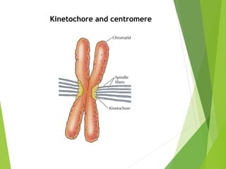
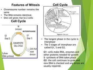
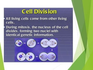
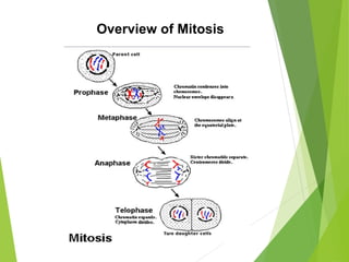
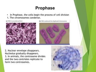
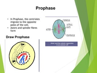
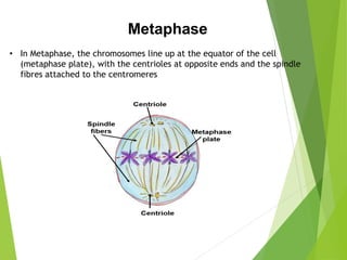
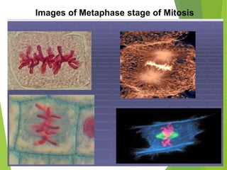
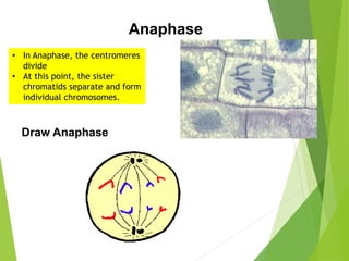
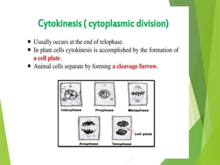
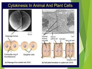
THE MITOTIC CELL CYCLE
PREPARED BY
Dr. S.SELVAKUMARI
5.1 Replication and division of nuclei and cells
a. Describe the structure of a chromosome, limited to DNA, histone proteins,
chromatids, centromere and telomeres.
b. Explain the importance of mitosis in the production of genetically identical cells,
growth, cell replacement, repair of tissues and asexual reproduction.
c. Outline the cell cycle, including interphase (growth in G1 and G2 phases and DNA
replication in S phase), mitosis and cytokinesis.
d. Outline the significance of telomeres in permitting continued replication and
preventing the loss of genes.
e. Outline the significance of stem cells in cell replacement and tissue repair by
mitosis and state that uncontrolled cell division can result in the formation of a
tumor.
4.2 Chromosome behavior in mitosis
a. Describe, with the aid of photomicrographs and diagrams, the behavior of
chromosomes in plant and animal cells during the mitotic cell cycle and the
associated behavior of the nuclear envelope, cell surface membrane and the
spindle (names of the main stages of mitosis are expected).
b. Observe and draw the mitotic stages visible in temporary root tip squash
preparations and in prepared slides of root tips of species such as those of Vicia
faba and Allium cepa.
• Chromosomes controls cell activities
• Are copied from cell to cell when cells divide
Chromatin – at normal time Chromosome – at cell division time
Chromosome and DNA
• Position of centromere is fixed for any
chromosome and the length of a chromosome
is also characteristic
• The number of chromosomes per species is
fixed
• Chromosomes occurs in pairs
• Chromosomes are copied between nuclear
divisions.
Chromosome and DNA
1.8 meters of DNA
has to be packed
into a nucleus of 6
µm diameter.
A combination of
DNA and proteins is
called chromatin.
• DNA: DeoxyriboNucleic Acid RNA: RiboNucleic Acid
• Bases: Adenine (A) and Guanine (G) = Purines
Cytosine (C) & Thymine (T) = Pyrimidines
• RNA has Uracil instead of Thymine
• Base sugar = Nucleoside (Deoxyribonucleoside & Ribonucleoside)
• Base sugar and phosphate unit = nucleotide . Many form a polynucleotide
• Phosphodiester bonds = link nucleotides for nucleic acid polymer
• Hydrogen bonding between paired bases
• Each chain is complimentary
Pentose sugar
Ribose
Deoxyribose
Phosphate
group
Phosphodiester
bond
DNA Packaging
DNA replication
DNA Double Helix
Minor GrooveMajor Groove
RNA
• DNA transcribed into mRNA by RNA polymerase
• RNA used as a primer in DNA replication
• Ribosomal RNA
• tRNA aids in translation of mRNA into protein
• Chromatin –
DNA bound to
histones (DNA
packaging
proteins)
• DNA and
histones are
organised into
nucleosomes.
Eight histones
= histone
octamer
• These are
linked by
linker DNA
1. Double helix is unwound and the base-pairs are separated
by DNA helicase.
2. Semi-conservative replication
3. Nucleotides are linked covalently by DNA polymerase
4. Lagging and leading strands
5. DNA ligase for the shorts strands on the lagging strand
PACKAGING OF DNA IN CHROMOSOME
147 bps
Linker DNA- 53 bps
TELOMERE
Kinetochore and centromere
Cell Division Mitosis
Significance of Mitosis
There are three main reasons
A. growth
B. repair/healing
C. asexual reproduction
Cell Division – Two types
• Mitosis– Division of somatic cells
• Meiosis –Creation of new sex cells
Somatic cell – a body cell; a cell whose genes will not be passed on
to future
Germ cell - a cell that is destined to become a gamete (egg or
sperm); a cell whose genes can be passed on to future generations
Diploid (2N) – a cell with 2 chromosome sets in each of its cells; all
body (somatic) cells
Haploid (N) – a cell with 1 chromosome set in each of its cells; all
gametes (sperm, eggs)
Vocabulary
Homologous Chromosomes– A pair of chromosomes that code for
the same types of proteins.
Telomere – end of chromosome which serves to protect it by
permitting continued replication and preventing the loss of genes
Features of Mitosis
• Chromosome number remains the
same
• The DNA remains identical.
• One cell gives rise to 2 cells
Cell Cycle
• The longest phase in the cycle is
interphase
• The 3 stages of interphase are
called G1, S and G2.
G1- cells make RNA, enzymes and
other proteins needed for growth
S- synthesis of DNA takes place
G2- the cell continues to grow and
new DNA is checked and any errors are
usually repaired.
Cell Cycle
Cell Cycle
Overview of Mitosis
• In Prophase, the cells begin the process of cell division
1. The chromosomes condense.
Prophase
2. Nuclear envelope disappears.
Nucleolus gradually disappears.
3. In animals, the centrosome divides
and the two centrioles replicate to
form two centrosomes.
• In Prophase, the centrioles
migrate to the opposite
poles of the cell.
• Asters and spindle fibres
form
Prophase
Draw Prophase
• In Metaphase, the chromosomes line up at the equator of the cell
(metaphase plate), with the centrioles at opposite ends and the spindle
fibres attached to the centromeres
Metaphase
Images of Metaphase stage of Mitosis
• In Anaphase, the centromeres
divide
• At this point, the sister
chromatids separate and form
individual chromosomes.
Anaphase
Draw Anaphase
The Mitotic Cell Cycle

The Mitotic Cell Cycle

  • 1.
    THE MITOTIC CELLCYCLE PREPARED BY Dr. S.SELVAKUMARI
  • 2.
    5.1 Replication anddivision of nuclei and cells a. Describe the structure of a chromosome, limited to DNA, histone proteins, chromatids, centromere and telomeres. b. Explain the importance of mitosis in the production of genetically identical cells, growth, cell replacement, repair of tissues and asexual reproduction. c. Outline the cell cycle, including interphase (growth in G1 and G2 phases and DNA replication in S phase), mitosis and cytokinesis. d. Outline the significance of telomeres in permitting continued replication and preventing the loss of genes. e. Outline the significance of stem cells in cell replacement and tissue repair by mitosis and state that uncontrolled cell division can result in the formation of a tumor. 4.2 Chromosome behavior in mitosis a. Describe, with the aid of photomicrographs and diagrams, the behavior of chromosomes in plant and animal cells during the mitotic cell cycle and the associated behavior of the nuclear envelope, cell surface membrane and the spindle (names of the main stages of mitosis are expected). b. Observe and draw the mitotic stages visible in temporary root tip squash preparations and in prepared slides of root tips of species such as those of Vicia faba and Allium cepa.
  • 3.
    • Chromosomes controlscell activities • Are copied from cell to cell when cells divide Chromatin – at normal time Chromosome – at cell division time Chromosome and DNA
  • 4.
    • Position ofcentromere is fixed for any chromosome and the length of a chromosome is also characteristic • The number of chromosomes per species is fixed • Chromosomes occurs in pairs • Chromosomes are copied between nuclear divisions. Chromosome and DNA 1.8 meters of DNA has to be packed into a nucleus of 6 µm diameter. A combination of DNA and proteins is called chromatin.
  • 5.
    • DNA: DeoxyriboNucleicAcid RNA: RiboNucleic Acid • Bases: Adenine (A) and Guanine (G) = Purines Cytosine (C) & Thymine (T) = Pyrimidines • RNA has Uracil instead of Thymine • Base sugar = Nucleoside (Deoxyribonucleoside & Ribonucleoside) • Base sugar and phosphate unit = nucleotide . Many form a polynucleotide • Phosphodiester bonds = link nucleotides for nucleic acid polymer • Hydrogen bonding between paired bases • Each chain is complimentary Pentose sugar Ribose Deoxyribose Phosphate group Phosphodiester bond DNA Packaging DNA replication DNA Double Helix Minor GrooveMajor Groove RNA • DNA transcribed into mRNA by RNA polymerase • RNA used as a primer in DNA replication • Ribosomal RNA • tRNA aids in translation of mRNA into protein • Chromatin – DNA bound to histones (DNA packaging proteins) • DNA and histones are organised into nucleosomes. Eight histones = histone octamer • These are linked by linker DNA 1. Double helix is unwound and the base-pairs are separated by DNA helicase. 2. Semi-conservative replication 3. Nucleotides are linked covalently by DNA polymerase 4. Lagging and leading strands 5. DNA ligase for the shorts strands on the lagging strand
  • 6.
    PACKAGING OF DNAIN CHROMOSOME 147 bps Linker DNA- 53 bps
  • 7.
  • 10.
  • 11.
    Cell Division Mitosis Significanceof Mitosis There are three main reasons A. growth B. repair/healing C. asexual reproduction
  • 15.
    Cell Division –Two types • Mitosis– Division of somatic cells • Meiosis –Creation of new sex cells Somatic cell – a body cell; a cell whose genes will not be passed on to future Germ cell - a cell that is destined to become a gamete (egg or sperm); a cell whose genes can be passed on to future generations Diploid (2N) – a cell with 2 chromosome sets in each of its cells; all body (somatic) cells Haploid (N) – a cell with 1 chromosome set in each of its cells; all gametes (sperm, eggs) Vocabulary Homologous Chromosomes– A pair of chromosomes that code for the same types of proteins. Telomere – end of chromosome which serves to protect it by permitting continued replication and preventing the loss of genes
  • 16.
    Features of Mitosis •Chromosome number remains the same • The DNA remains identical. • One cell gives rise to 2 cells Cell Cycle • The longest phase in the cycle is interphase • The 3 stages of interphase are called G1, S and G2. G1- cells make RNA, enzymes and other proteins needed for growth S- synthesis of DNA takes place G2- the cell continues to grow and new DNA is checked and any errors are usually repaired. Cell Cycle
  • 17.
  • 19.
  • 20.
    • In Prophase,the cells begin the process of cell division 1. The chromosomes condense. Prophase 2. Nuclear envelope disappears. Nucleolus gradually disappears. 3. In animals, the centrosome divides and the two centrioles replicate to form two centrosomes.
  • 21.
    • In Prophase,the centrioles migrate to the opposite poles of the cell. • Asters and spindle fibres form Prophase Draw Prophase
  • 22.
    • In Metaphase,the chromosomes line up at the equator of the cell (metaphase plate), with the centrioles at opposite ends and the spindle fibres attached to the centromeres Metaphase
  • 23.
    Images of Metaphasestage of Mitosis
  • 24.
    • In Anaphase,the centromeres divide • At this point, the sister chromatids separate and form individual chromosomes. Anaphase Draw Anaphase