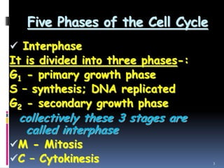
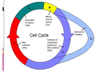
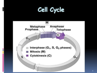
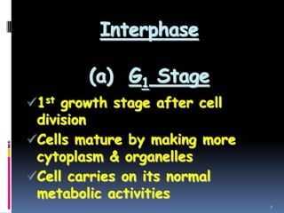
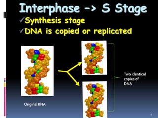
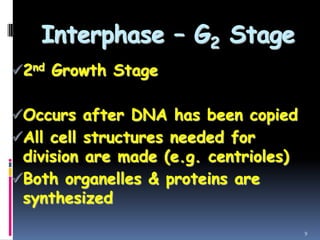
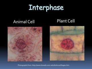
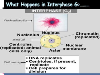
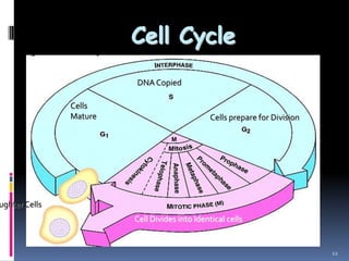
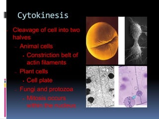
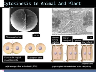
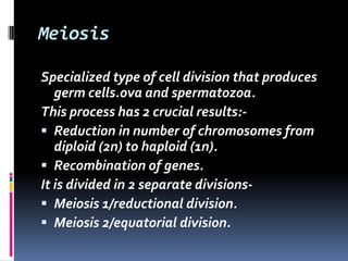
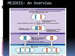
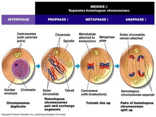
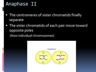
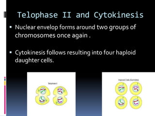
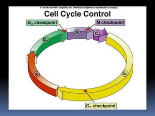
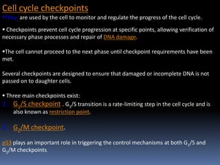
The document describes the cell cycle and its various phases. It begins by defining the cell cycle as the sequence of events a cell undergoes from formation after division of a parent cell until its own division into daughter cells. The cell cycle consists of interphase and the M phase. Interphase includes the G1, S, and G2 phases where the cell grows and duplicates its DNA. The M phase encompasses mitosis and cytokinesis where the cell divides into two daughter cells. Meiosis is also discussed, which produces gametes through two cell divisions and a reduction in chromosome number from diploid to haploid.
This slide describes the various stages of the Eukaryotic cell cycle. The diagrams included here explains the various changes that take place during the mitotic division of a eukaryotic cell.
Presentation include Nucleus and its components like nuclear envelope, nucleolus, chromatin fibers, ultra structure of nucleus and its general functions.
a deeply explained process of cell division, for understanding it thoroughly. i tried to put in all the information i knew and collected. i hope it is helpful or you.
Chapter-6Cell Cycle and DivisionCell Divisio.docxchristinemaritza
Chapter-6
Cell Cycle and Division
Cell Division
Cells reproduce by cell division, in which a parent cell normally gives rise to two daughter cells
Each daughter cell receives a complete set of hereditary information (DNA) from the parent cell and about half its cytoplasm
The hereditary information DNA is usually identical with that of the parent cell
The cell division of eukaryotic cells by which organisms grow or increase in number is called mitotic cell division
After cell division, the daughter cells may differentiate, becoming specialized for specific functions
The repeating pattern of divide, grow, and differentiate, then divide again is called the cell cycle
Most multicellular organisms have three categories of cells
1. stem cells
2. Other cells capable of dividing
3. Permanently differentiated cells
1.Stem cells :
- have two important characteristics: self-renewal, and the ability to differentiate into a variety of cell types
-Stem cells self-renew because they retain the ability to divide, perhaps for the entire life of the organism
-Some stem cells in early embryos can produce any of the specialized cell types of the entire body
2. Other cells capable of dividing
-Some cells other than stem cells are capable of continuing to divide, but typically differentiate into only one or two different cell types
-Dividing liver cells, for example, can only become more liver cells
3. Permanently differentiated cells
-Permanently differentiated cells differentiate and never divide again
-For example, most heart and brain cells cannot divide
CELL CYCLE
Both prokaryotic and eukaryotic cells have cell cycles that include growth, metabolic activity, DNA replication, and cell division
However, they have major structural and functional differences
Eukaryotic chromosome
Eukaryotic chromosomes are separated from the cytoplasm by a membrane-bound nucleus
Eukaryotic cells always have multiple chromosomes
Eukaryotic chromosomes are longer and have more DNA than prokaryotic chromosomes (human chromosomes are 10 to 80 times longer and have 10 to 50 times more DNA)
Genes
Genes are segments of the DNA of a chromosome
Genes are sequences of DNA from hundreds to thousands of nucleotides long
Each gene occupies a specific place, or locus (plural, loci) on the chromosome
Two important parts of chromosome
Two telomeres
One centromere
It temporarily holds two daughter DNA double helices together after DNA replication
It is the attachment site for microtubules that move the chromosomes during cell division
Homologous
11
Chromosomes that contain the same genes are called homologous chromosomes, or homologues
Cells with pairs of homologous chromosomes are called diploid, which means “double”
Cells with half the number of chromosomes are called haploid
Human Chromosomes
A typical human cell has ...
Infer the significance of cell division.
Differentiate a DNA molecule, a chromosome, and a chromatid.
Characterize the phases of the cell cycle and their control points.
Describe the major events associated with stages of mitosis.
Explain the process of cytokinesis.
Learning Objectives
Describe the role of apoptosis in the life cycle of a cell.
Relate cancer as a result of the malfunction of the cell during the cell cycle.
Title: Sense of Smell
Presenter: Dr. Faiza, Assistant Professor of Physiology
Qualifications:
MBBS (Best Graduate, AIMC Lahore)
FCPS Physiology
ICMT, CHPE, DHPE (STMU)
MPH (GC University, Faisalabad)
MBA (Virtual University of Pakistan)
Learning Objectives:
Describe the primary categories of smells and the concept of odor blindness.
Explain the structure and location of the olfactory membrane and mucosa, including the types and roles of cells involved in olfaction.
Describe the pathway and mechanisms of olfactory signal transmission from the olfactory receptors to the brain.
Illustrate the biochemical cascade triggered by odorant binding to olfactory receptors, including the role of G-proteins and second messengers in generating an action potential.
Identify different types of olfactory disorders such as anosmia, hyposmia, hyperosmia, and dysosmia, including their potential causes.
Key Topics:
Olfactory Genes:
3% of the human genome accounts for olfactory genes.
400 genes for odorant receptors.
Olfactory Membrane:
Located in the superior part of the nasal cavity.
Medially: Folds downward along the superior septum.
Laterally: Folds over the superior turbinate and upper surface of the middle turbinate.
Total surface area: 5-10 square centimeters.
Olfactory Mucosa:
Olfactory Cells: Bipolar nerve cells derived from the CNS (100 million), with 4-25 olfactory cilia per cell.
Sustentacular Cells: Produce mucus and maintain ionic and molecular environment.
Basal Cells: Replace worn-out olfactory cells with an average lifespan of 1-2 months.
Bowman’s Gland: Secretes mucus.
Stimulation of Olfactory Cells:
Odorant dissolves in mucus and attaches to receptors on olfactory cilia.
Involves a cascade effect through G-proteins and second messengers, leading to depolarization and action potential generation in the olfactory nerve.
Quality of a Good Odorant:
Small (3-20 Carbon atoms), volatile, water-soluble, and lipid-soluble.
Facilitated by odorant-binding proteins in mucus.
Membrane Potential and Action Potential:
Resting membrane potential: -55mV.
Action potential frequency in the olfactory nerve increases with odorant strength.
Adaptation Towards the Sense of Smell:
Rapid adaptation within the first second, with further slow adaptation.
Psychological adaptation greater than receptor adaptation, involving feedback inhibition from the central nervous system.
Primary Sensations of Smell:
Camphoraceous, Musky, Floral, Pepperminty, Ethereal, Pungent, Putrid.
Odor Detection Threshold:
Examples: Hydrogen sulfide (0.0005 ppm), Methyl-mercaptan (0.002 ppm).
Some toxic substances are odorless at lethal concentrations.
Characteristics of Smell:
Odor blindness for single substances due to lack of appropriate receptor protein.
Behavioral and emotional influences of smell.
Transmission of Olfactory Signals:
From olfactory cells to glomeruli in the olfactory bulb, involving lateral inhibition.
Primitive, less old, and new olfactory systems with different path
Knee anatomy and clinical tests 2024.pdfvimalpl1234
This includes all relevant anatomy and clinical tests compiled from standard textbooks, Campbell,netter etc..It is comprehensive and best suited for orthopaedicians and orthopaedic residents.
New Drug Discovery and Development .....NEHA GUPTA
The "New Drug Discovery and Development" process involves the identification, design, testing, and manufacturing of novel pharmaceutical compounds with the aim of introducing new and improved treatments for various medical conditions. This comprehensive endeavor encompasses various stages, including target identification, preclinical studies, clinical trials, regulatory approval, and post-market surveillance. It involves multidisciplinary collaboration among scientists, researchers, clinicians, regulatory experts, and pharmaceutical companies to bring innovative therapies to market and address unmet medical needs.
HOT NEW PRODUCT! BIG SALES FAST SHIPPING NOW FROM CHINA!! EU KU DB BK substit...GL Anaacs
Contact us if you are interested:
Email / Skype : kefaya1771@gmail.com
Threema: PXHY5PDH
New BATCH Ku !!! MUCH IN DEMAND FAST SALE EVERY BATCH HAPPY GOOD EFFECT BIG BATCH !
Contact me on Threema or skype to start big business!!
Hot-sale products:
NEW HOT EUTYLONE WHITE CRYSTAL!!
5cl-adba precursor (semi finished )
5cl-adba raw materials
ADBB precursor (semi finished )
ADBB raw materials
APVP powder
5fadb/4f-adb
Jwh018 / Jwh210
Eutylone crystal
Protonitazene (hydrochloride) CAS: 119276-01-6
Flubrotizolam CAS: 57801-95-3
Metonitazene CAS: 14680-51-4
Payment terms: Western Union,MoneyGram,Bitcoin or USDT.
Deliver Time: Usually 7-15days
Shipping method: FedEx, TNT, DHL,UPS etc.Our deliveries are 100% safe, fast, reliable and discreet.
Samples will be sent for your evaluation!If you are interested in, please contact me, let's talk details.
We specializes in exporting high quality Research chemical, medical intermediate, Pharmaceutical chemicals and so on. Products are exported to USA, Canada, France, Korea, Japan,Russia, Southeast Asia and other countries.
Ozempic: Preoperative Management of Patients on GLP-1 Receptor Agonists Saeid Safari
Preoperative Management of Patients on GLP-1 Receptor Agonists like Ozempic and Semiglutide
ASA GUIDELINE
NYSORA Guideline
2 Case Reports of Gastric Ultrasound
Flu Vaccine Alert in Bangalore Karnatakaaddon Scans
As flu season approaches, health officials in Bangalore, Karnataka, are urging residents to get their flu vaccinations. The seasonal flu, while common, can lead to severe health complications, particularly for vulnerable populations such as young children, the elderly, and those with underlying health conditions.
Dr. Vidisha Kumari, a leading epidemiologist in Bangalore, emphasizes the importance of getting vaccinated. "The flu vaccine is our best defense against the influenza virus. It not only protects individuals but also helps prevent the spread of the virus in our communities," he says.
This year, the flu season is expected to coincide with a potential increase in other respiratory illnesses. The Karnataka Health Department has launched an awareness campaign highlighting the significance of flu vaccinations. They have set up multiple vaccination centers across Bangalore, making it convenient for residents to receive their shots.
To encourage widespread vaccination, the government is also collaborating with local schools, workplaces, and community centers to facilitate vaccination drives. Special attention is being given to ensuring that the vaccine is accessible to all, including marginalized communities who may have limited access to healthcare.
Residents are reminded that the flu vaccine is safe and effective. Common side effects are mild and may include soreness at the injection site, mild fever, or muscle aches. These side effects are generally short-lived and far less severe than the flu itself.
Healthcare providers are also stressing the importance of continuing COVID-19 precautions. Wearing masks, practicing good hand hygiene, and maintaining social distancing are still crucial, especially in crowded places.
Protect yourself and your loved ones by getting vaccinated. Together, we can help keep Bangalore healthy and safe this flu season. For more information on vaccination centers and schedules, residents can visit the Karnataka Health Department’s official website or follow their social media pages.
Stay informed, stay safe, and get your flu shot today!
Basavarajeeyam is an important text for ayurvedic physician belonging to andhra pradehs. It is a popular compendium in various parts of our country as well as in andhra pradesh. The content of the text was presented in sanskrit and telugu language (Bilingual). One of the most famous book in ayurvedic pharmaceutics and therapeutics. This book contains 25 chapters called as prakaranas. Many rasaoushadis were explained, pioneer of dhatu druti, nadi pareeksha, mutra pareeksha etc. Belongs to the period of 15-16 century. New diseases like upadamsha, phiranga rogas are explained.
Title: Sense of Taste
Presenter: Dr. Faiza, Assistant Professor of Physiology
Qualifications:
MBBS (Best Graduate, AIMC Lahore)
FCPS Physiology
ICMT, CHPE, DHPE (STMU)
MPH (GC University, Faisalabad)
MBA (Virtual University of Pakistan)
Learning Objectives:
Describe the structure and function of taste buds.
Describe the relationship between the taste threshold and taste index of common substances.
Explain the chemical basis and signal transduction of taste perception for each type of primary taste sensation.
Recognize different abnormalities of taste perception and their causes.
Key Topics:
Significance of Taste Sensation:
Differentiation between pleasant and harmful food
Influence on behavior
Selection of food based on metabolic needs
Receptors of Taste:
Taste buds on the tongue
Influence of sense of smell, texture of food, and pain stimulation (e.g., by pepper)
Primary and Secondary Taste Sensations:
Primary taste sensations: Sweet, Sour, Salty, Bitter, Umami
Chemical basis and signal transduction mechanisms for each taste
Taste Threshold and Index:
Taste threshold values for Sweet (sucrose), Salty (NaCl), Sour (HCl), and Bitter (Quinine)
Taste index relationship: Inversely proportional to taste threshold
Taste Blindness:
Inability to taste certain substances, particularly thiourea compounds
Example: Phenylthiocarbamide
Structure and Function of Taste Buds:
Composition: Epithelial cells, Sustentacular/Supporting cells, Taste cells, Basal cells
Features: Taste pores, Taste hairs/microvilli, and Taste nerve fibers
Location of Taste Buds:
Found in papillae of the tongue (Fungiform, Circumvallate, Foliate)
Also present on the palate, tonsillar pillars, epiglottis, and proximal esophagus
Mechanism of Taste Stimulation:
Interaction of taste substances with receptors on microvilli
Signal transduction pathways for Umami, Sweet, Bitter, Sour, and Salty tastes
Taste Sensitivity and Adaptation:
Decrease in sensitivity with age
Rapid adaptation of taste sensation
Role of Saliva in Taste:
Dissolution of tastants to reach receptors
Washing away the stimulus
Taste Preferences and Aversions:
Mechanisms behind taste preference and aversion
Influence of receptors and neural pathways
Impact of Sensory Nerve Damage:
Degeneration of taste buds if the sensory nerve fiber is cut
Abnormalities of Taste Detection:
Conditions: Ageusia, Hypogeusia, Dysgeusia (parageusia)
Causes: Nerve damage, neurological disorders, infections, poor oral hygiene, adverse drug effects, deficiencies, aging, tobacco use, altered neurotransmitter levels
Neurotransmitters and Taste Threshold:
Effects of serotonin (5-HT) and norepinephrine (NE) on taste sensitivity
Supertasters:
25% of the population with heightened sensitivity to taste, especially bitterness
Increased number of fungiform papillae
Report Back from SGO 2024: What’s the Latest in Cervical Cancer?bkling
Are you curious about what’s new in cervical cancer research or unsure what the findings mean? Join Dr. Emily Ko, a gynecologic oncologist at Penn Medicine, to learn about the latest updates from the Society of Gynecologic Oncology (SGO) 2024 Annual Meeting on Women’s Cancer. Dr. Ko will discuss what the research presented at the conference means for you and answer your questions about the new developments.
TEST BANK for Operations Management, 14th Edition by William J. Stevenson, Ve...kevinkariuki227
TEST BANK for Operations Management, 14th Edition by William J. Stevenson, Verified Chapters 1 - 19, Complete Newest Version.pdf
MANAGEMENT OF ATRIOVENTRICULAR CONDUCTION BLOCK.pdfJim Jacob Roy
Cardiac conduction defects can occur due to various causes.
Atrioventricular conduction blocks ( AV blocks ) are classified into 3 types.
This document describes the acute management of AV block.
Tom Selleck Health: A Comprehensive Look at the Iconic Actor’s Wellness Journeygreendigital
Tom Selleck, an enduring figure in Hollywood. has captivated audiences for decades with his rugged charm, iconic moustache. and memorable roles in television and film. From his breakout role as Thomas Magnum in Magnum P.I. to his current portrayal of Frank Reagan in Blue Bloods. Selleck's career has spanned over 50 years. But beyond his professional achievements. fans have often been curious about Tom Selleck Health. especially as he has aged in the public eye.
Follow us on: Pinterest
Introduction
Many have been interested in Tom Selleck health. not only because of his enduring presence on screen but also because of the challenges. and lifestyle choices he has faced and made over the years. This article delves into the various aspects of Tom Selleck health. exploring his fitness regimen, diet, mental health. and the challenges he has encountered as he ages. We'll look at how he maintains his well-being. the health issues he has faced, and his approach to ageing .
Early Life and Career
Childhood and Athletic Beginnings
Tom Selleck was born on January 29, 1945, in Detroit, Michigan, and grew up in Sherman Oaks, California. From an early age, he was involved in sports, particularly basketball. which played a significant role in his physical development. His athletic pursuits continued into college. where he attended the University of Southern California (USC) on a basketball scholarship. This early involvement in sports laid a strong foundation for his physical health and disciplined lifestyle.
Transition to Acting
Selleck's transition from an athlete to an actor came with its physical demands. His first significant role in "Magnum P.I." required him to perform various stunts and maintain a fit appearance. This role, which he played from 1980 to 1988. necessitated a rigorous fitness routine to meet the show's demands. setting the stage for his long-term commitment to health and wellness.
Fitness Regimen
Workout Routine
Tom Selleck health and fitness regimen has evolved. adapting to his changing roles and age. During his "Magnum, P.I." days. Selleck's workouts were intense and focused on building and maintaining muscle mass. His routine included weightlifting, cardiovascular exercises. and specific training for the stunts he performed on the show.
Selleck adjusted his fitness routine as he aged to suit his body's needs. Today, his workouts focus on maintaining flexibility, strength, and cardiovascular health. He incorporates low-impact exercises such as swimming, walking, and light weightlifting. This balanced approach helps him stay fit without putting undue strain on his joints and muscles.
Importance of Flexibility and Mobility
In recent years, Selleck has emphasized the importance of flexibility and mobility in his fitness regimen. Understanding the natural decline in muscle mass and joint flexibility with age. he includes stretching and yoga in his routine. These practices help prevent injuries, improve posture, and maintain mobilit
2. CELL CYCLE
Described by Howard and Pele in 1953
Cell Cycle is defined as “The sequence of
events involving growth and division, a cell
undergoes from the time of its formation by
division of parent cell to its own division into
daughter cells .”
3. Five Phases of the Cell Cycle
Interphase
It is divided into three phases-:
G1 - primary growth phase
S – synthesis; DNA replicated
G2 - secondary growth phase
collectively these 3 stages are
called interphase
M - Mitosis
C – Cytokinesis 3
5. CONCEPT OF G0 PHASE-
Nonproliferative cells in eukaryotes generally enter
the quiescent G0 state from G1 and remain quiescent
for long periods of time, possibly indefinitely , e.g.
neurons
Cellular senescence occurs in response to DNA
damage or degradation that would make a cell's
progeny nonviable.
Some cells enter the G0 phase for a short period of
time e.g., liver and kidney cells.
Many cells do not enter G0 and continue to divide
throughout an organism's life, e.g. epithelial cells
7. Interphase
(a) G1 Stage
1st growth stage after cell
division
Cells mature by making more
cytoplasm & organelles
Cell carries on its normal
metabolic activities
7
8. Interphase –> S Stage
Synthesis stage
DNA is copied or replicated
8
Two identical
copies of
DNA
Original DNA
9. Interphase – G2 Stage
2nd Growth Stage
Occurs after DNA has been copied
All cell structures needed for
division are made (e.g. centrioles)
Both organelles & proteins are
synthesized
9
13. Chromosomes
All eukaryotic cells store genetic information
in chromosomes.
Human cells have 46 chromosomes.
23 nearly-identical pairs
14. Structure of Chromosomes
Chromosomes are composed of a complex of DNA
and protein called chromatin that condenses
during cell division
DNA exists as a single, long, double-stranded fiber
extending chromosome’s entire length.
Each unduplicated chromosome contains one DNA
molecule, which may be several inches long
15. After every 200 nucleotide pairs, the DNA wraps twice around a
group of 8 histone proteins to form a nucleosome.
Higher order coiling and supercoiling also helps in condensing
and packaging the chromatin inside the nucleus.
Structure of Chromosomes
16. Chromosomes
Non-homologous chromosomes
Look different
Control different traits
Sex chromosomes
Are distinct from each other in their
characteristics
Are represented as X andY
Determine the sex of the individual, XX being
female, XY being male
In a diploid cell, the chromosomes occur in pairs.
The 2 members of each pair are called
homologous chromosomes or homologues.
17. Chromosomes
A diploid cell has two sets of each of its chromosomes
In human being 46 chromosomes are present(2n = 46)
In a cell in which DNA synthesis has occurred all the chromosomes are
duplicated and thus each chromosome consists of two identical sister
chromatids
Maternal set of
chromosomes
Paternal set of
chromosomes
2n = 6
Two sister chromatids
of one replicated
chromosome
Two nonsister
chromatids in
a homologous pair
Pair of homologous
chromosomes
(one from each set)
Centromere
18. Homologues Chromosomes
Homologous chromosomes:
• Look the same
• Control the same traits
• May code for different forms of each trait
• Independent origin - each one was inherited from a
different parent
19. Chromosome Duplication
0.5 µm
Chromosome
duplication
(including DNA
synthesis)
Centromere
Separation
of sister
chromatids
Sister
chromatids
Centrometers
Sister chromatids
A eukaryotic cell has multiple
chromosomes, one of which is
represented here. Before
duplication, each chromosome
has a single DNA molecule.
Once duplicated, a chromosome
consists of two sister chromatids
connected at the centromere. Each
chromatid contains a copy of the
DNA molecule.
Mechanical processes separate
the sister chromatids into two
chromosomes and distribute
them to two daughter cells.
In preparation for cell division, DNA is replicated and the chromosomes condense
22. Structure of Chromosomes
Diploid - A cell possessing two copies of each chromosome
(human body cells).
Homologous chromosomes are made up of sister chromatids
joined at the centromere.
Haploid - A cell possessing a single copy of each chromosome
(human sex cells).
23. KARYOTYPE it is a
chromosome complement
of a cell or organism
depicting the number , size
and form of the
chromosome as seen in
metaphase of mitosis.
Chromosomes are
assembled as homologous
pairs in decreasing order of
length.
24. CELL DIVISION
Cell division involves a single cell (called a mother
cell) dividing into two daughter cells.This leads to
growth in multicellular organisms (the growth of
tissue) and to procreation (vegetative
reproduction) in unicellular organisms.
Prokaryotic cells divide by binary fission.
Eukaryotic cells usually undergo a process of
nuclear division, called mitosis, followed by
division of the cell, called cytokinesis.
A diploid cell may also undergo meiosis to
produce haploid cells, usually four. Haploid cells
serve as gametes in multicellular organisms, fusing
25. DNA replication, or the process of duplicating
a cell's genome, is required every time a cell
divides. Replication, like all cellular activities,
requires specialized proteins for carrying out
the job.
26. Protein synthesis
Cells are capable of synthesizing new
proteins, which are essential for the
modulation and maintenance of cellular
activities.
This process involves the formation of new
protein molecules from amino acid building
blocks based on information encoded in
DNA/RNA.
Protein synthesis generally consists of two
major steps: transcription and translation.
27. Transcription
Process by which genetic information encoded in DNA is
copied onto messenger RNA
Occurs in the nucleus
DNA mRNA
Translation
Process by which information encoded in mRNA is used
to assemble a protein at a ribosome
Occurs on a Ribosome
mRNA protein
29. AMITOSIS
Amitosis is very simple, it occurs without the
formation of spindle and appearance of
chromosome
Nuclear envelope remains intact.
E.g – Macromolecules of ciliates such as
paramecium.
In each case division of nucleus called
KARYOGENESIS, occurs before the division of
cytoplasm termed CYTOKINESIS
30. MITOSIS
MITOSIS was 1ST described by GERMAN BIOLOGIST
EDUARD STRASBURGER in plant cell in1875
and later by another GERMAN BIOLOGISTWALTHER
FLEMMING in animal cell 1879.
TERMED MITOSIS byWALTHER FLEMMING in 1882.
DEFINITION
It is defined as" the division of a parent cell into 2
identical daughter cells each with a nucleus having
the same amount of DNA, the same number and
kind of chromosomes and same hereditary
instructions as parent cell.”
Hence also known as EQUATIONAL DIVISION.
31. INTERPHASE
Period between two successive division.
Greater part of interphase is called G1 stage.
S Stage: - Synthesis of DNA takes place.
G2: - Protein synthesis takes placed required
for cell division.
32. MECHANISM OF MITOSIS
It involve a series of changes in nucleus as
well as cytoplasm
Therefore often called as indirect division
Two main events in mitosis -:
Karyokinesis
Cytokinesis
33. Karyokinesis may be divided into four stages
PROPHASE
METAPHASE
ANAPHASE
TELOPHASE
34.
35. STAGES OF MITOSIS
EARLY PROPHASE
Early in the prophase stage the chromatin fibers shorten into
chromosomes
Each prophase chromosome consists of a pair of identical double-
stranded chromatids.
36. LATE PROPHASE
Nucleolus disappears, the nuclear envelope breaks down,
and the two centrosomes begin to form the mitotic spindle
(which is an assembly of microtubules).
As the microtubules extend in length between the
centrosomes, the centrosomes are pushed to opposite "poles"
(extremes) of the cell.
Eventually, the spindle extends between two opposite poles
of the cell.
39. METAPHASE
CHROMOSOME CONDENSATIONCONTINUES INTO
METAPHASE.
MOST STRIKING FEATURE-CHROMOSOMES BECOME
ALIGNEDWITHTHERECENTOMERES IN A SINGLE
TRANSVERSE PLANE.
THIS PLANE LIES PERPENDICULARTOTHE LONG AXIS
OF SPINDLE AND IS KNOWN AS EQUTORIAL PLATE.
OFTEN KNOWN AS METAPHASE PLATE.
41. ANAPHASE
The centromeres split separating the two members of
each chromatid pair - which then move to the
opposite poles of the cell.
When they are separated , the chromatids are
now called as chromosomes.
As the chromosomes are pulled by the microtubules
during anaphase, they appear to be "V"-shaped
because the centromeres lead the way, dragging the
trailing arms of the chromosomes towards the poles
44. TELOPHASE
Telophase begins after the chromosomal movement stops.
The identical sets of chromosomes - which are by this stage at
opposite poles of the cell, uncoil and revert to the long, thin,
thread-like chromatin form.
A new nuclear envelope forms around each chromatin mass.
Nucleoli appear.
Eventually the miotic spindle breaks-up.
45. Cytokinesis
• Cleavage of cell into two
halves
– Animal cells
Constriction belt of
actin filaments
– Plant cells
Cell plate
– Fungi and protozoa
Mitosis occurs
within the nucleus
46. Cytokinesis In Animal And Plant
Cells
Daughter cells
Cleavage furrow
Contractile ring of
microfilaments
Daughter cells
100 µm
1 µmVesicles
forming
cell plate
Wall of
patent cell Cell plate
New cell wall
(a) Cleavage of an animal cell (SEM) (b) Cell plate formation in a plant cell (SEM)
47. Meiosis
Specialized type of cell division that produces
germ cells.ova and spermatozoa.
This process has 2 crucial results:-
Reduction in number of chromosomes from
diploid (2n) to haploid (1n).
Recombination of genes.
It is divided in 2 separate divisions-
Meiosis 1/reductional division.
Meiosis 2/equatorial division.
49. MEIOSIS I
Amount of DNA is doubled to
4n and the chromosome
number is also doubled to 4n
during S phase.
50. Prophase 1 -:
PROPHASE 1 is subdivided into following 5
phases -:
1. Leptotene
2. Zygotene
3. Pachytene
4. Diplotene
5. Diakinesis
51. 1. LEPTOTENE
Chromosomes condense forming
long strands.
Chromosome become visible each
chromosomes consists of two
chromatids.
At first the chromosomes are seen as
threads bearing bead thickening
(chromosome) along their length.
52. 2.ZYGOTENE -:
Pairing of chromosome also referred as
synapsis, takes place.
Synaptonemal complex is formed by a pair of
synapsed homologous chromosomes i called a
bivalent or a tetrad.
53. 3. PACHYTENE
Chiasmata (crossing over sites) start
forming
2 chromatid of each chromosomes
become distinct.The bivalent now has
formed chromatids in it called aTetrad
which is clearly visible at this stage.
Two central chromatids becomes
coiled over each other so that they
cross at number of points called
crossing over.
54. 4.DIPLOTENE
Dissolution of the synaptonemal complex
Chromosome continue to condense and then begin
to separate, revealing X shaped structures, called
chiasmata.
2 chromosomes of bivalent now try to move apart
as they do so chromatid break at the point of
crossing.
55. DIAKINESIS
The final stage of meiotic prophase I.
Marked by terminalisation of chiasmata
Nucleolus and nuclear envelop disappear, freeing
the chromosomes into the cytoplasm.
Diakinesis represents transition to metaphase- 1
57. METAPHASE 1-
Homologus chromosomes align
as pairs on the equatorial plate of
spindle apparatus.
Spindle Equatorial
58. ANAPHASE 1-
Homologous chromosomes migrate away from each
other going to opposite poles.
Sister chromatids still remain associated at their
centromeres
59. TELOPHASE 1-
Nuclear membrane and
nucleolus reappear .
Nuclei are formed
Cytokinesis occur giving rise
to 2 daughter cells.
60.
61. The stage between the two meiotic divisions
is called interkinesis and is generally short
lived.
Interkinesis is followed by prophase II, a
much simpler prophase than prophase I.
63. Meiosis II : Separates sister
chromatids
Proceeds similar to mitosis
THERE IS NO INTERPHASE II !
64. Prophase II
A spindle apparatus starts forming
The nuclear membrane disappears by the
end of prophase II .
The chromosomes again become
compact.
65. Metaphase II
The chromosomes are positioned on the
metaphase plate in a mitosis-like fashion
Kinetochores of sister chromatids of each
chromosome pointing toward opposite poles
66. Anaphase II
The centromeres of sister chromatids finally
separate
The sister chromatids of each pair move toward
opposite poles
(Now individual chromosomes)
67. Telophase II and Cytokinesis
Nuclear envelop forms around two groups of
chromosomes once again .
Cytokinesis follows resulting into four haploid
daughter cells.
Haploid cells (n)
68.
69. Cell cycle checkpoints
They are used by the cell to monitor and regulate the progress of the cell cycle.
Checkpoints prevent cell cycle progression at specific points, allowing verification of
necessary phase processes and repair of DNA damage.
The cell cannot proceed to the next phase until checkpoint requirements have been
met.
Several checkpoints are designed to ensure that damaged or incomplete DNA is not
passed on to daughter cells.
Three main checkpoints exist:
1. G1/S checkpoint . G1/S transition is a rate-limiting step in the cell cycle and is
also known as restriction point.
2. G2/M checkpoint.
p53 plays an important role in triggering the control mechanisms at both G1/S and
G2/M checkpoints.
70. 3. Spindle assembly checkpoint, between
metaphase and anaphase
Cell cycle can be arrested if spindle fibers are
not attached properly to chromatids
71.
72. Regulation of the Cell Cycle
Cyclin: major control switch for the cell cycle
Cdk (Cyclin-dependent kinase (phospohorylation)): major control switch; activated by cyclin;
causesG1 S or G2 M.
Cyclins form the regulatory subunit and Cdks the catalytic subunit
Checkpoints
DNA damage checkpoints, including tumor suppressor genes
p53: protein that blocks the cell cycle if DNA is damaged and can cause apoptosis.A p53 mutation is the most
frequent mutation leading to cancer.
Spindle checkpoints
73. Two families of proteins are involved in involved in the regulation
process—
i) Cyclin-dependent protein kinases (Cdks)
ii) Cyclins.
a)Cyclin-Dependent Protein Kinase (Cdks)
A Cdks is an enzyme that adds negatively charged phosphate groups to other molecules in a
process called phosphorylation.
Through phosphorylation, Cdks signal the cell that it is ready to pass into the next stage of the cell
cycle.
Cyclin-Dependent Protein Kinases are dependent on cyclins & Cyclins bind to Cdks, activating the
Cdks to phosphorylate other molecules.
b)Cyclins
Cyclins are named such because they undergo a constant cycle of synthesis and degradation
during cell division.
When cyclins are synthesized, they act as an activating protein and bind to Cdks forming a cyclin-
Cdk complex.
This complex then acts as a signal to the cell to pass to the next cell cycle phase.
Eventually, the cyclin degrades, deactivating the Cdk, thus signaling exit from a particular phase.