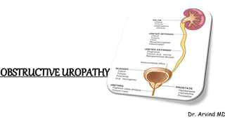
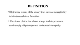
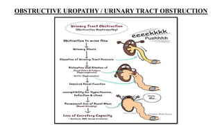
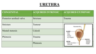
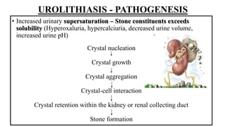
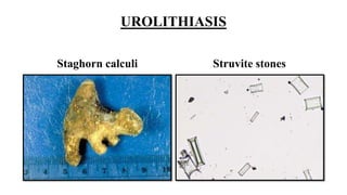
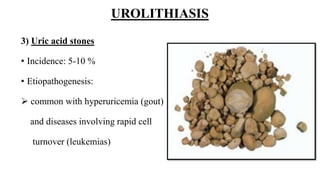
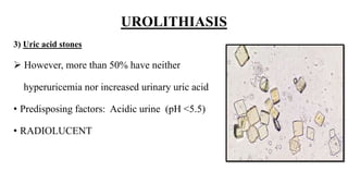
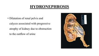
OBSTRUCTIVE UROPATHY
Dr. Arvind MD
OBJECTIVES
Define obstructive uropathy
List the etiology of obstructive uropathy
Discuss the pathogenesis of urolithiasis and hydronephrosis
List the types of renal stones and discuss the etiopathogenesis
Describe the morphology of renal stone
Define hydronephrosis and describe its pathogenesis
DEFINITION
Obstructive lesions of the urinary tract increase susceptibility
to infection and stone formation.
 Unrelieved obstruction almost always leads to permanent
renal atrophy - Hydronephrosis or obstructive uropathy.
OBSTRUCTIVE UROPATHY / URINARY TRACT OBSTRUCTION
OBSTRUCTION
• Sudden/ Insidious
• Partial/ complete
• Unilateral/ Bilateral
• At any level of the urinary
tract
ETIOLOGY - OBSTRUCTIVE LESIONS
URETER/ PELVIS
CONGENITAL ACQUIRED INTRINSIC ACQUIRED EXTRINSIC
PUJ narrowing/obstruction Calculi Pregnant uterus
UVJ narrowing/obstruction Inflammation Retroperitoneal fibrosis
Ureterocele Infection Aortic aneurysm
Retrocaval ureter Trauma Uterine leiomyoma
Sloughed papillae Carcinoma uterus, prostate,
bladder, rectum
Tumour Lymphoma
PID
Accidental surgical ligation
BLADDER OUTLET
CONGENITAL ACQUIRED INTRINSIC ACQUIRED EXTRINSIC
Bladder neck obstruction Benign prostatic hyperplasia Carcinoma cervix
Ureterocele Cancer of prostate, bladder Carcinoma colon
Calculi Trauma
Diabetic neuropathy
Spinal cord disease
Anticholinergic drugs and
Alpha adrenergic
antagonists
URETHRA
CONGENITAL ACQUIRED INTRINSIC ACQUIRED EXTRINSIC
Posterior urethral valve Stricture Trauma
Stricture Tumour
Meatal stenosis Calculi
Phimosis Trauma
Phimosis
UROLITHIASIS
Formed anywhere in the urinary tract, mostly in kidney
M>F
Age: Peak between 20 and 30 years
 Familial and hereditary predisposition; Many inborn errors of
metabolism (like cystinuria & primary hyperoxaluria)
UROLITHIASIS - PATHOGENESIS
• Increased urinary supersaturation – Stone constituents exceeds
solubility (Hyperoxaluria, hypercalciuria, decreased urine volume,
increased urine pH)
Crystal nucleation
Crystal growth
Crystal aggregation
Crystal-cell interaction
Crystal retention within the kidney or renal collecting duct
Stone formation
UROLITHIASIS - PATHOGENESIS
• Stone formation - enhanced by deficiency of inhibitors of crystal
formation in urine.
• These inhibitors for crystal formation are pyrophosphate,
diphosphonate, citrate, glycosaminoglycans, osteopontin and
glycoprotein called nephrocalcin.
UROLITHIASIS
TYPES:
1) Calcium oxalate stones
• Incidence: 70%
• Composition: calcium oxalate (OR)
calcium oxalate + calcium phosphate
UROLITHIASIS -Calcium oxalate stones
• Etiopathogenesis:
Hypercalciuria without hypercalcemia
(55%)
Hypercalciuria and hypercalcemia (5%)
Hyperuricosuria
Hyperoxaluria: primary, enteric
UROLITHIASIS
2) Struvite stones or Triple stones
• Incidence: 15%
• Composition: Magnesium, Ammonium, Phosphate stones
• Etiopathogenesis:
• Urea splitting bacteria (protease, staphylococci) –
convert urea  ammonia  resultant alkaline urine causes precipitation of
magnesium ammonium phosphate salts  Staghorn calculi
UROLITHIASIS
Staghorn calculi Struvite stones
UROLITHIASIS
3) Uric acid stones
• Incidence: 5-10 %
• Etiopathogenesis:
 common with hyperuricemia (gout)
and diseases involving rapid cell
turnover (leukemias)
UROLITHIASIS
3) Uric acid stones
 However, more than 50% have neither
hyperuricemia nor increased urinary uric acid
• Predisposing factors: Acidic urine (pH <5.5)
• RADIOLUCENT
UROLITHIASIS
4) Cystine stones
• Incidence: 1-2%
• Etiopathogenesis: genetic defects
in renal reabsorption of
aminoacids (cystine) 
cystinuria
• Form at low urinary pH
UROLITHIASIS – Clinical features
• Asymptomatic (OR) severe renal colic and abdominal pain (OR)
significant renal damage
• Small stones: may pass through (OR) ureteral obstruction
“loin to groin” pain
• Larger stones: often Haematuria
UROLITHIASIS – Complications
• Hematuria
• Hydronephrosis
• Pyelonephritis and pyonephrosis
HYDRONEPHROSIS
• Dilatation of renal pelvis and
calyces associated with progressive
atrophy of kidney due to obstruction
to the outflow of urine
HYDRONEPHROSIS
HYDRONEPHROSIS
• OBSTRUCTION
-sudden/insidious
- partial/complete
-unilateral/bilateral
This obstruction can occur at any level of the urinary tract.
HYDRONEPHROSIS - Pathogenesis
Obstruction in urinary tract + continued GF(Glomerular filtration)
Dilatation of pelvis and calyces
High pressure in the pelvis that transmits back through collecting duct
Initially: renal vasculature of medulla Later: Cortical atrophy & decreased GFR
is compressed
Diminished inner medullary blood flow
Deranged tubular function in the medulla
(medullary vascular defects initially reversible)
Manifested as: impaired concentrating ability
Dilatation of pelvis and calyces and parenchymal atrophy
HYDRONEPHROSIS
HYDRONEPHROSIS – Clinical features
• Asymptomatic
• Compensated by other kidney
U/L -partial/complete hydronephrosis
• Polyuria & Nocturia
• Due to inability to concentrate the urine (tubular dysfunction)
B/L partial obstruction
• Oliguria or Anuria
B/L complete obstruction
THANK YOU

OBSTRUCTIVE UROPATHY PPT.pptx

  • 1.
  • 2.
    OBJECTIVES Define obstructive uropathy Listthe etiology of obstructive uropathy Discuss the pathogenesis of urolithiasis and hydronephrosis List the types of renal stones and discuss the etiopathogenesis Describe the morphology of renal stone Define hydronephrosis and describe its pathogenesis
  • 3.
    DEFINITION Obstructive lesions ofthe urinary tract increase susceptibility to infection and stone formation.  Unrelieved obstruction almost always leads to permanent renal atrophy - Hydronephrosis or obstructive uropathy.
  • 4.
    OBSTRUCTIVE UROPATHY /URINARY TRACT OBSTRUCTION
  • 5.
    OBSTRUCTION • Sudden/ Insidious •Partial/ complete • Unilateral/ Bilateral • At any level of the urinary tract
  • 6.
  • 7.
    URETER/ PELVIS CONGENITAL ACQUIREDINTRINSIC ACQUIRED EXTRINSIC PUJ narrowing/obstruction Calculi Pregnant uterus UVJ narrowing/obstruction Inflammation Retroperitoneal fibrosis Ureterocele Infection Aortic aneurysm Retrocaval ureter Trauma Uterine leiomyoma Sloughed papillae Carcinoma uterus, prostate, bladder, rectum Tumour Lymphoma PID Accidental surgical ligation
  • 8.
    BLADDER OUTLET CONGENITAL ACQUIREDINTRINSIC ACQUIRED EXTRINSIC Bladder neck obstruction Benign prostatic hyperplasia Carcinoma cervix Ureterocele Cancer of prostate, bladder Carcinoma colon Calculi Trauma Diabetic neuropathy Spinal cord disease Anticholinergic drugs and Alpha adrenergic antagonists
  • 9.
    URETHRA CONGENITAL ACQUIRED INTRINSICACQUIRED EXTRINSIC Posterior urethral valve Stricture Trauma Stricture Tumour Meatal stenosis Calculi Phimosis Trauma Phimosis
  • 10.
    UROLITHIASIS Formed anywhere inthe urinary tract, mostly in kidney M>F Age: Peak between 20 and 30 years  Familial and hereditary predisposition; Many inborn errors of metabolism (like cystinuria & primary hyperoxaluria)
  • 11.
    UROLITHIASIS - PATHOGENESIS •Increased urinary supersaturation – Stone constituents exceeds solubility (Hyperoxaluria, hypercalciuria, decreased urine volume, increased urine pH) Crystal nucleation Crystal growth Crystal aggregation Crystal-cell interaction Crystal retention within the kidney or renal collecting duct Stone formation
  • 12.
    UROLITHIASIS - PATHOGENESIS •Stone formation - enhanced by deficiency of inhibitors of crystal formation in urine. • These inhibitors for crystal formation are pyrophosphate, diphosphonate, citrate, glycosaminoglycans, osteopontin and glycoprotein called nephrocalcin.
  • 13.
    UROLITHIASIS TYPES: 1) Calcium oxalatestones • Incidence: 70% • Composition: calcium oxalate (OR) calcium oxalate + calcium phosphate
  • 14.
    UROLITHIASIS -Calcium oxalatestones • Etiopathogenesis: Hypercalciuria without hypercalcemia (55%) Hypercalciuria and hypercalcemia (5%) Hyperuricosuria Hyperoxaluria: primary, enteric
  • 15.
    UROLITHIASIS 2) Struvite stonesor Triple stones • Incidence: 15% • Composition: Magnesium, Ammonium, Phosphate stones • Etiopathogenesis: • Urea splitting bacteria (protease, staphylococci) – convert urea  ammonia  resultant alkaline urine causes precipitation of magnesium ammonium phosphate salts  Staghorn calculi
  • 16.
  • 17.
    UROLITHIASIS 3) Uric acidstones • Incidence: 5-10 % • Etiopathogenesis:  common with hyperuricemia (gout) and diseases involving rapid cell turnover (leukemias)
  • 18.
    UROLITHIASIS 3) Uric acidstones  However, more than 50% have neither hyperuricemia nor increased urinary uric acid • Predisposing factors: Acidic urine (pH <5.5) • RADIOLUCENT
  • 19.
    UROLITHIASIS 4) Cystine stones •Incidence: 1-2% • Etiopathogenesis: genetic defects in renal reabsorption of aminoacids (cystine)  cystinuria • Form at low urinary pH
  • 20.
    UROLITHIASIS – Clinicalfeatures • Asymptomatic (OR) severe renal colic and abdominal pain (OR) significant renal damage • Small stones: may pass through (OR) ureteral obstruction “loin to groin” pain • Larger stones: often Haematuria
  • 21.
    UROLITHIASIS – Complications •Hematuria • Hydronephrosis • Pyelonephritis and pyonephrosis
  • 22.
    HYDRONEPHROSIS • Dilatation ofrenal pelvis and calyces associated with progressive atrophy of kidney due to obstruction to the outflow of urine
  • 23.
  • 24.
  • 25.
    HYDRONEPHROSIS - Pathogenesis Obstructionin urinary tract + continued GF(Glomerular filtration) Dilatation of pelvis and calyces High pressure in the pelvis that transmits back through collecting duct Initially: renal vasculature of medulla Later: Cortical atrophy & decreased GFR is compressed Diminished inner medullary blood flow Deranged tubular function in the medulla (medullary vascular defects initially reversible) Manifested as: impaired concentrating ability Dilatation of pelvis and calyces and parenchymal atrophy HYDRONEPHROSIS
  • 26.
    HYDRONEPHROSIS – Clinicalfeatures • Asymptomatic • Compensated by other kidney U/L -partial/complete hydronephrosis • Polyuria & Nocturia • Due to inability to concentrate the urine (tubular dysfunction) B/L partial obstruction • Oliguria or Anuria B/L complete obstruction
  • 27.

Editor's Notes

  • #3 WT is obstructive uropathy ?
  • #4 Obstruction in urinary tract and important because it increases the susceptibility to infection……chronic obstruction Any age
  • #5 Urinary tract obstruction leads to impairment in normal flow of urine-urinary stasis & increased pressure in part proximal to obstruction- increased pressure impairs renal and UT functions- increased susceptibility to hypertension, infection, and stone formation – chronic obstruction-cortical thinning -permentant loss of renal mass(renal atrophy) and excretory capacity lead to end stage disease
  • #6 Obstruction can be Complete bilateral obs.. Result in irreversible renal failure Long standing / partial – functional abnormalities & anatomical changes Level urinary tract – renal pelvis, ureters, urinary bladder, urethra
  • #7 Intrinsic inside Extrinsic – outside
  • #8 Pelvic uretric junction narrowing – portion of collecting system that connects the renal pelvis to ureter Ureterovesical junction- blockage area where ureter meets bladder Ureterocele – swelling of ureter at bottom near bladder- birth defect – prevents flow of urine into urethra Urinary calculi Prostatitis, ureteritis, urethritis,
  • #11 Renal calculi /stones Hyperoxaluria provide examples of heridary disease – characterized by excessive production and excretion of stone forming substance
  • #12 There are many causes for initation and propagation of stone Most important determinant is increase urinary concentration of stone constituents such that it exceeds their solubility- supersaturation Nucleation of calcium oxalate by uric acid crystals in collecting ducts
  • #15 Hy[percalciuria- too much of calcium in urine 1- hyperabsorption of calcium from intestine (absortive hypercalcuria)- intrinsic impairement of renal tubular reabsorption of calcium 2-hyperparathyroidism – diffuse bone disease-sarcoidosis 3-20% stone ass with increased uric acid secretion 4- hereditary (primary)- acquired by interstinal overabsorption with enteric disease
  • #16 Formed after largely after infections by urea spitting bacteria (proteus and staphylococci) that converts urea to ammonia Result alkaline urine causes precipitation of magnesium ammonium phosphate salts. – large stone
  • #19 Uric acid stone will be radiolucent Calcium stone will be radioopaque
  • #22 Hematuria – blood in urine Hydronephrosis – dilation of renal pelvis and calyces Pyelonephritis-inflammation of kidney due to infection Pyonephrosis – collection of pus in renal pelvis - obstruction
  • #24 Hydronephrosis of kidney – marked dilation of the pelvis and calyces and thinning of renal parenchyma
  • #26 Affects pelvis and calyces – dilated High….. Collecting ducts into the cortex causing renal atrophy