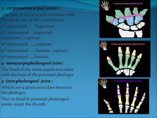
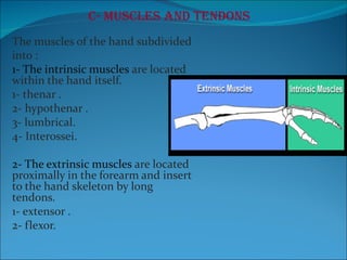
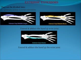
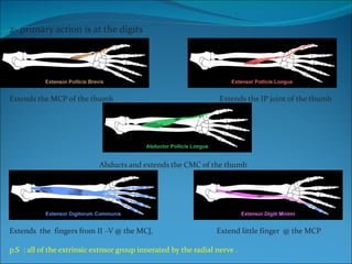
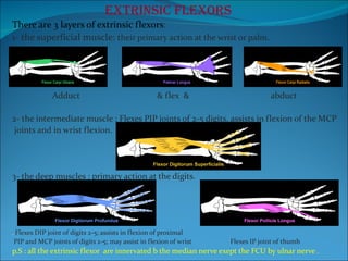
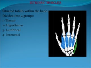
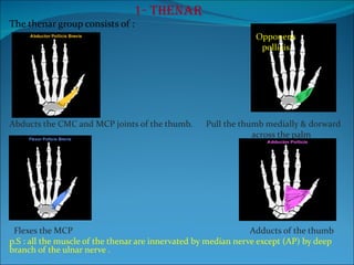
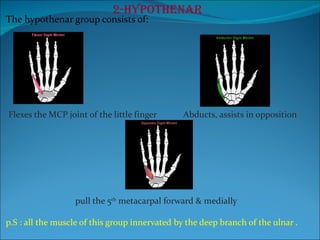
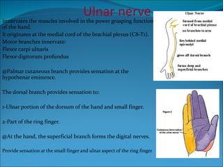
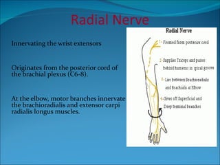
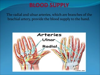
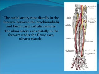
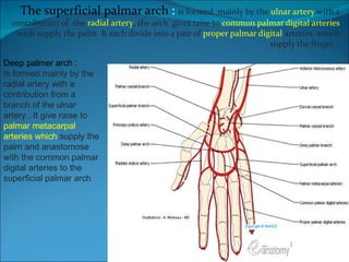
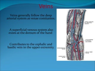
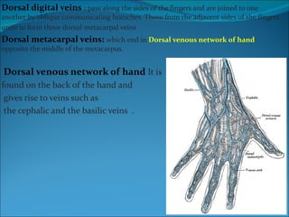
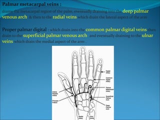
The anatomy of the wrist & hand  Done by: Mohad mubarak  Sameer esawi      DOWNLOAD THIS SLIDE For more slides click here
A- Bones 8 bones form the wrist or carpus;  5 bones form the metacarpus or palm. The remaining 14 are digital bones.
These eight bones of the wrist are arranged in two rows of four ( proximal and distal rows). The proximal raw :  pisiform –tiquetrum-lunate-scaphoid The distal raw : hamate – capitate-trapezoid-trapezium
metacarpal bone & phalangeal bone divide into : Base- shaft – head the 14 phalnges are divided to  Proximal-Middle – distal  . Exept the the thumb which contain only the proximal & the distal
B- The joints of the hand. 1- wrist : the distal end of the radius articulates with two bones of the proximal raw of the carbal bone which are the lunate & the scaopoid & triquetrum to form the wrist joint  2-  Intercarpal joints :  Articulation between the carpal bones .
3- carpometacarpal joints :  The base of metacarpals articulate with the distal  raw of the carpal bones . 1 st  metacarpal ….. Trapezium 2 nd  metacarpal ….trapezoid , trapezium ., capitate . 3 rd  metacarpal …….capitate. 4 th  metacarpal ……hamate , capitate . 5 th  metacarpal …..hamate. 4- metacarpophalangeal joint : The head of the metacarpals articulate with the base of thr proximal phalnges  5- interphalangeal  joint : Which are 9 joints articulate between the phalnges . Ther is distal & proximal phalangeal joints .exept the thumb .
C- Muscles and tendons The muscles of the hand subdivided into : 1- The intrinsic muscles  are located within the hand itself. 1- thenar . 2- hypothenar . 3- lumbrical. 4- Interossei. 2- The extrinsic muscles  are located proximally in the forearm and insert to the hand skeleton by long tendons. 1- extensor . 2- flexor.
Extrinsic extensors They can be divided into: A - primary action is wrist extension Extend & abduct the hand @ wrist joint Extend & adduct the hand @ the wrist joint
2- primary action is at the digits Extends the MCP of the thumb  Extends the IP joint of the thumb  Abducts and extends the CMC of the thumb Extends  the  fingers from II –V @ the MCJ,  Extend little finger  @ the MCP p.S  : all of the extrinsic extnsor group innerated by the radial nerve .
Extrinsic flexors There are 3 layers of extrinsic flexors :  1- the superficial muscle:  their primary action at the wrist or palm. Adduct  & flex  &  abduct  2- the intermediate muscle : Flexes PIP joints of 2-5 digits, assists in flexion of the MCP joints and in wrist flexion. 3- the deep muscles : primary action at the digits. .  Flexes DIP joint of digits 2-5; assists in flexion of proximal PIP and MCP joints of digits 2-5; may assist in flexion of wrist  Flexes IP joint of thumb  p.S : all the extrinsic flexor  are innervated b the median nerve exept the FCU by ulnar nerve  .
Intrinsic muscles Situated totally within the hand.  Divided into 4 groups: 1 -Thenar 2- Hypothenar 3- Lumbrical  4- Interossei
1- Thenar The thenar group consists of : . Abducts the CMC and MCP joints of the thumb.  Pull the thumb medially & dorward  across the palm  Flexes the MCP  Adducts of the thumb p.S : all the muscle of the thenar are innervated by median nerve except (AP) by deep branch of the ulnar nerve .  Opponens pollicis
2-Hypothenar The hypothenar group consists of: Flexes the MCP joint of the little finger  Abducts, assists in opposition pull the 5 th  metacarpal forward & medially  p.S : all the muscle of this group innervated by the deep branch of the ulnar .
Lumbricals The lumbrical muscles contribute to: Flexion of the MCP joints  & Extension of the interphalangeal joints.  p.s  Nerve to Lumbricals I, II,  Median Nerve to Lumbricals III, IV: deep branch of the Ulnar
Interossei The interossei group consists of : 3 palmar  muscles 4 dorsal muscles Innervated by thedeep branch of the  ulnar nerve Dorsal interossei abduct the fingers Palmar  interossei adduct the fingers to the hand axis.
End of the 1 st  part  By Mohad Mubarak
Nerves The hand is innervated by 3 nerves:  1- median nerve.  2- ulnar nerve. 3- radial nerve. Each one has a sensory and motor components.
Median nerve #Originates from the lateral and medial cords of the brachial plexus (C5-T1). #Palmar cutaneous branch provides sensation at the thenar eminence.  #Recurrent motor branch innervates the thenar and lumbrical muscles.   Sensory digital branches provide sensation to: 1-Thumb 2-Index 3-Ring finger (radial side)
Innervates the muscles involved in the power grasping function of the hand.  It originates at the medial cord of the brachial plexus (C8-T1). Motor branches innervate: Flexor carpi ulnaris  Flexor digitorum profundus @Palmar cutaneous branch provides sensation at the hypothenar eminence. The dorsal branch provides sensation to: 1-Ulnar portion of the dorsum of the hand and small finger. 2-Part of the ring finger.  @At the hand, the superficial branch forms the digital nerves. Provide sensation at the small finger and ulnar aspect of the ring finger Ulnar nerve
Radial Nerve Innervating the wrist extensors Originates from the posterior cord of the brachial plexus (C6-8). At the elbow, motor branches innervate the brachioradialis and extensor carpi radialis longus muscles.
BLOOD SUPPLY The radial and ulnar arteries, which are branches of the brachial artery, provide the blood supply to the hand.
The radial artery runs distally in the forearm between the brachioradialis and flexor carpi radialis muscles. The ulnar artery runs distally in the forearm under the flexor carpi ulnaris muscle.
The superficial palmar arch  :  is formed  mainly by the  ulnar artery  with a contribution of  the  radial artery , the arch  gives raise to  common palmar digital arteries  wich supply the palm  & each divide into a pair of  proper palmar digital  arteries  which supply the finger  Deep palmer arch  : Is formed mainly by the radial artery with a contribution from a branch of the ulnar artery . It give raise to  palmar metacarpal arteries which  supply the palm and anastomose with the common palmar digital arteries to the superficial palmar arch
Veins Veins generally follow the deep arterial system as venae comitantes. A superficial venous system also exists at the dorsum of the hand  Contributes to the cephalic and basilic vein in the upper extremity.
Dorsal digital veins  :  pass along the sides of the fingers and are joined to one another by oblique communicating branches. Those from the adjacent sides of the fingers unite to form three   dorsal metacarpal veins  Dorsal metacarpal veins:  which end in  Dorsal venous network of hand  opposite the middle of the metacarpus.  Dorsal venous network of hand  It is  found on the back of the hand and gives rise to veins such as the cephalic and the basilic veins  .
Palmar metacarpal veins :  drains the metacarpal region of the palm, eventually draining into the  deep palmar venous arch . & then to the  radial veins  which drain the lateral aspect of the arm  Proper palmar digital  :  which drain into the  common palmar digital veins  then drain to the  superficial palmar venous arch  and eventually draining to the  ulnar veins   which drain the medial aspect of the arm .
 

The Hand Anatomy Part 1

  • 1.
    The anatomy ofthe wrist & hand Done by: Mohad mubarak Sameer esawi DOWNLOAD THIS SLIDE For more slides click here
  • 2.
    A- Bones 8bones form the wrist or carpus; 5 bones form the metacarpus or palm. The remaining 14 are digital bones.
  • 3.
    These eight bonesof the wrist are arranged in two rows of four ( proximal and distal rows). The proximal raw : pisiform –tiquetrum-lunate-scaphoid The distal raw : hamate – capitate-trapezoid-trapezium
  • 4.
    metacarpal bone &phalangeal bone divide into : Base- shaft – head the 14 phalnges are divided to Proximal-Middle – distal . Exept the the thumb which contain only the proximal & the distal
  • 5.
    B- The jointsof the hand. 1- wrist : the distal end of the radius articulates with two bones of the proximal raw of the carbal bone which are the lunate & the scaopoid & triquetrum to form the wrist joint 2- Intercarpal joints : Articulation between the carpal bones .
  • 6.
    3- carpometacarpal joints: The base of metacarpals articulate with the distal raw of the carpal bones . 1 st metacarpal ….. Trapezium 2 nd metacarpal ….trapezoid , trapezium ., capitate . 3 rd metacarpal …….capitate. 4 th metacarpal ……hamate , capitate . 5 th metacarpal …..hamate. 4- metacarpophalangeal joint : The head of the metacarpals articulate with the base of thr proximal phalnges 5- interphalangeal joint : Which are 9 joints articulate between the phalnges . Ther is distal & proximal phalangeal joints .exept the thumb .
  • 7.
    C- Muscles andtendons The muscles of the hand subdivided into : 1- The intrinsic muscles are located within the hand itself. 1- thenar . 2- hypothenar . 3- lumbrical. 4- Interossei. 2- The extrinsic muscles are located proximally in the forearm and insert to the hand skeleton by long tendons. 1- extensor . 2- flexor.
  • 8.
    Extrinsic extensors Theycan be divided into: A - primary action is wrist extension Extend & abduct the hand @ wrist joint Extend & adduct the hand @ the wrist joint
  • 9.
    2- primary actionis at the digits Extends the MCP of the thumb Extends the IP joint of the thumb Abducts and extends the CMC of the thumb Extends the fingers from II –V @ the MCJ, Extend little finger @ the MCP p.S : all of the extrinsic extnsor group innerated by the radial nerve .
  • 10.
    Extrinsic flexors Thereare 3 layers of extrinsic flexors : 1- the superficial muscle: their primary action at the wrist or palm. Adduct & flex & abduct 2- the intermediate muscle : Flexes PIP joints of 2-5 digits, assists in flexion of the MCP joints and in wrist flexion. 3- the deep muscles : primary action at the digits. . Flexes DIP joint of digits 2-5; assists in flexion of proximal PIP and MCP joints of digits 2-5; may assist in flexion of wrist Flexes IP joint of thumb p.S : all the extrinsic flexor are innervated b the median nerve exept the FCU by ulnar nerve .
  • 11.
    Intrinsic muscles Situatedtotally within the hand. Divided into 4 groups: 1 -Thenar 2- Hypothenar 3- Lumbrical 4- Interossei
  • 12.
    1- Thenar Thethenar group consists of : . Abducts the CMC and MCP joints of the thumb. Pull the thumb medially & dorward across the palm Flexes the MCP Adducts of the thumb p.S : all the muscle of the thenar are innervated by median nerve except (AP) by deep branch of the ulnar nerve . Opponens pollicis
  • 13.
    2-Hypothenar The hypothenargroup consists of: Flexes the MCP joint of the little finger Abducts, assists in opposition pull the 5 th metacarpal forward & medially p.S : all the muscle of this group innervated by the deep branch of the ulnar .
  • 14.
    Lumbricals The lumbricalmuscles contribute to: Flexion of the MCP joints & Extension of the interphalangeal joints. p.s Nerve to Lumbricals I, II, Median Nerve to Lumbricals III, IV: deep branch of the Ulnar
  • 15.
    Interossei The interosseigroup consists of : 3 palmar muscles 4 dorsal muscles Innervated by thedeep branch of the ulnar nerve Dorsal interossei abduct the fingers Palmar interossei adduct the fingers to the hand axis.
  • 16.
    End of the1 st part By Mohad Mubarak
  • 17.
    Nerves The handis innervated by 3 nerves: 1- median nerve. 2- ulnar nerve. 3- radial nerve. Each one has a sensory and motor components.
  • 18.
    Median nerve #Originatesfrom the lateral and medial cords of the brachial plexus (C5-T1). #Palmar cutaneous branch provides sensation at the thenar eminence. #Recurrent motor branch innervates the thenar and lumbrical muscles. Sensory digital branches provide sensation to: 1-Thumb 2-Index 3-Ring finger (radial side)
  • 19.
    Innervates the musclesinvolved in the power grasping function of the hand. It originates at the medial cord of the brachial plexus (C8-T1). Motor branches innervate: Flexor carpi ulnaris Flexor digitorum profundus @Palmar cutaneous branch provides sensation at the hypothenar eminence. The dorsal branch provides sensation to: 1-Ulnar portion of the dorsum of the hand and small finger. 2-Part of the ring finger. @At the hand, the superficial branch forms the digital nerves. Provide sensation at the small finger and ulnar aspect of the ring finger Ulnar nerve
  • 20.
    Radial Nerve Innervatingthe wrist extensors Originates from the posterior cord of the brachial plexus (C6-8). At the elbow, motor branches innervate the brachioradialis and extensor carpi radialis longus muscles.
  • 21.
    BLOOD SUPPLY Theradial and ulnar arteries, which are branches of the brachial artery, provide the blood supply to the hand.
  • 22.
    The radial arteryruns distally in the forearm between the brachioradialis and flexor carpi radialis muscles. The ulnar artery runs distally in the forearm under the flexor carpi ulnaris muscle.
  • 23.
    The superficial palmararch : is formed mainly by the ulnar artery with a contribution of the radial artery , the arch gives raise to common palmar digital arteries wich supply the palm & each divide into a pair of proper palmar digital arteries which supply the finger Deep palmer arch : Is formed mainly by the radial artery with a contribution from a branch of the ulnar artery . It give raise to palmar metacarpal arteries which supply the palm and anastomose with the common palmar digital arteries to the superficial palmar arch
  • 24.
    Veins Veins generallyfollow the deep arterial system as venae comitantes. A superficial venous system also exists at the dorsum of the hand Contributes to the cephalic and basilic vein in the upper extremity.
  • 25.
    Dorsal digital veins : pass along the sides of the fingers and are joined to one another by oblique communicating branches. Those from the adjacent sides of the fingers unite to form three dorsal metacarpal veins Dorsal metacarpal veins: which end in Dorsal venous network of hand opposite the middle of the metacarpus. Dorsal venous network of hand It is found on the back of the hand and gives rise to veins such as the cephalic and the basilic veins .
  • 26.
    Palmar metacarpal veins: drains the metacarpal region of the palm, eventually draining into the deep palmar venous arch . & then to the radial veins which drain the lateral aspect of the arm Proper palmar digital : which drain into the common palmar digital veins then drain to the superficial palmar venous arch and eventually draining to the ulnar veins which drain the medial aspect of the arm .
  • 27.