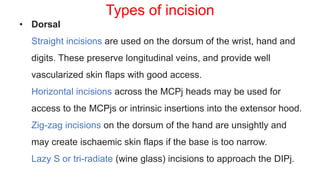
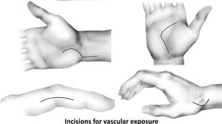
The document provides an overview of hand anatomy including terminology, skin, fascia, muscles, tendons, bones, joints, nerves and vasculature. Key points include descriptions of various hand incisions and their purposes, fascial layers and spaces of the hand, flexor tendon anatomy and zones of injury, and anatomy of important muscles like the thenar and hypothenar muscles. The document covers the detailed functional anatomy of the hand.
The document provides an in-depth summary of the anatomy of the hand. It discusses the extensor mechanism including the extrinsic muscles, intrinsic muscles, and fibrous structures. It describes the anatomy of the extensor tendons including the juncturae tendinae, sagittal bands, central slip, lateral bands, and terminal tendon. It also discusses the lumbrical and interosseous muscles and their functions. Finally, it covers the embryology, nerves, and clinical implications of hand anatomy.
1. The proper collateral ligaments at the PIP joints are under relatively uniform tension in flexion and extension and therefore are not a factor in irreversible contracture. However, the check-rein ligaments at the proximal end of the palmar plate at the PIP joint may hypertrophy and contract, resulting in a fixed flexion contracture.
2. The oblique pulley is the most important pulley in the thumb because the FPB can provide adequate and independent MCP joint flexion, and the A1 pulley often is released for stenosing tenosynovitis without apparent loss of function.
3. When the MP joint is hyperextended, the palmar plate moves distally
The document summarizes the anatomy of the hand, including:
1) The skin, fascia, muscles, blood vessels, and nerves of the palm and dorsum. Key structures include the thenar and hypothenar muscles innervated by the median and ulnar nerves respectively.
2) The flexor tendons in the hand divide into zones as they pass through the carpal tunnel and palm. Extensor tendons are held in place by the extensor retinaculum.
3) Bones and joints of the hand include the carpals that make up the carpal tunnel, the metacarpals, and the interphalangeal joints between phalanges.
The document summarizes the anatomy of the wrist and hand. It describes the bones that make up the wrist (carpal bones), palm (metacarpal bones), and fingers (phalanges). It then discusses the joints between these bones, including the wrist, intercarpal, carpometacarpal, metacarpophalangeal, and interphalangeal joints. Next, it outlines the muscles of the hand, dividing them into intrinsic and extrinsic muscles. Finally, it briefly reviews the nerves, blood supply, and veins of the hand.
Flexor tendon repair requires protecting the tendon repair while allowing early controlled motion to minimize adhesions. This involves splinting the fingers in flexion after primary repair or tendon grafting, followed by progressive range of motion exercises. Complications can include infection, scarring and joint contractures, but good outcomes are achieved with protocols emphasizing early motion like Duran or Kleinert methods.
This document provides information on claw hand deformities, including definitions, anatomy, classifications, evaluation, and surgical reconstruction techniques. It begins with defining claw hand as a flattening of the transverse metacarpal arch with hyperextension of the MCP joints and flexion of the PIP and DIP joints. It then discusses the anatomy and biomechanics involved in normal versus paralytic claw hands. Various classification systems for claw hands are presented based on etiology, pattern of nerve injury, degree of involvement, and physical characteristics. Evaluation techniques such as specific tests and angle measurements are outlined. Both static and dynamic surgical reconstruction methods are then described in detail, including tendon transfers, capsulotomies, and tenode
The document discusses various local flap options for reconstructing fingertip injuries. It describes the anatomy of the fingertip and goals of reconstruction which are to close wounds, maximize sensation, preserve length and function. Common local flaps include the volar V-Y flap, bilateral V-Y flaps, cross-finger flap, thenar flap and lateral island flaps. Choice depends on wound orientation and configuration.
1) Median nerve injuries can result in loss of motor function to key muscles in the forearm and hand. Tendon transfers can help restore function by transferring muscles still innervated to replace lost functions.
2) Common tendon transfers include using the superficialis or EIP tendon to restore thumb opposition. The BR or ECRL can be used to restore thumb or index finger flexion.
3) Rehabilitation after tendon transfer focuses initially on splinting the transfers and mobilizing other joints, followed by gentle active and assisted range of motion exercises to strengthen and condition the transfers.
The hand has fine motor control due to its complex anatomy. It contains bones like the carpals, metacarpals and phalanges. Muscles originate from the forearm and act on the hand. The median, ulnar and radial nerves innervate muscles and provide sensation. These nerves are vulnerable to compression at specific points in the arm, elbow, forearm and wrist. The document provides an overview of the hand's embryology, bones, joints, muscles, vessels and nerves.
1. The gastrocnemius muscle consists of medial and lateral heads that are supplied by the medial and lateral sural arteries respectively.
2. The medial gastrocnemius flap is most commonly used due to its large size and reliable vascular pedicle. It can be raised as a muscle or musculocutaneous flap to cover defects of the upper leg and knee.
3. The lateral gastrocnemius flap is smaller but can be used for smaller defects of the upper lateral leg and knee. Both flaps have consistent anatomy and can be reliably elevated based on the dominant sural artery pedicles.
The document summarizes the anterolateral thigh (ALT) flap, which has become a popular reconstructive option. It describes the history and indications of the ALT flap, including head and neck and extremity reconstruction. The key aspects of evaluating patients, raising the flap, and post-operative care and complications are covered. The advantages include a long vascular pedicle and ability to harvest a large skin paddle, while disadvantages include a bulky flap and potential donor site morbidity. Variations in anatomy are also discussed.
This document discusses flexor tendon injuries of the hand. It covers flexor tendon anatomy, zones of injury, examination techniques, types of repairs, and considerations for treatment. Primary repair is generally preferred if possible within 12 hours of injury. The McLarney stitch is described as a strong yet simple repair technique. Partial tendon lacerations under 60% may not require repair while larger injuries are treated similarly to complete ruptures.
This document discusses anatomy and reconstruction techniques for the heel. It describes the layers of the sole, including muscles, tendons, and nerves. The medial and lateral plantar nerves and arteries are examined in detail. Reconstruction options for the anterior and posterior heel are provided, such as local flaps, skin grafting, and free flaps. The medial plantar and sural flaps are highlighted as examples. In summary, this document reviews the anatomy of the foot sole and discusses approaches for reconstructing soft tissue injuries of the heel region.
Thumb reconstruction by microvascular methodsDr. Suiyibangbe
This document discusses microvascular reconstruction of the thumb using toe transfer techniques. It begins by outlining the importance of the thumb in hand function. Reconstruction of the thumb is challenging due to its unique position. Microsurgical toe transfer meets the requirements of providing stable, sensitive mobility. The great toe is well-suited anatomically and can be transferred with its vessels, nerves, tendons and skin. A thorough preoperative evaluation of the recipient site and donor toe is important for planning. The goals of reconstruction are to restore painless stability, sensibility and mobility. Careful surgical planning and technique along with postoperative therapy can achieve excellent results.
The document discusses ulnar nerve palsy and tendon transfers used to treat it. It begins by describing the anatomy of the ulnar nerve and its motor and sensory functions. It then discusses clinical findings associated with ulnar nerve injuries at different locations. Various tendon transfers are summarized that aim to restore small and ring finger flexion, key pinch, correct clawing, and improve grip strength for patients with ulnar nerve palsy. These include transferring forearm muscles like the ECRB to restore key pinch or correct clawing. The modified Stiles-Bunnell procedure is also summarized, which uses the middle finger superficialis tendon to dynamically correct clawing during finger flexion.
This document discusses hand reconstruction techniques following injuries. It introduces relevant hand anatomy and the goals of reconstruction, which are to restore functional, sensate hands that are aesthetically acceptable. Various local and regional flap options for covering dorsal and palmar hand defects ranging from fingertips to multiple fingers are described, including considerations for selecting the appropriate flap based on defect size and location. Post-operative care and potential complications are also mentioned.
This document describes the scapular flap procedure. It details the relevant surgical landmarks and arterial system around the scapula. The flap can include skin, muscle, and bone from the scapula region. The flap is designed and harvested by following the path of the circumflex scapular artery. For a bone flap, the periosteal branch is dissected to access bone from the lateral border of the scapula. The scapular flap provides a large volume of soft tissue and bone to reconstruct significant defects, such as after extensive maxillectomies or mandibular resections.
The document provides an overview of the anatomy of the hand. It describes the 27 bones in the hand and wrist, including the carpals, metacarpals, and phalanges. It outlines the extrinsic muscles that flex and extend the wrist and digits, as well as the intrinsic muscles of the hand including the thenar, hypothenar, lumbricals, and interossei groups. It also briefly discusses the arterial and venous blood supply, as well as the innervation of the hand by the median and ulnar nerves.
Algorithm to approach the lower extremity defect and to select appropriate fl...Binh Phuoc
1. The document provides an algorithm for selecting appropriate flaps to reconstruct lower extremity defects, discussing various options from primary closure to free flaps.
2. It begins by covering primary and secondary wound closure, then skin grafts including split-thickness and full-thickness grafts.
3. Next it discusses flaps in general and provides classifications. It then details various local, regional, and free flap options for reconstruction, including musculocutaneous flaps like the gastrocnemius and latissimus dorsi flaps.
Hand anatomy 2017 new microsoft power point presentationessameahady
The document summarizes the anatomy of the hand and wrist. It describes the bones that make up the skeleton of the hand including the carpus, metacarpals, and phalanges. It then discusses the muscles, ligaments, tendons, blood vessels and nerves of the hand and wrist. In particular, it outlines the structures that pass through the carpal tunnel and extensor retinaculum.
At the end of this lecture, you should be able to:-
Understand and describe the anatomy of the flexor tendon system in the hand and its relevance in surgery
Describe and relate the normal physiology of the tendon and its implications in injury
Understand the reaction of tendon to injury and its healing process and its implications in surgery and rehabilitation
The fascial compartments of thigh are the three fascial compartments that divide and contain the thigh muscles. The fascia lata is the strong and deep fascia of the thigh that surrounds the thigh muscles and forms the outer limits of the compartments. Internally the muscle compartments are divided by the lateral and medial intermuscular septa.
This document describes the arterial blood supply and potential flaps in the lower limb. It discusses various fasciocutaneous and musculocutaneous flaps that can be raised based on named arteries in the lower limb, including the femoral, lateral circumflex femoral, profunda femoris, popliteal, and posterior tibial arteries. Specific flaps are described such as the anteromedial thigh flap, gracilis flap, and gastrocnemius flap. The angiosome concept and variations in vascular anatomy are also covered.
The document discusses various types of skin grafts and flaps used in orthopedics. It describes split thickness skin grafts which contain part of the epidermis and dermis and are useful for covering defects when the area is too wide for a full thickness graft. It also discusses axial pattern flaps which contain a direct cutaneous artery and allow coverage of the hand while preserving movement. Local flaps are preferred over distant flaps when possible due to better color and texture matching and reduced risk of complications.
The ulnar nerve originates from the medial cord of the brachial plexus. It runs down the arm and enters the forearm between the two heads of the flexor carpi ulnaris muscle. In the hand, it passes through the ulnar tunnel, dividing into superficial and deep branches that provide sensory and motor innervation to portions of the fourth and fifth fingers. Lesions of the ulnar nerve can occur at different locations, with varying effects depending on whether the nerve is damaged proximally in the arm, at the elbow, in the forearm, or distally in the hand or wrist.
This document discusses various techniques for pollicization, which is a hand surgery to create a functioning thumb from another finger. It begins by defining pollicization and describing the anatomy and function of the thumb. It then discusses different conditions that may require pollicization, including thumb hypoplasia, absence due to trauma, macrodactyly, multifinger hands, and mirror hands. For each condition, it provides details on the surgical procedure for pollicization, including steps like isolating nerves/vessels, shortening bones, transferring muscles/tendons, and positioning the new thumb. Post-operative casting is also addressed. The document contains many diagrams illustrating thumb anatomy and different pollicization procedures.
Detailed Hand surgical anatomy by mohamed abdelhadyMohamed Abdelhady
Detailed Hand surgical anatomy including bones , blood and nerve supply and special structures eg flexor retiniculum , extensor compartments and anatomical snaff box
This document provides an overview of the surgical anatomy of the hand. It describes the surface landmarks and structures of the palmar aspect of the hand such as the tubercles and bones that can be felt. It details the layers of the palm including the skin, superficial and deep fascia, muscles such as the palmaris brevis, nerves, blood vessels, and other structures. It also describes the dorsal aspect including fascia and extensor retinaculum. Key areas covered include the carpal tunnel, flexor retinaculum, fibrous flexor sheaths, intrinsic hand muscles, and fascial spaces of the palm.
Dr.MD.Monsur Rahman,PT
MPT-Musculoskeletal Disorders
Maharishi Markandeshwar Institute Of Physiotherapy And Rehabilitation, Maharishi Markandeshwar (Deemed to be University), Mullana - Ambala,133-207 (Haryana)
1.Anatomy
a.Course
b.Motor distribution
c.Sensory distribution
2.Common sites affected
3.Level of median nerve injury
4.Clinical feature with various test performed
5.Various syndromes related to median nerve
6.Treatment
7.Summary
This document provides an overview of the anatomy of the palm. It describes the palmar skin as thick, glabrous, and rich in sweat glands. It is firmly attached to the underlying palmar aponeurosis by fibrous bands. The document outlines the creases and lines of the palmar skin, as well as the layers of fascia in the palm including the palmar aponeurosis and flexor retinaculum. It also describes the muscles of the palm including the thenar, hypothenar, interossei and lumbrical muscles. Additionally, it discusses the arteries, nerves and fascial spaces of the palm.
Apply gentle pressure proximally
Surgeon: Check distal pulses and capillary refill
If no improvement:
Consider temporary arteriotomy or venous shunt
Delay closure and observe
Flap or graft may be needed
Flexor tendons - enclosed by synovial sheaths.
Tendons - blood supply through synovial folds known as vincula, each
tendon having two, vincula longa and vincula brevia.
The sheath of the little finger is continuous with the ulnar bursa covering
the flexor tendons in the palm.
The flexor pollicis longus is covered by a single sheath throughout, the
radial bursa.
Synovial sheaths can be infected producing tenosynovitis. Infection can
spread throughout the sheath. Infection of the sheath of the little finger can thus spread up the distal aspect of the forearm into the space of Parona.
The document provides an overview of the anatomy of the forearm, including its osteology, fascial compartments, muscles, nerves, blood vessels, and other structures. It describes the objectives as outlining the osteology, cutaneous nerve supply, fascial compartments, muscles within each compartment, blood supply, and compartment syndrome of the forearm. Key points include the forearm being divided into anterior, lateral, and posterior compartments by fascia, each with their own muscles, nerves and blood vessels. The median and ulnar nerves and arteries are discussed along with the muscles in the various compartments.
200427 Examination of compressive neuropathies of ulnar nerveDr MADAN MOHAN
The document summarizes clinical examination of ulnar nerve compressive neuropathies. It describes the anatomy of the ulnar nerve from the arm to the hand. Key points of physical examination are discussed, including signs like Duchenne sign (clawing of ring and little fingers), Wartenberg sign (abduction of little finger on extension), and Froment's sign (inability to clasp paper between thumb and index finger). Common sites of ulnar nerve compression, like the cubital tunnel at the elbow and Guyon's canal at the wrist, are explained. Specific tests for evaluating these sites, such as the cubital tunnel compression test and scratch collapse test, are outlined.
This document discusses median nerve injuries, including:
- The anatomy and functions of the median nerve in the forearm and hand.
- Clinical assessment of median nerve function through specific muscle tests.
- Common median nerve compression syndromes like carpal tunnel syndrome.
- Classification of nerve injuries and management options for median nerve injuries.
6. fascial spaces and arterial anastomoses of the upper limbDr. Mohammad Mahmoud
The document summarizes the fascial spaces and arterial anatomy of the upper limb. It describes the boundaries and contents of fascial spaces in the palm, fingers, forearm, and elbow. It also outlines important arterial anastomoses around the shoulder, elbow, wrist, and hand that help ensure adequate blood flow, including the palmar and dorsal carpal arches and the superficial and deep palmar arches.
The document provides information on the wrist joint, hand bones, joints of the hand, nerves and muscles of the hand. It describes that the wrist joint is an ellipsoid joint between the lower end of the radius, articular disc and three carpal bones. The hand contains 8 carpal bones, 5 metacarpals and 14 phalanges. It also details the intrinsic muscles of the hand, nerves including the median and ulnar nerves, synovial sheaths and spaces of the hand.
This slide gives you information regarding the Types of Palmar spaces, their contents & boundaries. Also certain aspects of Applied anatomy has been enlightened in the interest of Integrated teaching.
1. The document describes the anatomy and infections of the hand. It details the bones, muscles, nerves and blood supply of the hand.
2. Common hand infections include paronychia, felon, web space infections, and tenosynovitis. These infections can involve the skin, fascial spaces or tendon sheaths of the hand.
3. Treatment of hand infections involves drainage of pus, antibiotics, elevation and immobilization of the hand. Early recognition and treatment of infections is important to prevent complications and preserve hand function.
This document provides information on radial, median, and ulnar nerve injuries, including anatomy, causes, examination findings, and treatment options. It describes the anatomy and branches of each nerve. Common causes of injury include lacerations, fractures, and entrapment syndromes. Examination involves testing specific muscles innervated by each nerve. Surgical treatment may include nerve repair or reconstruction using tendon transfers to restore function when nerve recovery is not possible. The document outlines various tendon transfer procedures used to treat different patterns of nerve injury.
The document summarizes the anatomy of the hand. It describes the intrinsic muscles of the hand including the thenar and hypothenar muscles supplied by the median and ulnar nerves respectively. It discusses the tendons in the hand including the long flexors passing through fibrous flexor sheaths. The document also describes the neurovascular supply to the hand including the superficial and deep palmar arches and digital nerves from the median and ulnar nerves. Finally, it briefly discusses anatomical structures like the anatomical snuff box on the radial side of the wrist.
The radial nerve is the largest terminal branch of the posterior cord. It arises from spinal cord segments C5-T1 and innervates all muscles in the posterior arm and forearm compartment as well as skin on the posterior arm and forearm. In the arm, it passes between the triceps muscles before entering the spiral groove on the humerus. It continues down the humerus, piercing the lateral intermuscular septum and supplying muscles of the anterior arm. In the forearm, it divides into superficial and deep branches, with the deep branch becoming the posterior interosseous nerve. Radial nerve injuries are commonly caused by fractures of the humerus. Nonoperative treatment focuses on preventing contract
This document discusses the anatomy, motor and sensory innervation, and injuries of the median nerve. It provides detailed information on:
1) The anatomy of the median nerve from its origins in the brachial plexus through the arm, forearm, and hand.
2) The muscles innervated by the median nerve in the forearm and hand.
3) The sensory distribution of the median nerve in the hand and fingers.
4) Types of median nerve injuries including high injuries proximal to the elbow and low injuries in the distal forearm, and the clinical signs associated with each.
5) Tests to assess median nerve function including testing specific muscles and sensory areas.
This document discusses syndactyly, a congenital hand anomaly where adjacent digits are fused. It describes the typical surgical techniques used to correct syndactyly, including the use of local flaps and skin grafts. Recently, some reconstructive techniques have been developed that do not require skin grafts. The document outlines the steps of syndactyly release surgery, including flap design and elevation, separation of digits, and use of skin grafts if needed. Postoperative complications are also discussed, such as web creep caused by scar contracture.
हिंदी वर्णमाला पीपीटी, hindi alphabet PPT presentation, hindi varnamala PPT, Hindi Varnamala pdf, हिंदी स्वर, हिंदी व्यंजन, sikhiye hindi varnmala, dr. mulla adam ali, hindi language and literature, hindi alphabet with drawing, hindi alphabet pdf, hindi varnamala for childrens, hindi language, hindi varnamala practice for kids, https://www.drmullaadamali.com
Executive Directors Chat Leveraging AI for Diversity, Equity, and InclusionTechSoup
Let’s explore the intersection of technology and equity in the final session of our DEI series. Discover how AI tools, like ChatGPT, can be used to support and enhance your nonprofit's DEI initiatives. Participants will gain insights into practical AI applications and get tips for leveraging technology to advance their DEI goals.
The simplified electron and muon model, Oscillating Spacetime: The Foundation...RitikBhardwaj56
Discover the Simplified Electron and Muon Model: A New Wave-Based Approach to Understanding Particles delves into a groundbreaking theory that presents electrons and muons as rotating soliton waves within oscillating spacetime. Geared towards students, researchers, and science buffs, this book breaks down complex ideas into simple explanations. It covers topics such as electron waves, temporal dynamics, and the implications of this model on particle physics. With clear illustrations and easy-to-follow explanations, readers will gain a new outlook on the universe's fundamental nature.
This slide is special for master students (MIBS & MIFB) in UUM. Also useful for readers who are interested in the topic of contemporary Islamic banking.
How to Setup Warehouse & Location in Odoo 17 InventoryCeline George
In this slide, we'll explore how to set up warehouses and locations in Odoo 17 Inventory. This will help us manage our stock effectively, track inventory levels, and streamline warehouse operations.
How to Make a Field Mandatory in Odoo 17Celine George
In Odoo, making a field required can be done through both Python code and XML views. When you set the required attribute to True in Python code, it makes the field required across all views where it's used. Conversely, when you set the required attribute in XML views, it makes the field required only in the context of that particular view.
বাংলাদেশের অর্থনৈতিক সমীক্ষা ২০২৪ [Bangladesh Economic Review 2024 Bangla.pdf] কম্পিউটার , ট্যাব ও স্মার্ট ফোন ভার্সন সহ সম্পূর্ণ বাংলা ই-বুক বা pdf বই " সুচিপত্র ...বুকমার্ক মেনু 🔖 ও হাইপার লিংক মেনু 📝👆 যুক্ত ..
আমাদের সবার জন্য খুব খুব গুরুত্বপূর্ণ একটি বই ..বিসিএস, ব্যাংক, ইউনিভার্সিটি ভর্তি ও যে কোন প্রতিযোগিতা মূলক পরীক্ষার জন্য এর খুব ইম্পরট্যান্ট একটি বিষয় ...তাছাড়া বাংলাদেশের সাম্প্রতিক যে কোন ডাটা বা তথ্য এই বইতে পাবেন ...
তাই একজন নাগরিক হিসাবে এই তথ্য গুলো আপনার জানা প্রয়োজন ...।
বিসিএস ও ব্যাংক এর লিখিত পরীক্ষা ...+এছাড়া মাধ্যমিক ও উচ্চমাধ্যমিকের স্টুডেন্টদের জন্য অনেক কাজে আসবে ...
Walmart Business+ and Spark Good for Nonprofits.pdfTechSoup
"Learn about all the ways Walmart supports nonprofit organizations.
You will hear from Liz Willett, the Head of Nonprofits, and hear about what Walmart is doing to help nonprofits, including Walmart Business and Spark Good. Walmart Business+ is a new offer for nonprofits that offers discounts and also streamlines nonprofits order and expense tracking, saving time and money.
The webinar may also give some examples on how nonprofits can best leverage Walmart Business+.
The event will cover the following::
Walmart Business + (https://business.walmart.com/plus) is a new shopping experience for nonprofits, schools, and local business customers that connects an exclusive online shopping experience to stores. Benefits include free delivery and shipping, a 'Spend Analytics” feature, special discounts, deals and tax-exempt shopping.
Special TechSoup offer for a free 180 days membership, and up to $150 in discounts on eligible orders.
Spark Good (walmart.com/sparkgood) is a charitable platform that enables nonprofits to receive donations directly from customers and associates.
Answers about how you can do more with Walmart!"
1. Anatomy of The Hand
Prepared by
Mahmoud Elsayed Gouda Mohamed
Plastic Surgery Unit – Zagazig University
2. Agenda
Part 1 Part 2
• Terminology & Hand Motion
• Skin
• Fascia
• Musclo-Tendious Unit
• Embryology
• Bones & joints
• Neurovascular Unit
• Carpal tunnel
• Nail bed
3. Terminology
• Forearm and Hand
Radial and ulnar, dorsal and volar (or palmar). Avoid medial/lateral, anterior/posterior
• Digits
Thumb
Index finger
Long or middle finger
Ring finger
Small or little finger
• Palm
Thenar eminence
Hypothenar eminence
Midpalm: Area between thenar and hypothenar eminences
4.
5. • Thumb
• Abduction:
• Movement out of plane of palm (i.e., volar abduction)
or in plane of palm (i.e., planar or radial abduction)
• Abduction occurs at the CMC joint and refers to
metacarpal motion.
• Flexion/extension: Occurs at MP joint or IP joint of
thumb—it is important to specify (i.e., IP joint extension)
• Opposition: Combination of movements, including CMC
joint rotation, resulting in the thumb pulp directly opposing
the pulp of another finger
Hand Motion
6.
7. Fingers
• Abduction: Movement is away from the long
finger.
• Adduction: Movement is toward the long finger.
Occurs at MP joints during Extension.
(reference point is sagittal line through third ray)
Flexion/extension/Hyperextension: Occurs at MP
joints and IP joints
8. Wrist
• Flexion, extension, radial and ulnar deviation:
at mdicarpal and radiocarpal joints
• Pronosupination: at distal radioulnar joint
9.
10.
11. • Volar skin is thicker, less mobile, and has papillary ridges for
grasping.
• Dorsal skin is thinner and more mobile, and the subcutaneous
tissue contains veins and lymphatics.
Skin
13. Skin incisions
• Surgical incisions should:
• Be extensible
• Provide access to deep structures
• Provide vascularized skin flaps (avoid very long, narrow flaps)
• Avoid formation of scar contractures. Straight incisions across
flexor creases (digits, palm, wrist,) lead to scar contracture.
• Avoid unnecessary dissection (leading to oedema and scarring)
• Scars should be placed on the non-dependent sides of the
digits (e.g. radial side of little finger) where possible.
14. (A, B) Schematic representation of the joint axes. The longitudinal dimensions in the
midpalmar and middorsal aspect of the digits change maximally. The midaxial line
through the three joint axes does not change in length with flexion and extension.
Palmar incisions placed longitudinally produce contracture if they pass across the
palmar diamonds delineated by lines joining the joint axes (after Littler). Transverse
incisions avoid the occurrence of flexion scar contractures. The same principle
applies at the wrist.
15. • Dorsal
Straight incisions are used on the dorsum of the wrist, hand and
digits. These preserve longitudinal veins, and provide well
vascularized skin flaps with good access.
Horizontal incisions across the MCPj heads may be used for
access to the MCPjs or intrinsic insertions into the extensor hood.
Zig-zag incisions on the dorsum of the hand are unsightly and
may create ischaemic skin flaps if the base is too narrow.
Lazy S or tri-radiate (wine glass) incisions to approach the DIPj.
Types of incision
16. • Mid-lateral
This runs along the side of the digit, dorsal to the neurovascular
bundle, and therefore safe.
It is designed by
flexing the fingertip into the palm,
and marking dots at the apices
of the DIPj, PIPj and MCPj creases.
The digit is then extended and a line drawn connecting the dots.
17. Palmar
Bruner: These zig-zag type incisions are used in the volar aspect of
the palm and digits. In the digits, they run from the mid-lateral line
across the span of a phalanx to the contralateral mid-lateral line. In
the palm they should be 1-2cm long, zig-zagging between palmar
creases
Half Bruner: These are similar to Bruner incisions, but zig-zag
across half of the width of the digit.
22. • Volar fascia
• Anchors volar skin to bone for grasping, in contrast to loose skin
on dorsum.
• The deep fascia :covers the interosseous muscles and is not
involved in Dupuytren’s disease .
• Superfcial volar fascia: Triangular-shaped fascia attached
proximally to PL tendon; composed of longitudinal fibers, vertical
fibers, transverse fibers.
Fascia of The Hand:
23. The longitudinal fibers
course superfcial to the flexor retinaculum, forming pretendinous
bands. These bands travel distally and insert into the deep surface
of the dermis at the distal palmar crease,
bifurcate around the flexor tendon sheath to insert on the radial
and ulnar sides of the MCP joint
24.
25. The transverse fibers are characterized by two distinct bands, one
proximal and one distal.
• The proximal transverse fibers, located at the level of the distal
palmar crease, course deep to the longitudinal pretendinous
bands and are not typically affected by Dupuytren’s disease.
Radially, these fibers form the proximal commissural ligament
of the frst webspace.
26. • The distal transverse fibers, alternately referred to as the
natatory ligament, course superfcial to the longitudinal
pretendinous bands and are affected by Dupuytren’s disease.
The natatory ligament extends from the radial border of the
index finger to the ulnar border of the small finger. Ulnarly, the
natatory ligament divides to envelop the abductor digiti minimi
(ADM) and the ulnar neurovascular bundle. Radially, the
natatory ligament is continuous with the distal commissural
ligament of Grapow within the frst webspace.
27.
28. • The vertical fibers connect the superficial palmar aponeurosis to the
deep fascia. These fibers forma series of eight vertical septa on the
radial and ulnar sides of the flexor digital apparatus. These septa
divide longitudinal compartments containing the flexor tendons from
those containing the lumbricals and digital neurovascular bundles.
• Additional vertical fibers connect the superficial palmar fascia
to the overlying dermis, providing resistance to shear forces
within the palm
29.
30. • Retaining ligaments of fingers ( Digital Fascia)
• Stabilize skin and extensor mechanism of digits and support
neurovascular bundles
• more variable in the anatomy
• Grayson’s ligament
• Cleland’s ligament
• Transverse retinacular ligament: originate from the volar capsule of
the PIP joint and course dorsally to insert into the lateral margin of the
extensor mechanism
• Oblique retinacular ligament (ligament of Landsmeer):
Originates on volar aspect of middle phalanx and inserts on dorsal aspect
of distal phalanx. Helps coordinate PIP joint and DIP joint motion
31.
32. Deep Fascial Spaces
• Potential spaces that can be sites of infection
• Midpalmar space
• Thenar space
• Hypothenar space
• Interdigital web space
• Parona’s space
34. Synovial sheaths
The synovial sheaths
are closed sacs
around the tendons
composed of a visceral
layer on the tendon
surface and a parietal
layer on the fibrous
sheath surface.
35. • The thumb synovial sheath is continuous from the wrist to the
distal extreme of the flexor pollicis longus.
• The digital synovial sheaths for the index, long, and ring fingers
usually start at the level of the distal palmar crease and extend to
the distal interphalangeal joints.
• Often the little-finger sheath extends more proximally to
communicate with a common sheath around the finger flexors and
then across the wrist to the distal forearm, where tendons pass
through the carpal tunnel.
37. In the thumb and little finger, the infection can spread within the
sheath into the distal forearm because of the continuation of the
sheath (radial bursa around FPL, ulnar bursa around little finger
tendons)
38. Muscle Tendon Units
• Extrinsic Flexors
• Pulley system
• Thenar and Hypothenar Muscles
• Extensor mechanism
39. • There are 12 flexor tendons in the hand and forearm. Common
flexor group arises near the medial epicondyle .
• Nine flexors passes under the flexor retinaculum to reach the hand
• The long flexors to the fingers ( FDP, FDS) are responsible
for flexion of the interphalangeal joints and are supplements
to active flexion of the metacarpophalangeal joints and the
wrist joint
40. Finger flexors
▪ Flexor digitorum superficialis (FDS)
○ Separate muscle belly origin, allowing independent finger
motion
○ Tendons superficial to the flexor digitorum profundus (FDP)
tendons up to their bifurcation into slips at the
metacarpophalangeal (MCP) joint, where they travel around the
FDP tendon, dive deep, rejoin to form Camper's chiasm, and
insert onto the middle phalanx
○ Flexes the proximal interphalangeal (PIP) joint
41. ▪ FDP
○ Common muscle belly origin
♦ Because of this common origin of the middle ,ring and little,
shortening of FDP tendon or overtightening of repair can lead
to decreased grip strength and decreased flexion of the
uninjured digits (“quadrigia” effect).
○ Inserts into the volar aspect of the distal phalanx
○ Flexes the distal interphalangeal (DIP) joint
45. ▪ Flexor pollicis longus (FPL)
○ Arises from the midaspect of the
radial shaft and interosseous
membrane
○ The only tendon inside the flexor
sheath of the thumb; inserts onto
the distal phalanx
○ Flexes the thumb interphalangeal
(IP) joint
50. • Muscle excursion is defined as the distance a muscle can shorten
and is proportional to fiber length.
• The joint moment arm is the perpendicular distance between the
joint center rotation and the central longitudinal axis of the tendon
• The larger the moment arm, the greater the tendon excursion must
be to produce a given angle of joint rotation
51. Wrist Flexors
Flexor carpi radialis (FCR)
○ Inserts onto the base of the 2nd and 3rd metacarpals
Prime Wrist Flexor
▪ Flexor carpi ulnaris (FCU)
○ Inserts onto the base of the 5th metacarpal, hook of hamate, and
pisiform
Stability of DRUJ
○ Overlies ulnar artery and nerve
♦ Laceration to FCU is concerning for injury to the ulnar a. and n.
▪ Palmaris longus (PL)
○ Absent in ~15% to 20%
○ Ends in the fan-shaped palmar fascia
○ Lies volar to median nerve traveling within carpal canal
○ Lacerations to PL are concerning for median nerve laceration.