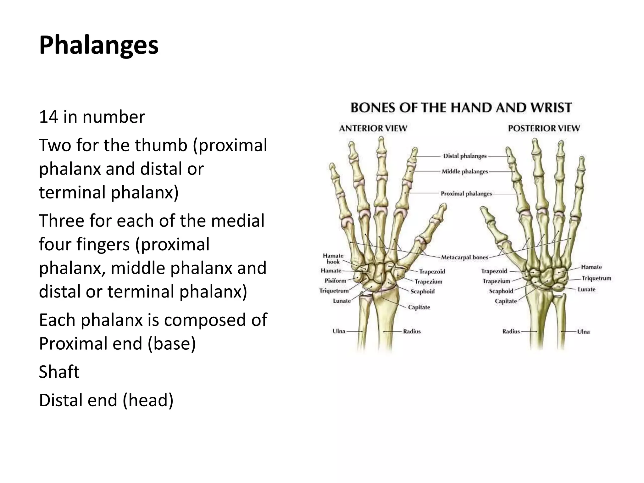
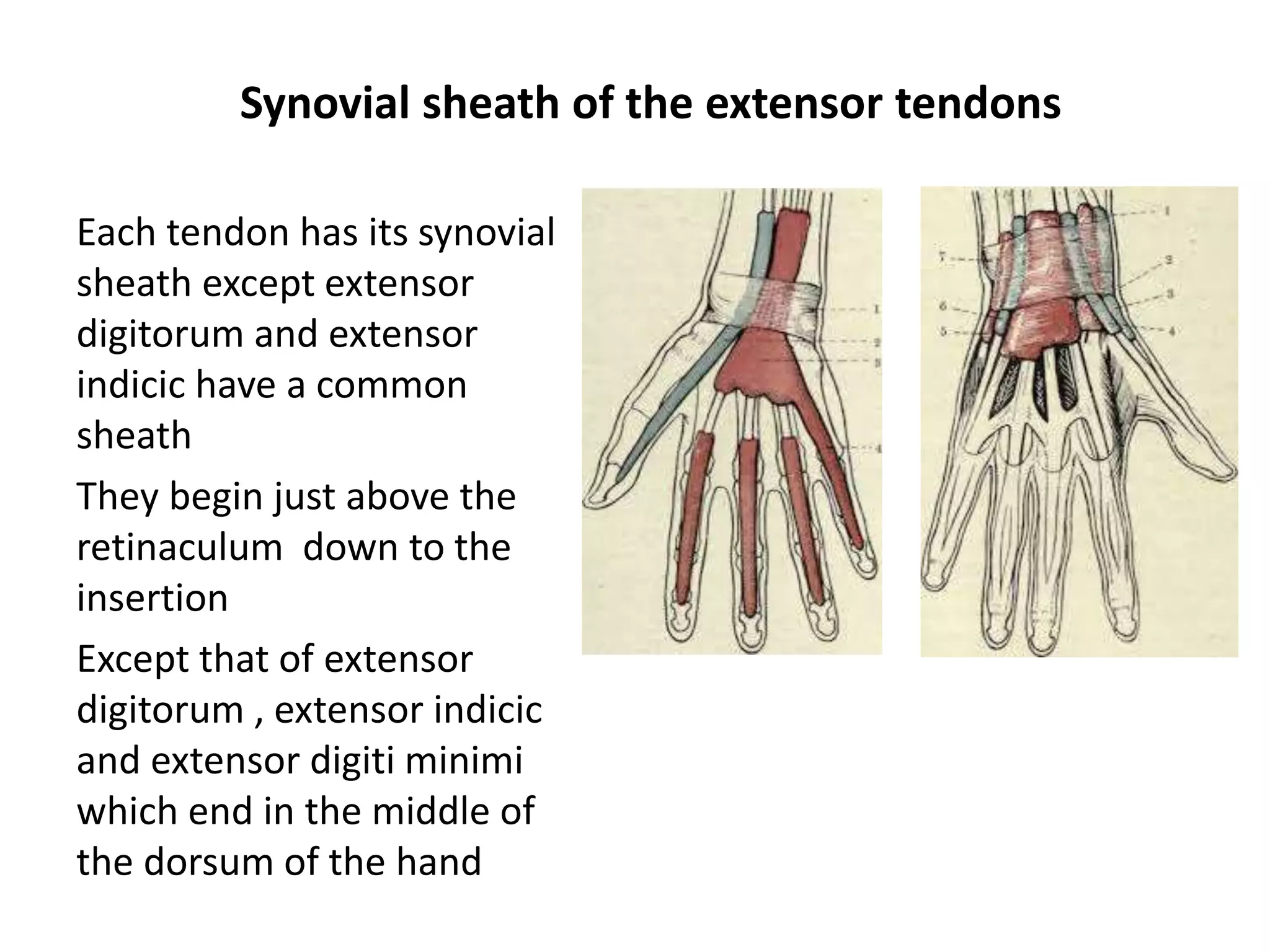
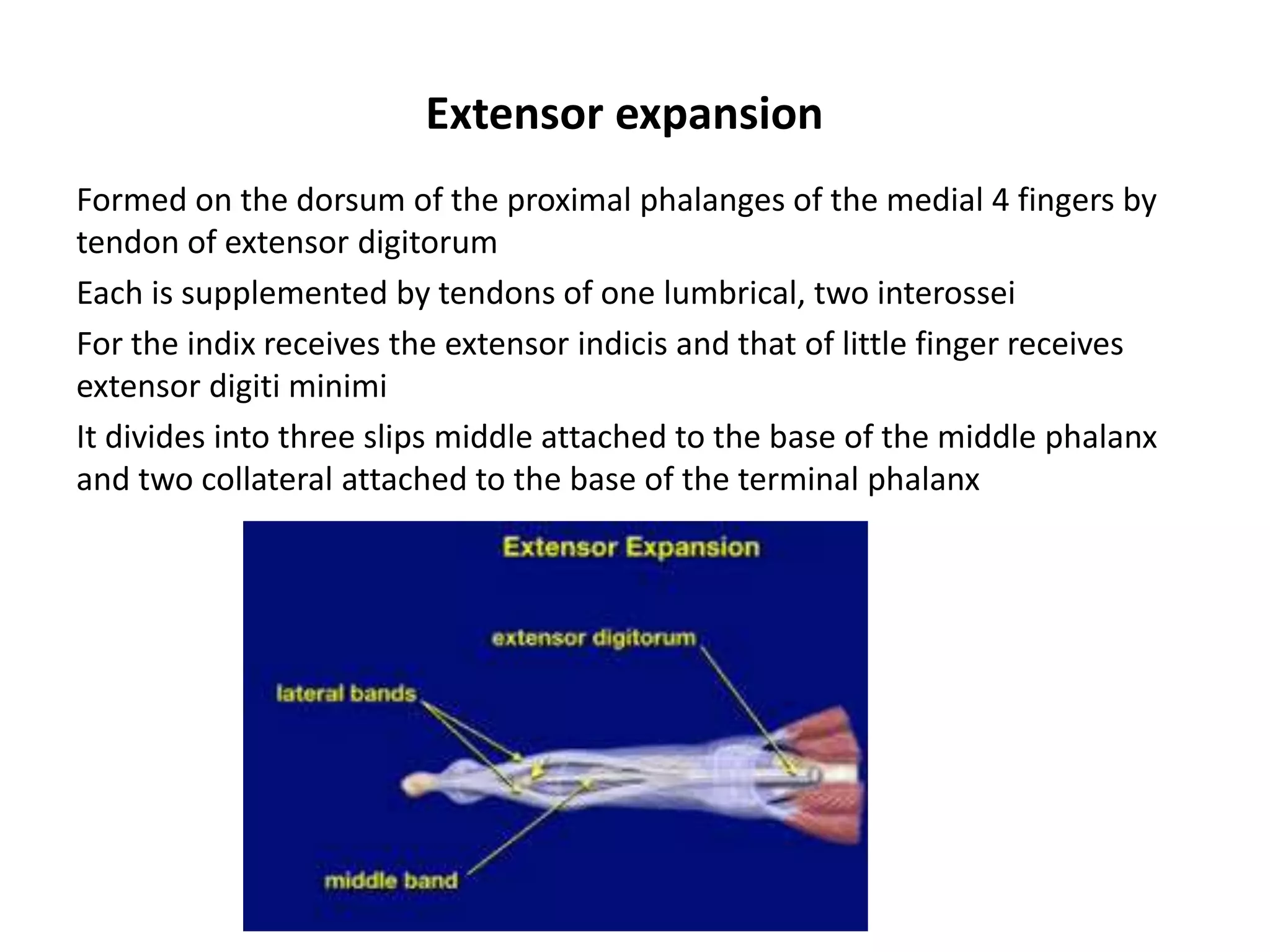
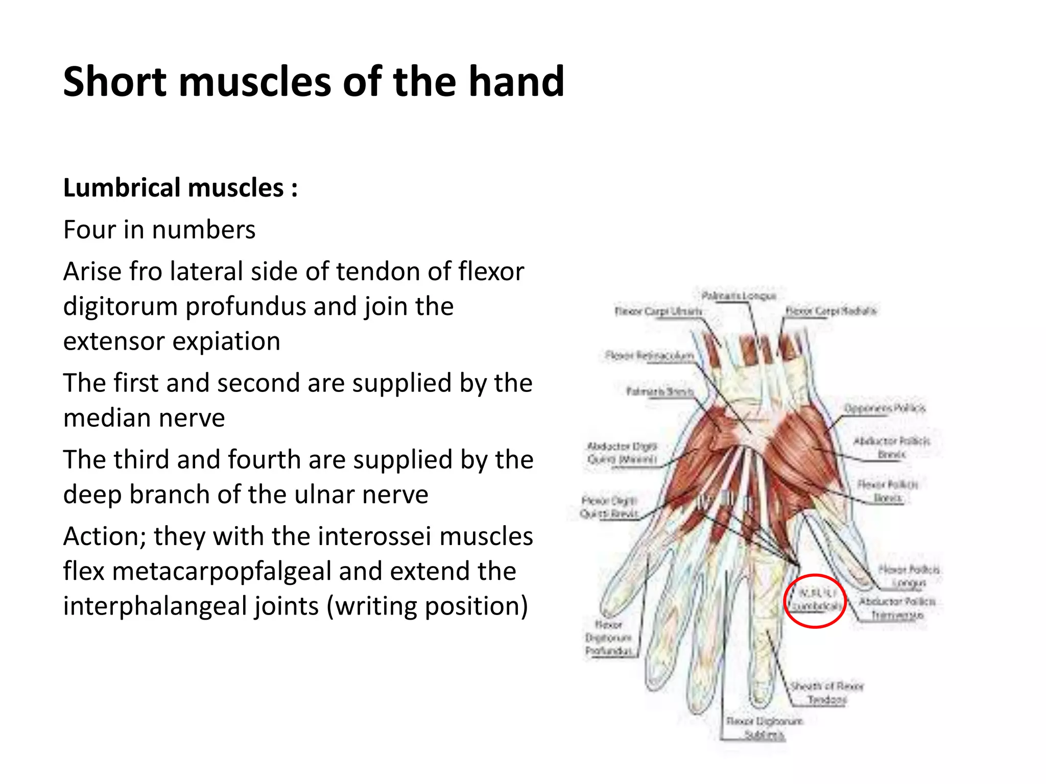
HAND AND
WRIST JOINT
Essam Eldin Abdel Hady Salama
Skeleton of the hand
• Composed of
• Carpus
• Metacarpus
• Phalanges
Carpus
Proximal row
From lateral to medial
Scaphoid
Lunate
Triquitral
Pisiform
Carpus
Distal row
From lateral to medial
trapezium
Trapezoid
Capitate
Hamate
Metacarpus
5 metacarpus
From lateral to medial;
1st,2nd ,3rd,4th,and 5th
Each one is composed of
Proximal end (base)
Shaft
Distal end (head)
Phalanges
14 in number
Two for the thumb (proximal
phalanx and distal or
terminal phalanx)
Three for each of the medial
four fingers (proximal
phalanx, middle phalanx and
distal or terminal phalanx)
Each phalanx is composed of
Proximal end (base)
Shaft
Distal end (head)
Dorsum of the hand
Extensor retinaculum
Attachments:
Laterally; lower part
anterior part of the
radius
Medially; triquetral and
pisiform bones
Structures superficial to the retinaculum
Beginning of cephalic
vein
Beginning of basilic
vein
Terminal branches of
the radial nerve
Structures passing deep to the retinaculum
1st compartment;
Abductor pollicis longus
Extensor pollicis brevis.
2nd compartment;
Extensor carpi radialis longus
3rd compartment;
Extensor pollicis longus
4th compartment;
1. Extensor digitorum
2. Extensor indicis
3. Extensor part of posterior interosseous nerve
4. Extensor part of anterior interosseous artery.
5th compartment;
Extensor digiti minimi.
6th compartment;
Extensor carpi ulnaris.
Synovial sheath of the extensor tendons
Each tendon has its synovial
sheath except extensor
digitorum and extensor
indicic have a common
sheath
They begin just above the
retinaculum down to the
insertion
Except that of extensor
digitorum , extensor indicic
and extensor digiti minimi
which end in the middle of
the dorsum of the hand
Extensor expansion
Formed on the dorsum of the proximal phalanges of the medial 4 fingers by
tendon of extensor digitorum
Each is supplemented by tendons of one lumbrical, two interossei
For the indix receives the extensor indicis and that of little finger receives
extensor digiti minimi
It divides into three slips middle attached to the base of the middle phalanx
and two collateral attached to the base of the terminal phalanx
Palm of the hand
Palmar aponeurosis
Triangular thickening
in deep facial of the
palm
Flexor retinaculum and carpal tunnel
Attachments:
Medially to pisiform and hook of
hamate
Laterally; to tubercle of scaphoid and
crest of trapezium
Structures passing in front of the
retinaculum
Ulnar nerve
Ulnar Artery
Palmar cutaneous branch of Ulnar
nerve
Tendon of palmaris longus
Palmar cutaneous branch of median
nerve
Flexor retinaculum and carpal tunnel
Structures passing deep to the
retinaculum (carpal tunnel)
Tendon of flexor carbi radialis and its
synovial sheath
Tendon of flexor digitorum superficialis
Tendon of flexor digitorum profundus
Tendon of flexor pollicis longus
Median nerve
Synovial sheath of Tendon of flexor pollicis
longus
Common flexor sheath
Short muscles of the thumb
Abductor pollicis brevis
Flexor pollicis brevis
Opponens pollicis
adductor pollicis
Short muscles of the little finger
Abductor digiti minimi
Flexor digiti minimi brevis
Opponens digiti minimi
Short muscles of the hand
Lumbrical muscles :
Four in numbers
Arise fro lateral side of tendon of flexor
digitorum profundus and join the
extensor expiation
The first and second are supplied by the
median nerve
The third and fourth are supplied by the
deep branch of the ulnar nerve
Action; they with the interossei muscles
flex metacarpopfalgeal and extend the
interphalangeal joints (writing position)
Short muscles of the hand
Palmar interossei muscles :
Four in numbers
Arise from the metacarpal bone
on which it acts, and join the
extensor expiation
They are supplied by the deep
branch of the ulnar nerve.
Action; they adduct the fingers to
the middle line of the middle
finger
Short muscles of the hand
Dorsal interossei muscles :
Four in numbers
Arise from the adjacent sides of two
metacarpal bone,
The 1st is inserted in the proximal phalanx
of the index and the extensor expiation.
The 2tnd and 3rd reach the middle finger.
The 4th reaches the little finger
They are supplied by the deep branch of
the ulnar nerve.
Action; they abduct the fingers from the
middle line of the middle finger (fanning
of the fingers).
Ulnar artery
Enters the hand superficial to
the flexor retinaculum
Gives deep branch to
complete the deep palmar
arch
Curves laterally to form the
superficial palmar arch
Superficial palmar arch
Its formed by continuation of the
ulnar artery
It is completed by the superficial
palmar branch of the radial artery.
It lies deep to the palmar
aponeurosis in front of the branches
of median nerve, the long flexor
tendons and lumbrical muscles.
The arch gives four palmar digital
arteries which supply the medical
three and half fingers.
Radial artery
Above the wrist it curves backwards
to run in the floor of the anatomical
snuff box on the lateral ligament of
the wrist.
It is crossed by the tendons of
abductor pollicis longus, beginning of
the cephalic vein and terminal
branches of radial nerve.
It dips between the two heads of the
first dorsal intersseous to reach the
palm where it forms the deep palmar
arch.
Branches
Posterior carpal artery
First dorsal metcarpal artery
Princeps pollicis
Radialis indicis
Deep palmar arch;
It is formed by continaution of the
radial artyey
Lies on inerossi muscles infront of
bases of metacarpal bones deep to
flexor tendons and lumbricals
It is completed by deep branch of ulnar
aretry
It gives 3 palmar metacarpal arteries
3 perforating arteries
Recurrent branches
Median nerve
Enters the palm deep to the flexor
retinaculum
Divides into; medial and lateral
branches
It supplies;
muscles of the thenar eminence
1st and 2nd lumbricals
Lateral three and half fingers
Ulnar nerve
Enters the palm superficial to the
flexor retinaculum
Divides into:
Superficial branch; gives digital
branches to medial one and half
fingers
Deep branches; gives muscular to the
hypothenar muscles
3rd and 4th lumbricals
All palmar and dorsal interossei
Abductor pollicis
Articular to wrist, intermetacarpal,
carpometacarpal, and intercarpal
joints
Wrist joint
It is an ellipsoid synovial joint
Articular surfaces; carpal (inferior)
surface of radius
Inferior surface of the articular disc
Proximal row articular srfaces
Capsular attachment; to the
margins of the articular surfaces
Thickened by palmar, dorsal, ulnar
collateral and radial collateral
ligaments.
Arterial and nerve supply
Arterial supply; anterior
and posterior carpal
arches and deep palmar
arch
Nerve supply; anterior
and posterior interosseis
nerves
Movements of the wrist joint
THANK YOU
and
GOOD LUCK

Hand anatomy 2017 new microsoft power point presentation

  • 1.
    HAND AND WRIST JOINT EssamEldin Abdel Hady Salama
  • 2.
    Skeleton of thehand • Composed of • Carpus • Metacarpus • Phalanges
  • 3.
    Carpus Proximal row From lateralto medial Scaphoid Lunate Triquitral Pisiform
  • 4.
    Carpus Distal row From lateralto medial trapezium Trapezoid Capitate Hamate
  • 5.
    Metacarpus 5 metacarpus From lateralto medial; 1st,2nd ,3rd,4th,and 5th Each one is composed of Proximal end (base) Shaft Distal end (head)
  • 6.
    Phalanges 14 in number Twofor the thumb (proximal phalanx and distal or terminal phalanx) Three for each of the medial four fingers (proximal phalanx, middle phalanx and distal or terminal phalanx) Each phalanx is composed of Proximal end (base) Shaft Distal end (head)
  • 7.
  • 8.
    Extensor retinaculum Attachments: Laterally; lowerpart anterior part of the radius Medially; triquetral and pisiform bones
  • 9.
    Structures superficial tothe retinaculum Beginning of cephalic vein Beginning of basilic vein Terminal branches of the radial nerve
  • 10.
    Structures passing deepto the retinaculum 1st compartment; Abductor pollicis longus Extensor pollicis brevis. 2nd compartment; Extensor carpi radialis longus Extensor carpi radialis longus 3rd compartment; Extensor pollicis longus 4th compartment; 1. Extensor digitorum 2. Extensor indicis 3. Extensor part of posterior interosseous nerve 4. Extensor part of anterior interosseous artery. 5th compartment; Extensor digiti minimi. 6th compartment; Extensor carpi ulnaris.
  • 11.
    Synovial sheath ofthe extensor tendons Each tendon has its synovial sheath except extensor digitorum and extensor indicic have a common sheath They begin just above the retinaculum down to the insertion Except that of extensor digitorum , extensor indicic and extensor digiti minimi which end in the middle of the dorsum of the hand
  • 12.
    Extensor expansion Formed onthe dorsum of the proximal phalanges of the medial 4 fingers by tendon of extensor digitorum Each is supplemented by tendons of one lumbrical, two interossei For the indix receives the extensor indicis and that of little finger receives extensor digiti minimi It divides into three slips middle attached to the base of the middle phalanx and two collateral attached to the base of the terminal phalanx
  • 13.
  • 14.
  • 15.
    Flexor retinaculum andcarpal tunnel Attachments: Medially to pisiform and hook of hamate Laterally; to tubercle of scaphoid and crest of trapezium Structures passing in front of the retinaculum Ulnar nerve Ulnar Artery Palmar cutaneous branch of Ulnar nerve Tendon of palmaris longus Palmar cutaneous branch of median nerve
  • 16.
    Flexor retinaculum andcarpal tunnel Structures passing deep to the retinaculum (carpal tunnel) Tendon of flexor carbi radialis and its synovial sheath Tendon of flexor digitorum superficialis Tendon of flexor digitorum profundus Tendon of flexor pollicis longus Median nerve Synovial sheath of Tendon of flexor pollicis longus Common flexor sheath
  • 17.
    Short muscles ofthe thumb Abductor pollicis brevis Flexor pollicis brevis Opponens pollicis adductor pollicis
  • 18.
    Short muscles ofthe little finger Abductor digiti minimi Flexor digiti minimi brevis Opponens digiti minimi
  • 19.
    Short muscles ofthe hand Lumbrical muscles : Four in numbers Arise fro lateral side of tendon of flexor digitorum profundus and join the extensor expiation The first and second are supplied by the median nerve The third and fourth are supplied by the deep branch of the ulnar nerve Action; they with the interossei muscles flex metacarpopfalgeal and extend the interphalangeal joints (writing position)
  • 20.
    Short muscles ofthe hand Palmar interossei muscles : Four in numbers Arise from the metacarpal bone on which it acts, and join the extensor expiation They are supplied by the deep branch of the ulnar nerve. Action; they adduct the fingers to the middle line of the middle finger
  • 21.
    Short muscles ofthe hand Dorsal interossei muscles : Four in numbers Arise from the adjacent sides of two metacarpal bone, The 1st is inserted in the proximal phalanx of the index and the extensor expiation. The 2tnd and 3rd reach the middle finger. The 4th reaches the little finger They are supplied by the deep branch of the ulnar nerve. Action; they abduct the fingers from the middle line of the middle finger (fanning of the fingers).
  • 22.
    Ulnar artery Enters thehand superficial to the flexor retinaculum Gives deep branch to complete the deep palmar arch Curves laterally to form the superficial palmar arch
  • 23.
    Superficial palmar arch Itsformed by continuation of the ulnar artery It is completed by the superficial palmar branch of the radial artery. It lies deep to the palmar aponeurosis in front of the branches of median nerve, the long flexor tendons and lumbrical muscles. The arch gives four palmar digital arteries which supply the medical three and half fingers.
  • 24.
    Radial artery Above thewrist it curves backwards to run in the floor of the anatomical snuff box on the lateral ligament of the wrist. It is crossed by the tendons of abductor pollicis longus, beginning of the cephalic vein and terminal branches of radial nerve. It dips between the two heads of the first dorsal intersseous to reach the palm where it forms the deep palmar arch.
  • 25.
    Branches Posterior carpal artery Firstdorsal metcarpal artery Princeps pollicis Radialis indicis Deep palmar arch; It is formed by continaution of the radial artyey Lies on inerossi muscles infront of bases of metacarpal bones deep to flexor tendons and lumbricals It is completed by deep branch of ulnar aretry It gives 3 palmar metacarpal arteries 3 perforating arteries Recurrent branches
  • 26.
    Median nerve Enters thepalm deep to the flexor retinaculum Divides into; medial and lateral branches It supplies; muscles of the thenar eminence 1st and 2nd lumbricals Lateral three and half fingers
  • 27.
    Ulnar nerve Enters thepalm superficial to the flexor retinaculum Divides into: Superficial branch; gives digital branches to medial one and half fingers Deep branches; gives muscular to the hypothenar muscles 3rd and 4th lumbricals All palmar and dorsal interossei Abductor pollicis Articular to wrist, intermetacarpal, carpometacarpal, and intercarpal joints
  • 28.
    Wrist joint It isan ellipsoid synovial joint Articular surfaces; carpal (inferior) surface of radius Inferior surface of the articular disc Proximal row articular srfaces Capsular attachment; to the margins of the articular surfaces Thickened by palmar, dorsal, ulnar collateral and radial collateral ligaments.
  • 29.
    Arterial and nervesupply Arterial supply; anterior and posterior carpal arches and deep palmar arch Nerve supply; anterior and posterior interosseis nerves
  • 30.
    Movements of thewrist joint
  • 31.