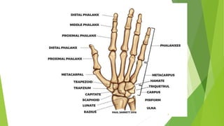
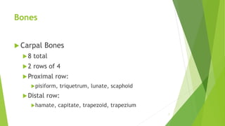
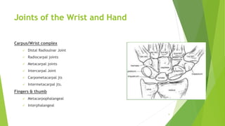
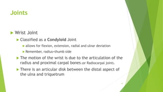
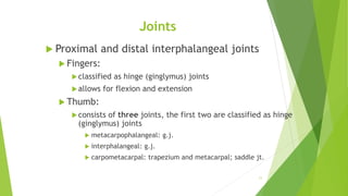
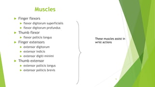
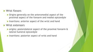
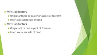
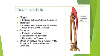
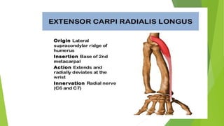
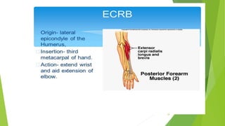
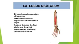
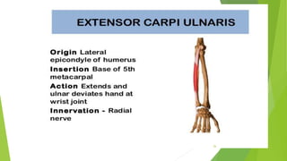
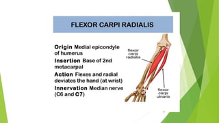
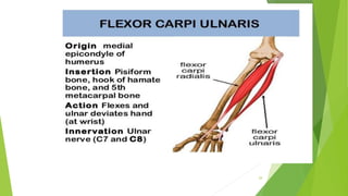
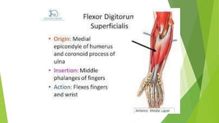
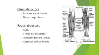
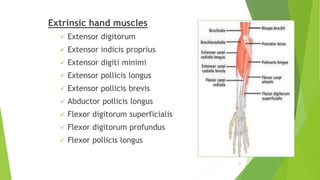
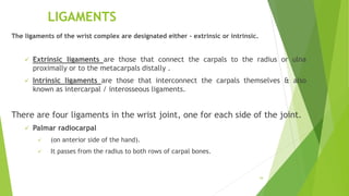
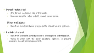
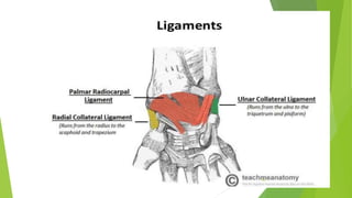
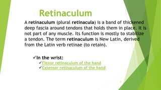
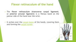
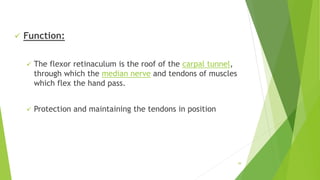
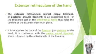
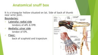
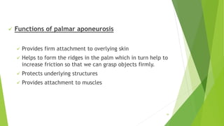
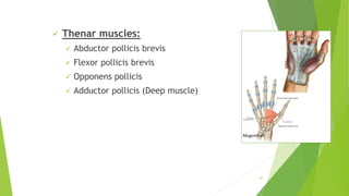
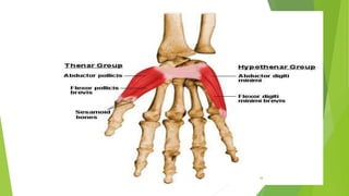
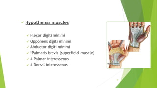
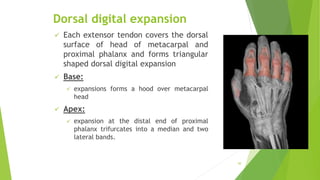
Wrist and Hand
1
Contents
 Wrist joint
 Hand
 Bones
 Joints
 Ligaments
 Muscles
 Retinaculum
 Palmer aponeurois
 Dorsal digital expansion
2
Terminology
 Wrist:
 as a joint, it is the articulation between the forearm and
hand.
 Hand:
 the bones distal to the wrist joint.
 As a region, the wrist is part of the hand.
3
4
Bones
 29 bones including:
 (2 from the forearm, 27 from the hand)
 distal aspect of the radius and ulna
 8 carpal (bulk of the hand)
 14 phalanges (the fingers)
 5 metacarpals
 total: 2+8+14+5=29
5
Bones
 Radius and Ulna
 the radius is more massive distally than the ulna
 the ulna is more massive proximally than the radius
 Remember, forearm pronation and supination are the result of the
radius rotating about the ulna
6
Bones
 Carpal Bones
8 total
2 rows of 4
Proximal row:
pisiform, triquetrum, lunate, scaphoid
Distal row:
hamate, capitate, trapezoid, trapezium
7
Bones
Metacarpals
5
Phalanges
3 per finger
2 per thumb
8
Joints of the Wrist and Hand
Carpus/Wrist complex
 Distal Radioulnar Joint
 Radiocarpal joints
 Metacarpal joints
 Intercarpal Joint
 Carpometacarpal jts
 Intermetacarpal jts.
Fingers & thumb
 Metacarpophalangeal
 Interphalangeal
9
Joints
 Wrist Joint
 Classified as a Condyloid Joint
 allows for flexion, extension, radial and ulnar deviation
 Remember, radius=thumb side
 The motion of the wrist is due to the articulation of the
radius and proximal carpal bones (or Radiocarpal joint).
 There is an articular disk between the distal aspect of
the ulna and triquetrum
10
Joints
 Proximal and distal interphalangeal joints
 Fingers:
classified as hinge (ginglymus) joints
allows for flexion and extension
 Thumb:
consists of three joints, the first two are classified as hinge
(ginglymus) joints
 metacarpophalangeal: g.j.
 interphalangeal: g.j.
 carpometacarpal: trapezium and metacarpal; saddle jt.
11
Movements of the wrist and hand
 Wrist (remember start from anatomical position)
 Flexion
 (or palmer flexion): anterior deviation from anatomical position.
 Extension: dorsal flexion
 Abduction
 (or radial deviation or radial flexion)
 Adduction
 (or ulnar deviation or ulnar flexion)
12
Movements
 Fingers
 Flexion: anteriorly directed movement
 Extension
 Abduction: movement of the fingers away from the midline of the
hand.
 Adduction
 Thumb opposition: movement of the thumb to oppose any of the
phalanges
13
Muscles of the Wrist and Fingers
 There are 15 critical muscles
 6 muscles are group as primary movers for wrist flexion and extension actions
 9 muscles are group as primary movers for hand (finger)
 of the 15 muscles
 6 are primary movers for wrist ulnar deviation
 2 are primary movers for wrist radial deviation
14
Muscles causing wrist actions
 Wrist flexors
 flexor carpi radialis
 flexor carpi ulnaris
 palmaris longus
 Wrist extensors
 extensor carpi radialis longus
 extensor carpi radialis brevis
 extensor carpi ulnaris
15
These muscles do not cause
finger movements
Muscles
 Finger flexors
 flexor digitorum superficialis
 flexor digitorum profundus
 Thumb flexor
 flexor pollicis longus
 Finger extensors
 extensor digitorum
 extensor indicis
 extensor digiti minimi
 Thumb extensor
 extensor pollicis longus
 extensor pollicis brevis
16
These muscles assist in
wrist actions
Muscles
 Wrist radial flexors
 flexor carpi radialis
 extensor carpi radialis longus
 extensor carpi radialis brevis
 abductor pollicis longus
 extensor pollicis longus
 extensor pollicis brevis
17
Muscles
 Wrist ulnar flexors
 flexor carpi ulnaris
 extensor carpi ulnaris
18
 Wrist flexors
 Origins generally on the anteromedial aspect of the
proximal aspect of the forearm and medial epicondyle
 Insertions: anterior aspect of the wrist and hand
 Wrist extensors
 origins: posterolateral aspect of the proximal forearm &
lateral humeral epicondyle
 insertions: posterior aspect of wrist and hand
19
 Wrist abductors
 Origin: anterior or posterior aspect of forearm
 Insertion: radial side of hand
 Wrist adductors
 Origin: ant or post aspect of forearm
 Insertion: ulnar side of hand
20
Muscles of the Wrist and Hand
Wrist extensors
 Brachioradialis
 ECRL
 ECRB
 Extensor digitorum
 Extensor carpi ulnaris
Wrist flexors
 Palmaris longus
 Flexor carpi radialis
 Flexor carpi ulnaris
 Flexor digitorum superficialis 21
22
23
24
25
26
27
28
29
30
Ulnar abductors
 Extensor carpi ulnaris
 Flexor carpi ulnaris
Radial abductors
 ECRL
 Flexor carpi radialis
 Abductor pollicis longus
 Extensor pollicis brevis
31
Extrinsic hand muscles
 Extensor digitorum
 Extensor indicis proprius
 Extensor digiti minimi
 Extensor pollicis longus
 Extensor pollicis brevis
 Abductor pollicis longus
 Flexor digitorum superficialis
 Flexor digitorum profundus
 Flexor pollicis longus
32
JOINT CAPSULE
(Capsular Ligament)
 Like any synovial joint, the capsule is double
layered.
 The fibrous outer layer attaches to the distal ends
of radius, ulna and the proximal row of the carpal
bones.
 The internal layer, synovial membrane extends up
to the margins of the articular surfaces.
33
LIGAMENTS
The ligaments of the wrist complex are designated either – extrinsic or intrinsic.
 Extrinsic ligaments are those that connect the carpals to the radius or ulna
proximally or to the metacarpals distally .
 Intrinsic ligaments are those that interconnect the carpals themselves & also
known as intercarpal / interosseous ligaments.
There are four ligaments in the wrist joint, one for each side of the joint.
 Palmar radiocarpal
 (on anterior side of the hand).
 It passes from the radius to both rows of carpal bones.
34
 Dorsal radiocarpal
 (the dorsum (posterior) side of the hand).
 It passes from the radius to both rows of carpal bones.
Ulnar collateral
 Runs from the ulnar styloid process to the triquetrum and pisiform.
 Radial collateral
 Runs from the radial styloid process to the scaphoid and trapezium.
 Works in union with the other collateral ligament to prevent
excessive lateral joint displacement.
35
36
Retinaculum
37
A retinaculum (plural retinacula) is a band of thickened
deep fascia around tendons that holds them in place. It is
not part of any muscle. Its function is mostly to stabilize
a tendon. The term retinaculum is New Latin, derived
from the Latin verb retinae (to retain).
In the wrist:
Flexor retinaculum of the hand
Extensor retinaculum of the hand
Flexor retinaculum of the hand
 The flexor retinaculum (transverse carpal ligament,
or anterior annular ligament) is a fibrous band on the
palmar side of the hand near the wrist.
 It arches over the carpal bones of the hands, covering them
and forming the carpal tunnel.
38
 Function:
 The flexor retinaculum is the roof of the carpal tunnel,
through which the median nerve and tendons of muscles
which flex the hand pass.
 Protection and maintaining the tendons in position
39
Extensor retinaculum of the hand
 The extensor retinaculum (dorsal carpal ligament,
or posterior annular ligament) is an anatomical term for
the thickened part of the antebrachial fascia that holds the
tendons of the extensor muscles in place.
 It is located on the back of the forearm, just proximal to the
hand. It is continuous with the palmar carpal ligament,
which is located on the anterior side of the forearm.
40
Anatomical snuff box
It is a triangular hollow situated on lat. Side of back of thumb
near wrist joint.
Boundaries:
 Laterally: radial side
tendons of APL & EPB
 Medially: ulnar side
tendon of EPL
 Floor:
back of scaphoid and trapezium
41
PALMAR APONEUROSIS
42
PALMAR APONEUROSIS
 Also called palmar fascia
 invests the muscles of the palm
 consists of central, lateral, and medial portions.
43
 Functions of palmar aponeurosis
 Provides firm attachment to overlying skin
 Helps to form the ridges in the palm which in turn help to
increase friction so that we can grasp objects firmly.
 Protects underlying structures
 Provides attachment to muscles
44
Flexor Tendon Dorsal Digital
Expansion
45
dorsal digital expansion. a triangular
aponeurotic extension of
the digital extensor tendon on the dorsum
of the proximal phalanx of each digit, to
which the tendonsof the lumbrical and
interosseous muscles are also attached. It
forms a movable hood around the
metacarpophalangeal joint.
46
 Thenar muscles:
 Abductor pollicis brevis
 Flexor pollicis brevis
 Opponens pollicis
 Adductor pollicis (Deep muscle)
47
48
 Hypothenar muscles
 Flexor digiti minimi
 Opponens digiti minimi
 Abductor digiti minimi
 *Palmaris brevis (superficial muscle)
 4 Palmar interosseous
 4 Dorsal interosseous
49
Dorsal digital expansion
 Each extensor tendon covers the dorsal
surface of head of metacarpal and
proximal phalanx and forms triangular
shaped dorsal digital expansion
 Base:
 expansions forms a hood over metacarpal
head
 Apex:
 expansion at the distal end of proximal
phalanx trifurcates into a median and two
lateral bands.
50
Summary
 The wrist is a complex joint that bridges
the hand to the forearm. It is actually a
collection of multiple bones and joints. The
bones comprising the wrist include the distal
ends of the radius and ulna, 8 carpal bones,
and the proximal portions of the 5
metacarpal bones.
51

Lec#9-B.WRIST JOINT & HAND.presentation.

  • 1.
  • 2.
    Contents  Wrist joint Hand  Bones  Joints  Ligaments  Muscles  Retinaculum  Palmer aponeurois  Dorsal digital expansion 2
  • 3.
    Terminology  Wrist:  asa joint, it is the articulation between the forearm and hand.  Hand:  the bones distal to the wrist joint.  As a region, the wrist is part of the hand. 3
  • 4.
  • 5.
    Bones  29 bonesincluding:  (2 from the forearm, 27 from the hand)  distal aspect of the radius and ulna  8 carpal (bulk of the hand)  14 phalanges (the fingers)  5 metacarpals  total: 2+8+14+5=29 5
  • 6.
    Bones  Radius andUlna  the radius is more massive distally than the ulna  the ulna is more massive proximally than the radius  Remember, forearm pronation and supination are the result of the radius rotating about the ulna 6
  • 7.
    Bones  Carpal Bones 8total 2 rows of 4 Proximal row: pisiform, triquetrum, lunate, scaphoid Distal row: hamate, capitate, trapezoid, trapezium 7
  • 8.
  • 9.
    Joints of theWrist and Hand Carpus/Wrist complex  Distal Radioulnar Joint  Radiocarpal joints  Metacarpal joints  Intercarpal Joint  Carpometacarpal jts  Intermetacarpal jts. Fingers & thumb  Metacarpophalangeal  Interphalangeal 9
  • 10.
    Joints  Wrist Joint Classified as a Condyloid Joint  allows for flexion, extension, radial and ulnar deviation  Remember, radius=thumb side  The motion of the wrist is due to the articulation of the radius and proximal carpal bones (or Radiocarpal joint).  There is an articular disk between the distal aspect of the ulna and triquetrum 10
  • 11.
    Joints  Proximal anddistal interphalangeal joints  Fingers: classified as hinge (ginglymus) joints allows for flexion and extension  Thumb: consists of three joints, the first two are classified as hinge (ginglymus) joints  metacarpophalangeal: g.j.  interphalangeal: g.j.  carpometacarpal: trapezium and metacarpal; saddle jt. 11
  • 12.
    Movements of thewrist and hand  Wrist (remember start from anatomical position)  Flexion  (or palmer flexion): anterior deviation from anatomical position.  Extension: dorsal flexion  Abduction  (or radial deviation or radial flexion)  Adduction  (or ulnar deviation or ulnar flexion) 12
  • 13.
    Movements  Fingers  Flexion:anteriorly directed movement  Extension  Abduction: movement of the fingers away from the midline of the hand.  Adduction  Thumb opposition: movement of the thumb to oppose any of the phalanges 13
  • 14.
    Muscles of theWrist and Fingers  There are 15 critical muscles  6 muscles are group as primary movers for wrist flexion and extension actions  9 muscles are group as primary movers for hand (finger)  of the 15 muscles  6 are primary movers for wrist ulnar deviation  2 are primary movers for wrist radial deviation 14
  • 15.
    Muscles causing wristactions  Wrist flexors  flexor carpi radialis  flexor carpi ulnaris  palmaris longus  Wrist extensors  extensor carpi radialis longus  extensor carpi radialis brevis  extensor carpi ulnaris 15 These muscles do not cause finger movements
  • 16.
    Muscles  Finger flexors flexor digitorum superficialis  flexor digitorum profundus  Thumb flexor  flexor pollicis longus  Finger extensors  extensor digitorum  extensor indicis  extensor digiti minimi  Thumb extensor  extensor pollicis longus  extensor pollicis brevis 16 These muscles assist in wrist actions
  • 17.
    Muscles  Wrist radialflexors  flexor carpi radialis  extensor carpi radialis longus  extensor carpi radialis brevis  abductor pollicis longus  extensor pollicis longus  extensor pollicis brevis 17
  • 18.
    Muscles  Wrist ulnarflexors  flexor carpi ulnaris  extensor carpi ulnaris 18
  • 19.
     Wrist flexors Origins generally on the anteromedial aspect of the proximal aspect of the forearm and medial epicondyle  Insertions: anterior aspect of the wrist and hand  Wrist extensors  origins: posterolateral aspect of the proximal forearm & lateral humeral epicondyle  insertions: posterior aspect of wrist and hand 19
  • 20.
     Wrist abductors Origin: anterior or posterior aspect of forearm  Insertion: radial side of hand  Wrist adductors  Origin: ant or post aspect of forearm  Insertion: ulnar side of hand 20
  • 21.
    Muscles of theWrist and Hand Wrist extensors  Brachioradialis  ECRL  ECRB  Extensor digitorum  Extensor carpi ulnaris Wrist flexors  Palmaris longus  Flexor carpi radialis  Flexor carpi ulnaris  Flexor digitorum superficialis 21
  • 22.
  • 23.
  • 24.
  • 25.
  • 26.
  • 27.
  • 28.
  • 29.
  • 30.
  • 31.
    Ulnar abductors  Extensorcarpi ulnaris  Flexor carpi ulnaris Radial abductors  ECRL  Flexor carpi radialis  Abductor pollicis longus  Extensor pollicis brevis 31
  • 32.
    Extrinsic hand muscles Extensor digitorum  Extensor indicis proprius  Extensor digiti minimi  Extensor pollicis longus  Extensor pollicis brevis  Abductor pollicis longus  Flexor digitorum superficialis  Flexor digitorum profundus  Flexor pollicis longus 32
  • 33.
    JOINT CAPSULE (Capsular Ligament) Like any synovial joint, the capsule is double layered.  The fibrous outer layer attaches to the distal ends of radius, ulna and the proximal row of the carpal bones.  The internal layer, synovial membrane extends up to the margins of the articular surfaces. 33
  • 34.
    LIGAMENTS The ligaments ofthe wrist complex are designated either – extrinsic or intrinsic.  Extrinsic ligaments are those that connect the carpals to the radius or ulna proximally or to the metacarpals distally .  Intrinsic ligaments are those that interconnect the carpals themselves & also known as intercarpal / interosseous ligaments. There are four ligaments in the wrist joint, one for each side of the joint.  Palmar radiocarpal  (on anterior side of the hand).  It passes from the radius to both rows of carpal bones. 34
  • 35.
     Dorsal radiocarpal (the dorsum (posterior) side of the hand).  It passes from the radius to both rows of carpal bones. Ulnar collateral  Runs from the ulnar styloid process to the triquetrum and pisiform.  Radial collateral  Runs from the radial styloid process to the scaphoid and trapezium.  Works in union with the other collateral ligament to prevent excessive lateral joint displacement. 35
  • 36.
  • 37.
    Retinaculum 37 A retinaculum (pluralretinacula) is a band of thickened deep fascia around tendons that holds them in place. It is not part of any muscle. Its function is mostly to stabilize a tendon. The term retinaculum is New Latin, derived from the Latin verb retinae (to retain). In the wrist: Flexor retinaculum of the hand Extensor retinaculum of the hand
  • 38.
    Flexor retinaculum ofthe hand  The flexor retinaculum (transverse carpal ligament, or anterior annular ligament) is a fibrous band on the palmar side of the hand near the wrist.  It arches over the carpal bones of the hands, covering them and forming the carpal tunnel. 38
  • 39.
     Function:  Theflexor retinaculum is the roof of the carpal tunnel, through which the median nerve and tendons of muscles which flex the hand pass.  Protection and maintaining the tendons in position 39
  • 40.
    Extensor retinaculum ofthe hand  The extensor retinaculum (dorsal carpal ligament, or posterior annular ligament) is an anatomical term for the thickened part of the antebrachial fascia that holds the tendons of the extensor muscles in place.  It is located on the back of the forearm, just proximal to the hand. It is continuous with the palmar carpal ligament, which is located on the anterior side of the forearm. 40
  • 41.
    Anatomical snuff box Itis a triangular hollow situated on lat. Side of back of thumb near wrist joint. Boundaries:  Laterally: radial side tendons of APL & EPB  Medially: ulnar side tendon of EPL  Floor: back of scaphoid and trapezium 41
  • 42.
  • 43.
    PALMAR APONEUROSIS  Alsocalled palmar fascia  invests the muscles of the palm  consists of central, lateral, and medial portions. 43
  • 44.
     Functions ofpalmar aponeurosis  Provides firm attachment to overlying skin  Helps to form the ridges in the palm which in turn help to increase friction so that we can grasp objects firmly.  Protects underlying structures  Provides attachment to muscles 44
  • 45.
    Flexor Tendon DorsalDigital Expansion 45 dorsal digital expansion. a triangular aponeurotic extension of the digital extensor tendon on the dorsum of the proximal phalanx of each digit, to which the tendonsof the lumbrical and interosseous muscles are also attached. It forms a movable hood around the metacarpophalangeal joint.
  • 46.
  • 47.
     Thenar muscles: Abductor pollicis brevis  Flexor pollicis brevis  Opponens pollicis  Adductor pollicis (Deep muscle) 47
  • 48.
  • 49.
     Hypothenar muscles Flexor digiti minimi  Opponens digiti minimi  Abductor digiti minimi  *Palmaris brevis (superficial muscle)  4 Palmar interosseous  4 Dorsal interosseous 49
  • 50.
    Dorsal digital expansion Each extensor tendon covers the dorsal surface of head of metacarpal and proximal phalanx and forms triangular shaped dorsal digital expansion  Base:  expansions forms a hood over metacarpal head  Apex:  expansion at the distal end of proximal phalanx trifurcates into a median and two lateral bands. 50
  • 51.
    Summary  The wristis a complex joint that bridges the hand to the forearm. It is actually a collection of multiple bones and joints. The bones comprising the wrist include the distal ends of the radius and ulna, 8 carpal bones, and the proximal portions of the 5 metacarpal bones. 51