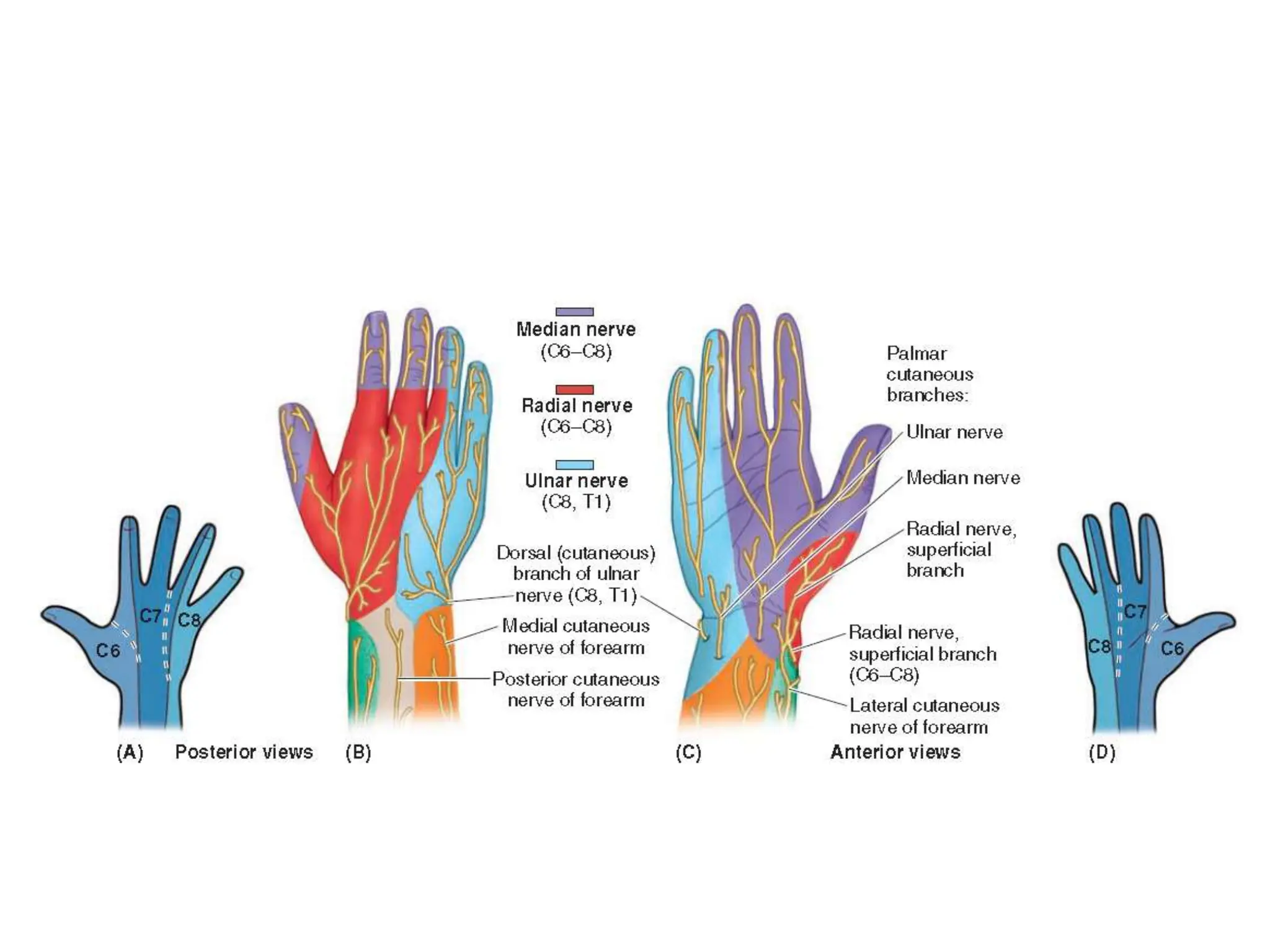
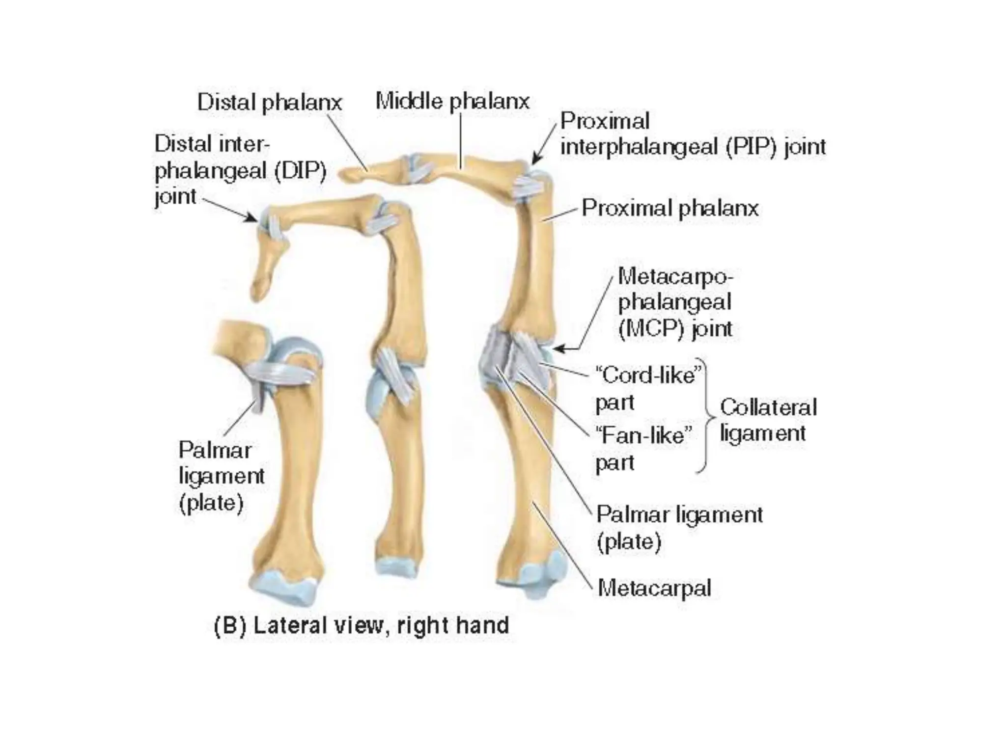
Hand, wrist and joints of the
hand
Introduction
• The hand is the manual part of the upper limb distal to the forearm.
• The wrist is located at the junction of the forearm and hand.
• The skeleton of the hand consists of carpals in the wrist, metacarpals in
the hand proper, and phalanges in the digits (fingers).
• The digits are numbered from one to five, beginning with the thumb:
digit 1 is the thumb; digit 2, the index finger; digit 3, the middle finger;
digit 4, the ring finger; and digit 5, the little finger.
• The hand has palmar aspect and dorsal aspect.
• The palmar aspect of the hand features a central concavity that, with the
crease proximal to it (over the wrist bones) separates two eminences: a
lateral, larger and more prominent thenar eminence at the base of the
thumb, and a medial, smaller hypothenar eminence proximal to the
base of the 5th finger.
• Function include free motion, power grasping, precision handling, and
pinching.
Wrist
• Has two parts, anterior and posterior
• Anterior part has the flexor retinaculum
– flexor carpi ulnaris tendon, ulnar nerve, ulnar
artery, palmar cutaneous branch of the ulnar
nerve, palmaris longus tendon, palmar cutaneous
branch of the median nerve. Structures passing
deep to the flexor retinaculum: flexor digitorum
superficialis tendon, median nerve, flexor pollicis
longus tendon, flexor carpi radialis tendon.
• Posterior Compartment has the extensor
compartment
Bones of the wrist
• Has two rows: proximal
and distal
– Carpal bones (bones of
the wrist or carpus)
• Proximal row: Scaphoid,
lunate, triquetrum,
pisiform,
• Distal row: Trapezium,
trapezoid, capitate,
hamate
Palmer surface of the hand
• Has skin
• Superficial
fascia
• Deep fascia
(flexor
retinaculum,
flexor
sheath,
palmar
aponeuroses
Muscles of Hand
The intrinsic muscles of the hand are located in five
compartments
1. Thenar muscles in the thenar compartment: abductor
pollicis brevis, flexor pollicis brevis, and opponens pollicis.
2. Adductor pollicis in the adductor compartment.
3. Hypothenar muscles in the hypothenar compartment:
abductor digiti minimi, flexor digiti minimi brevis, and
opponens digiti minimi.
4. Short muscles of the hand, the lumbricals, in the central
compartment with the long flexor tendons.
5. The interossei in separate interosseous compartments
between the metacarpals.
Thenar muscles
• Forms the thenar eminence on
lateral surface
• Muscles: opponens pollicis,
abductor pollicis brevis, flexor
pollicis brevis adductor pollicis
• Movements: extension, flexion,
abduction, adduction opposition
• Supplied by recurrent branch of
median nerve except deep head
of FPL and adductor policis which
is by Deep branch of ulnar nerve
Hypothenar muscles
• Forms the hypohenar
eminence on medial
surface
• Muscles: adbuctor digiti
minimi, flexor digiti
minimi brevis, opponens
digiti minimi, palmaris
brevis
• Movements: extension,
flexion, abduction,
adduction opposition
• Supplied by deep branch
of ulnar nerve
Short muscles
• Four slender
lumbricals flex the
fingers at MCP joints
and extendd IP joints
• Four dorsal interossei
and three palmar
interossei
• Dorsal abduct, palmar
adduct.
• Median nerve supplies
1 & 2 lumbricals, rest is
by deep branch of
ulnar
Arteries of the hand
• Two major arteries are ulnar and radial arteries
• Ulnar enters hand via Guyon’s canal (between pisiform and
hook of harmate)
• Divides into two to form superficial and deep palmar arches
• Superficial (main branch) gives 3 common palmar digital
arteries.
• Common palmar gives rise to a pair of proper digital along
adjacent sides of 2-5 digits
• Radial enters via antomical snuff box, forms deep palmar arch
with ulanr artery, gives 3 palmer metacarpal arteries, princeps
policis artery and radialis artery.
• Dorsal carpal arch gives dorsal metacarpal and dorsal digital
Veins
• Superficial and deep venous palmar arches
drain into deep veins of forearm
• Dorsal digital vein drain into three dorsal
metacarpal veins and forms dorsal venous
network
• This continues laterally as the cephalic vein
• Basilic vein arises from the medial side of the
dorsal venous network.
Nerves of the hand
• Three major nerves: ulnar, radial and median
• They give both motor and sensory innervation to the hand.
• Ulnar nerve
– Gives off palmar cutaneous branch proximal to the wrist
– Dorsal cutaneous branch supplies medial half of dorsum of the
hand, 5th finger and medial half of 4th finger
– At distal part of flexor retinaculum divides into superfical and
deep
– Deep suplies hypothenar muscles, 3 and 4 lumbricals adductor
policis, all interossei and deep head of FPL
– Superficial supplies anterior surfaces of medial one and half
digits
Nerves of the hand
• Median nerve
– Enters through carpal tunnel deep to flexor retinaculum
– Supplies 2 and half of thenar muscles, 1 & 2 lumbricals distal to
the tunnel
– Sensory innervation of entire palmar surface (palmar cutaneous
branch), sides of first three digits, lateral half of 4th digit
• Radial nerve
– Supplies no hand muscles
– Superficial branch is entirely sensory
– Supplies the skin, fascia over the lateral 2/3 of the dorsum of
hand, thumb and proximal portions of the dorsal aspects of
digits 2 and 3 and lateral half of digit 4
Joints of the hand
• Intercarpal joints
– Joints between carpal bones of first row and joints
between carpal bones of second row
– Midcarpal joint between first and second rows
– Supported by anterior, posterior, and interosseous
ligaments
– Function as a single unit
– Small gliding movements between carpal bones
Joints of the hand
• Carpometacarpal joints
– Plane type synovial joints, except for carpometacarpal of
thumb (saddle type)
– Medial four carpometacarpal joints in one fibrous joint
capsule
– Separate capsule for thumb
– Joint for thumb between the trapezium and first
metacarpal
• Allows flexion, extension, abduction, adduction, circumduction, and
opposition
• Loose joint capsule allows for free movement
Joints of the hand
• Metacarpophalangeal joints (MCP)
– Heads of metacarpals articulate with base of
proximal phalanx
– Deep transverse metacarpal ligaments hold heads
of metacarpals 2 through 5 together
– Separate joint capsule for each joint
– Movements: flexion, extension, abduction, and
adduction
– Because the collateral ligaments tighten during
flexion, abduction, and adduction are only
possible in the extended position
Joints of the hand
• Interphalangeal joints
– Proximal interphalangeal joint = PIP
– Distal interphalangeal joint = DIP
– Allow flexion and extension
Joints of the hand
• Intercarpal joints
• Carpometacarpal joints
• Metacarpophalangeal joints (MCP)
• Interphalangeal joints
Some clinical anatomy
• Dupuytren Contracture of Palmar fascia:
– disease of palmar fasica resulting in progressive
shortening, thickening and fibrosis of palmar
fascia and aponeurosis
• Carpal tunnel syndrome:
– affects median nerve
– reduction in size of carpal tunnel.
– May be caused by fluid retention, infection,
excessive exercise of finger leading to swelling of
synovial sheaths.
Some clinical anatomy
• Ulnar canal sydrome:
– Compression of ulnar nerve in Guyon’s canal
– Hyothesis may occur in medial one and half and
intrinsic muscle of hand.

Hand, wrist and forearm gross anatomy .pptx

  • 1.
    Hand, wrist andjoints of the hand
  • 2.
    Introduction • The handis the manual part of the upper limb distal to the forearm. • The wrist is located at the junction of the forearm and hand. • The skeleton of the hand consists of carpals in the wrist, metacarpals in the hand proper, and phalanges in the digits (fingers). • The digits are numbered from one to five, beginning with the thumb: digit 1 is the thumb; digit 2, the index finger; digit 3, the middle finger; digit 4, the ring finger; and digit 5, the little finger. • The hand has palmar aspect and dorsal aspect. • The palmar aspect of the hand features a central concavity that, with the crease proximal to it (over the wrist bones) separates two eminences: a lateral, larger and more prominent thenar eminence at the base of the thumb, and a medial, smaller hypothenar eminence proximal to the base of the 5th finger. • Function include free motion, power grasping, precision handling, and pinching.
  • 4.
    Wrist • Has twoparts, anterior and posterior • Anterior part has the flexor retinaculum – flexor carpi ulnaris tendon, ulnar nerve, ulnar artery, palmar cutaneous branch of the ulnar nerve, palmaris longus tendon, palmar cutaneous branch of the median nerve. Structures passing deep to the flexor retinaculum: flexor digitorum superficialis tendon, median nerve, flexor pollicis longus tendon, flexor carpi radialis tendon. • Posterior Compartment has the extensor compartment
  • 5.
    Bones of thewrist • Has two rows: proximal and distal – Carpal bones (bones of the wrist or carpus) • Proximal row: Scaphoid, lunate, triquetrum, pisiform, • Distal row: Trapezium, trapezoid, capitate, hamate
  • 7.
    Palmer surface ofthe hand • Has skin • Superficial fascia • Deep fascia (flexor retinaculum, flexor sheath, palmar aponeuroses
  • 8.
    Muscles of Hand Theintrinsic muscles of the hand are located in five compartments 1. Thenar muscles in the thenar compartment: abductor pollicis brevis, flexor pollicis brevis, and opponens pollicis. 2. Adductor pollicis in the adductor compartment. 3. Hypothenar muscles in the hypothenar compartment: abductor digiti minimi, flexor digiti minimi brevis, and opponens digiti minimi. 4. Short muscles of the hand, the lumbricals, in the central compartment with the long flexor tendons. 5. The interossei in separate interosseous compartments between the metacarpals.
  • 9.
    Thenar muscles • Formsthe thenar eminence on lateral surface • Muscles: opponens pollicis, abductor pollicis brevis, flexor pollicis brevis adductor pollicis • Movements: extension, flexion, abduction, adduction opposition • Supplied by recurrent branch of median nerve except deep head of FPL and adductor policis which is by Deep branch of ulnar nerve
  • 10.
    Hypothenar muscles • Formsthe hypohenar eminence on medial surface • Muscles: adbuctor digiti minimi, flexor digiti minimi brevis, opponens digiti minimi, palmaris brevis • Movements: extension, flexion, abduction, adduction opposition • Supplied by deep branch of ulnar nerve
  • 11.
    Short muscles • Fourslender lumbricals flex the fingers at MCP joints and extendd IP joints • Four dorsal interossei and three palmar interossei • Dorsal abduct, palmar adduct. • Median nerve supplies 1 & 2 lumbricals, rest is by deep branch of ulnar
  • 12.
    Arteries of thehand • Two major arteries are ulnar and radial arteries • Ulnar enters hand via Guyon’s canal (between pisiform and hook of harmate) • Divides into two to form superficial and deep palmar arches • Superficial (main branch) gives 3 common palmar digital arteries. • Common palmar gives rise to a pair of proper digital along adjacent sides of 2-5 digits • Radial enters via antomical snuff box, forms deep palmar arch with ulanr artery, gives 3 palmer metacarpal arteries, princeps policis artery and radialis artery. • Dorsal carpal arch gives dorsal metacarpal and dorsal digital
  • 14.
    Veins • Superficial anddeep venous palmar arches drain into deep veins of forearm • Dorsal digital vein drain into three dorsal metacarpal veins and forms dorsal venous network • This continues laterally as the cephalic vein • Basilic vein arises from the medial side of the dorsal venous network.
  • 15.
    Nerves of thehand • Three major nerves: ulnar, radial and median • They give both motor and sensory innervation to the hand. • Ulnar nerve – Gives off palmar cutaneous branch proximal to the wrist – Dorsal cutaneous branch supplies medial half of dorsum of the hand, 5th finger and medial half of 4th finger – At distal part of flexor retinaculum divides into superfical and deep – Deep suplies hypothenar muscles, 3 and 4 lumbricals adductor policis, all interossei and deep head of FPL – Superficial supplies anterior surfaces of medial one and half digits
  • 18.
    Nerves of thehand • Median nerve – Enters through carpal tunnel deep to flexor retinaculum – Supplies 2 and half of thenar muscles, 1 & 2 lumbricals distal to the tunnel – Sensory innervation of entire palmar surface (palmar cutaneous branch), sides of first three digits, lateral half of 4th digit • Radial nerve – Supplies no hand muscles – Superficial branch is entirely sensory – Supplies the skin, fascia over the lateral 2/3 of the dorsum of hand, thumb and proximal portions of the dorsal aspects of digits 2 and 3 and lateral half of digit 4
  • 21.
    Joints of thehand • Intercarpal joints – Joints between carpal bones of first row and joints between carpal bones of second row – Midcarpal joint between first and second rows – Supported by anterior, posterior, and interosseous ligaments – Function as a single unit – Small gliding movements between carpal bones
  • 22.
    Joints of thehand • Carpometacarpal joints – Plane type synovial joints, except for carpometacarpal of thumb (saddle type) – Medial four carpometacarpal joints in one fibrous joint capsule – Separate capsule for thumb – Joint for thumb between the trapezium and first metacarpal • Allows flexion, extension, abduction, adduction, circumduction, and opposition • Loose joint capsule allows for free movement
  • 23.
    Joints of thehand • Metacarpophalangeal joints (MCP) – Heads of metacarpals articulate with base of proximal phalanx – Deep transverse metacarpal ligaments hold heads of metacarpals 2 through 5 together – Separate joint capsule for each joint – Movements: flexion, extension, abduction, and adduction – Because the collateral ligaments tighten during flexion, abduction, and adduction are only possible in the extended position
  • 24.
    Joints of thehand • Interphalangeal joints – Proximal interphalangeal joint = PIP – Distal interphalangeal joint = DIP – Allow flexion and extension
  • 25.
    Joints of thehand • Intercarpal joints • Carpometacarpal joints • Metacarpophalangeal joints (MCP) • Interphalangeal joints
  • 28.
    Some clinical anatomy •Dupuytren Contracture of Palmar fascia: – disease of palmar fasica resulting in progressive shortening, thickening and fibrosis of palmar fascia and aponeurosis • Carpal tunnel syndrome: – affects median nerve – reduction in size of carpal tunnel. – May be caused by fluid retention, infection, excessive exercise of finger leading to swelling of synovial sheaths.
  • 29.
    Some clinical anatomy •Ulnar canal sydrome: – Compression of ulnar nerve in Guyon’s canal – Hyothesis may occur in medial one and half and intrinsic muscle of hand.