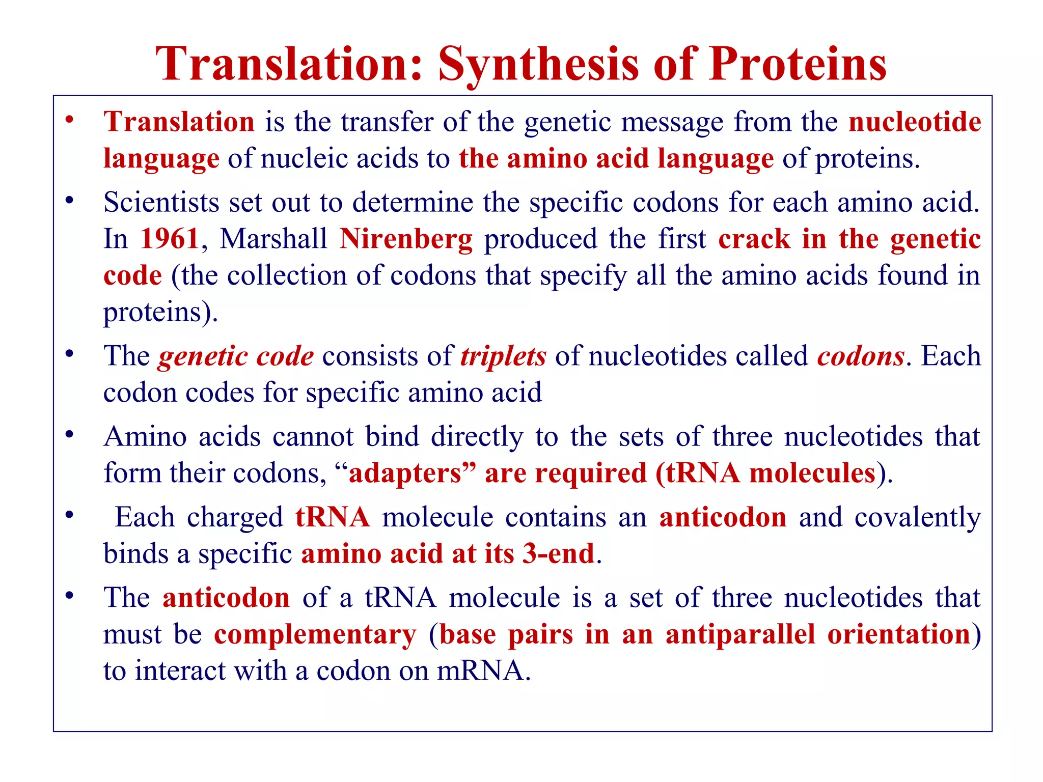
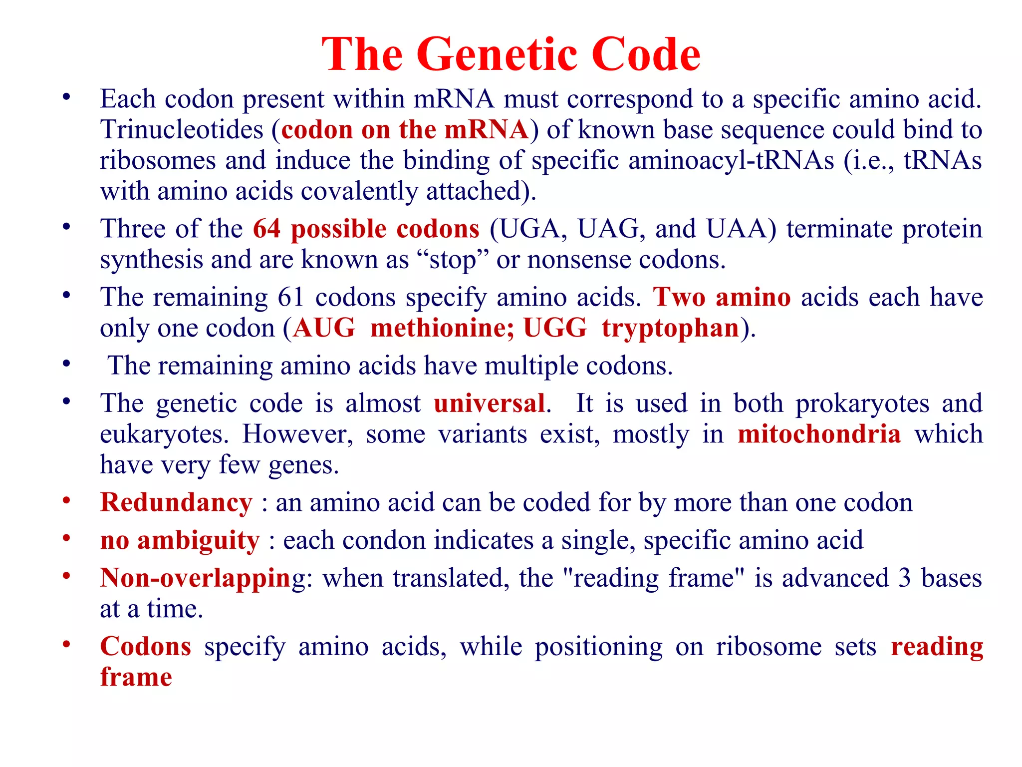
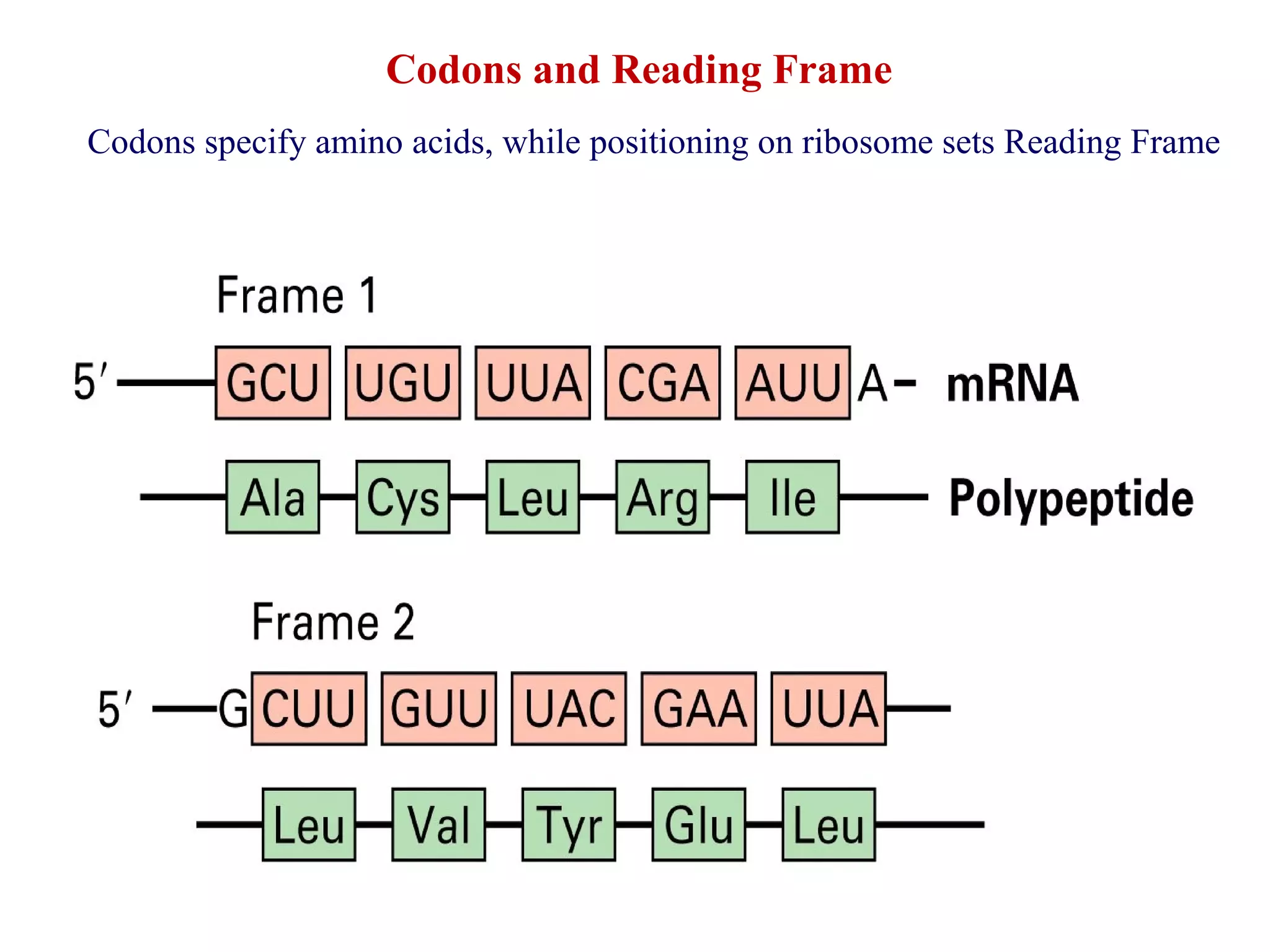
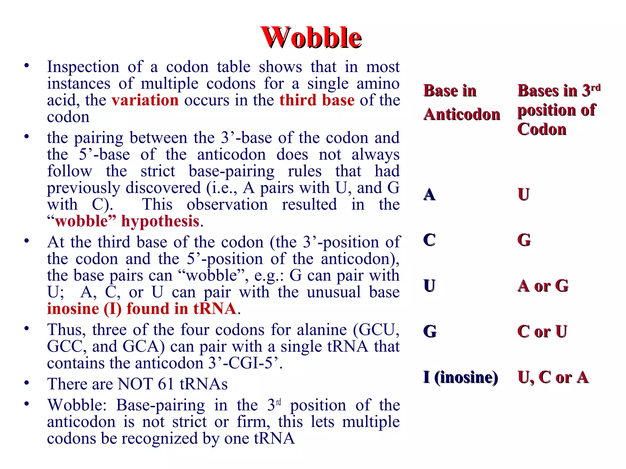
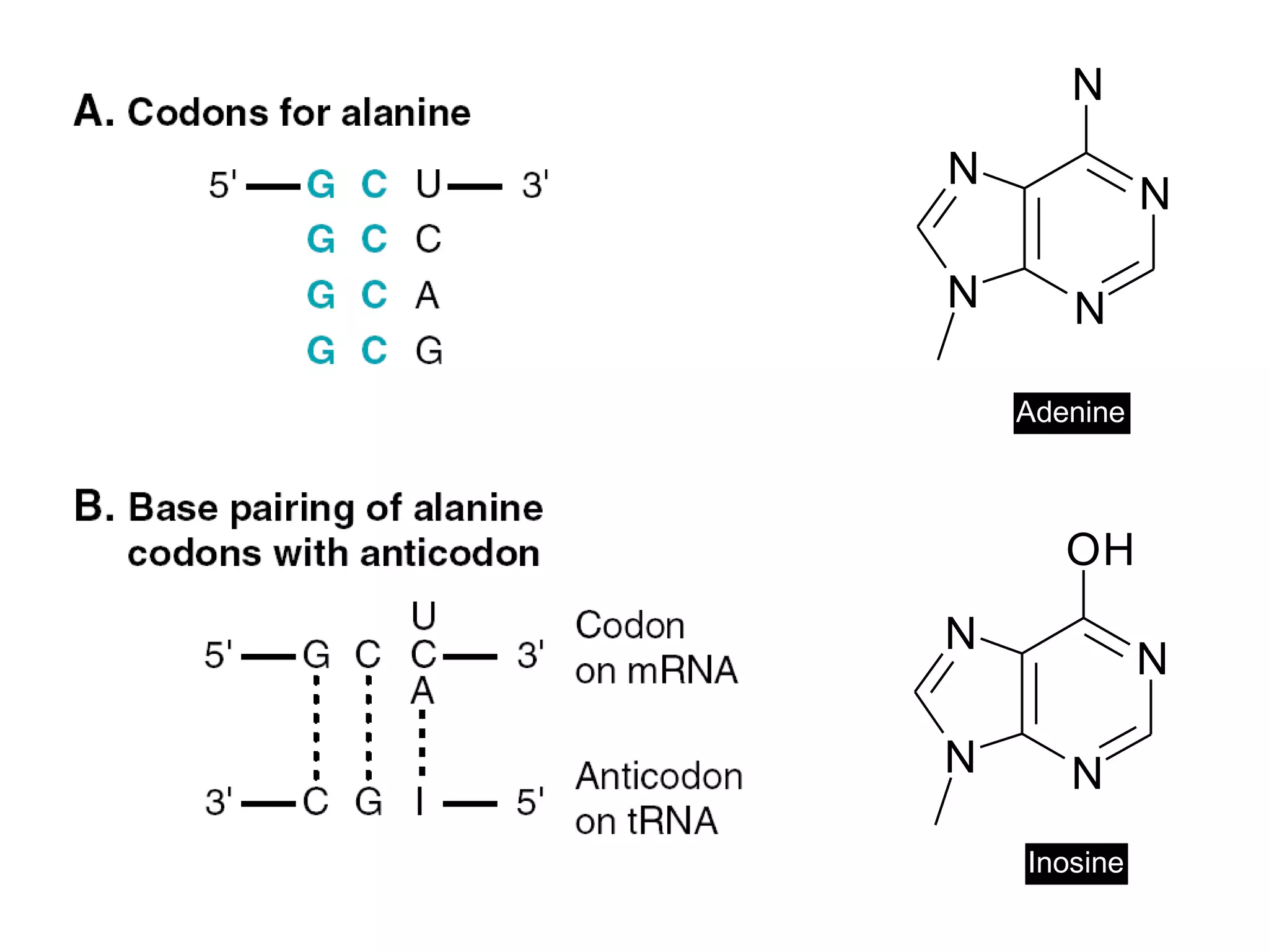
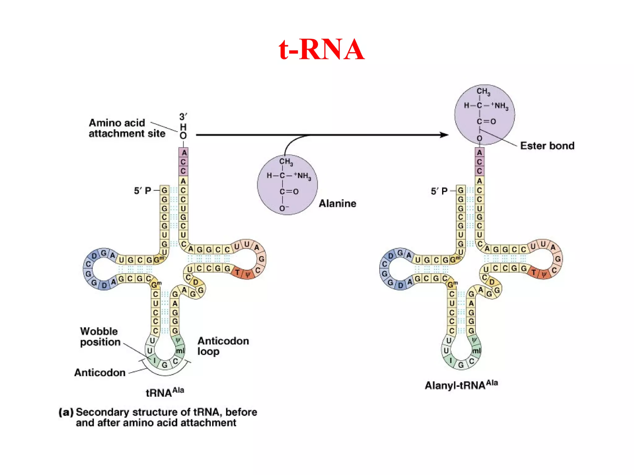
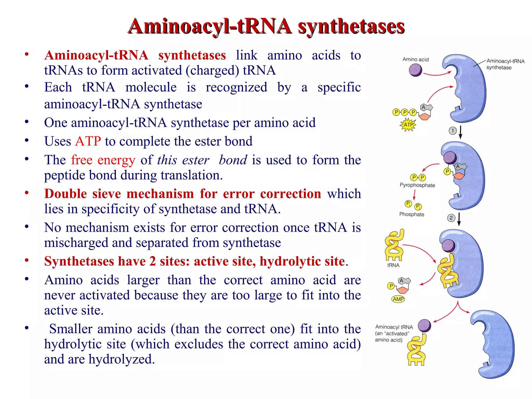
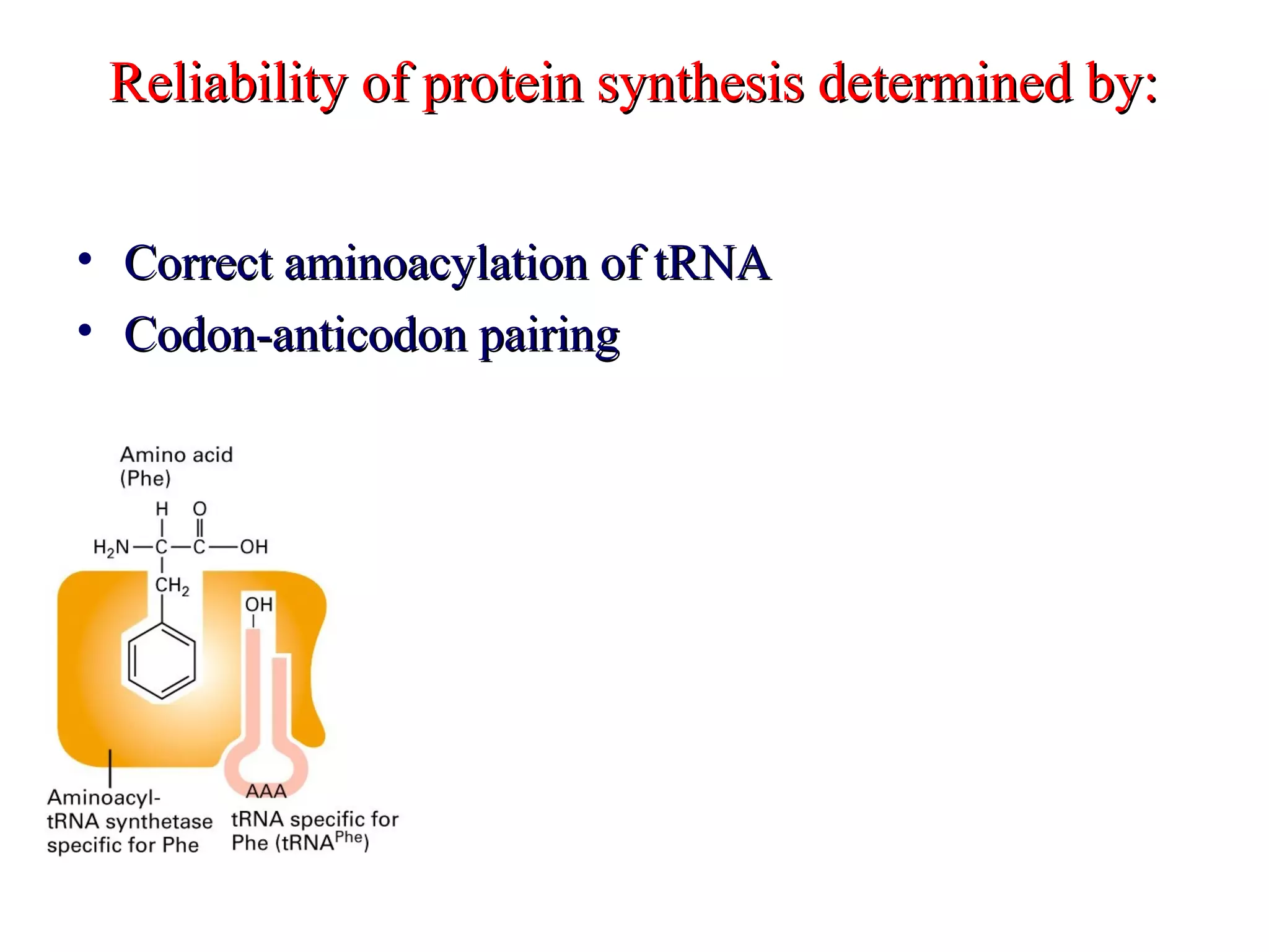
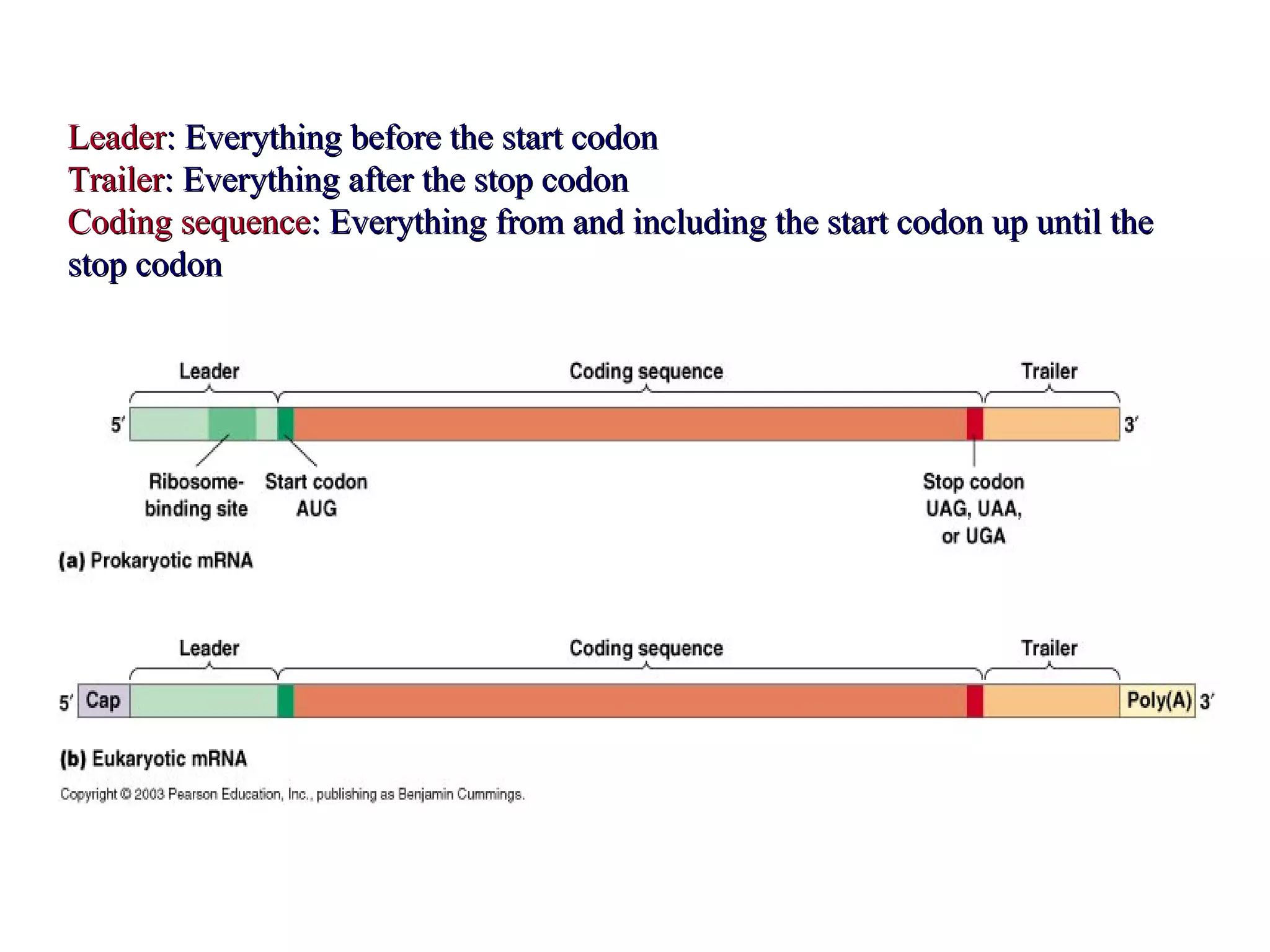
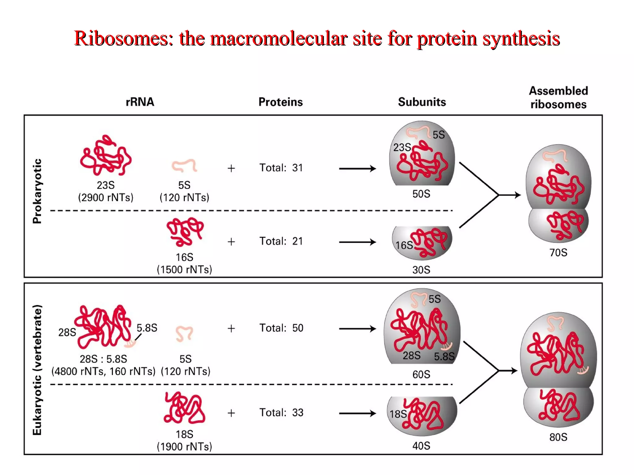
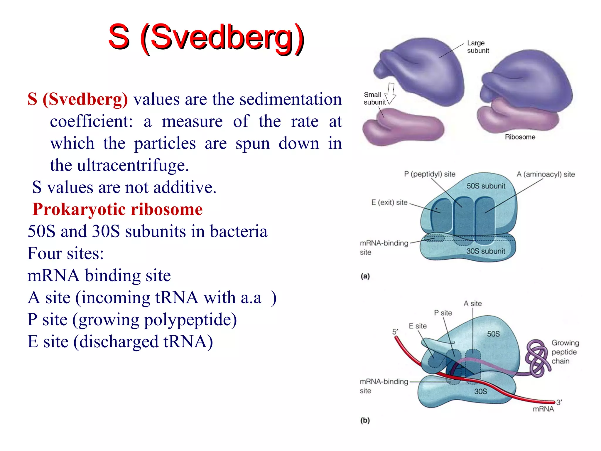
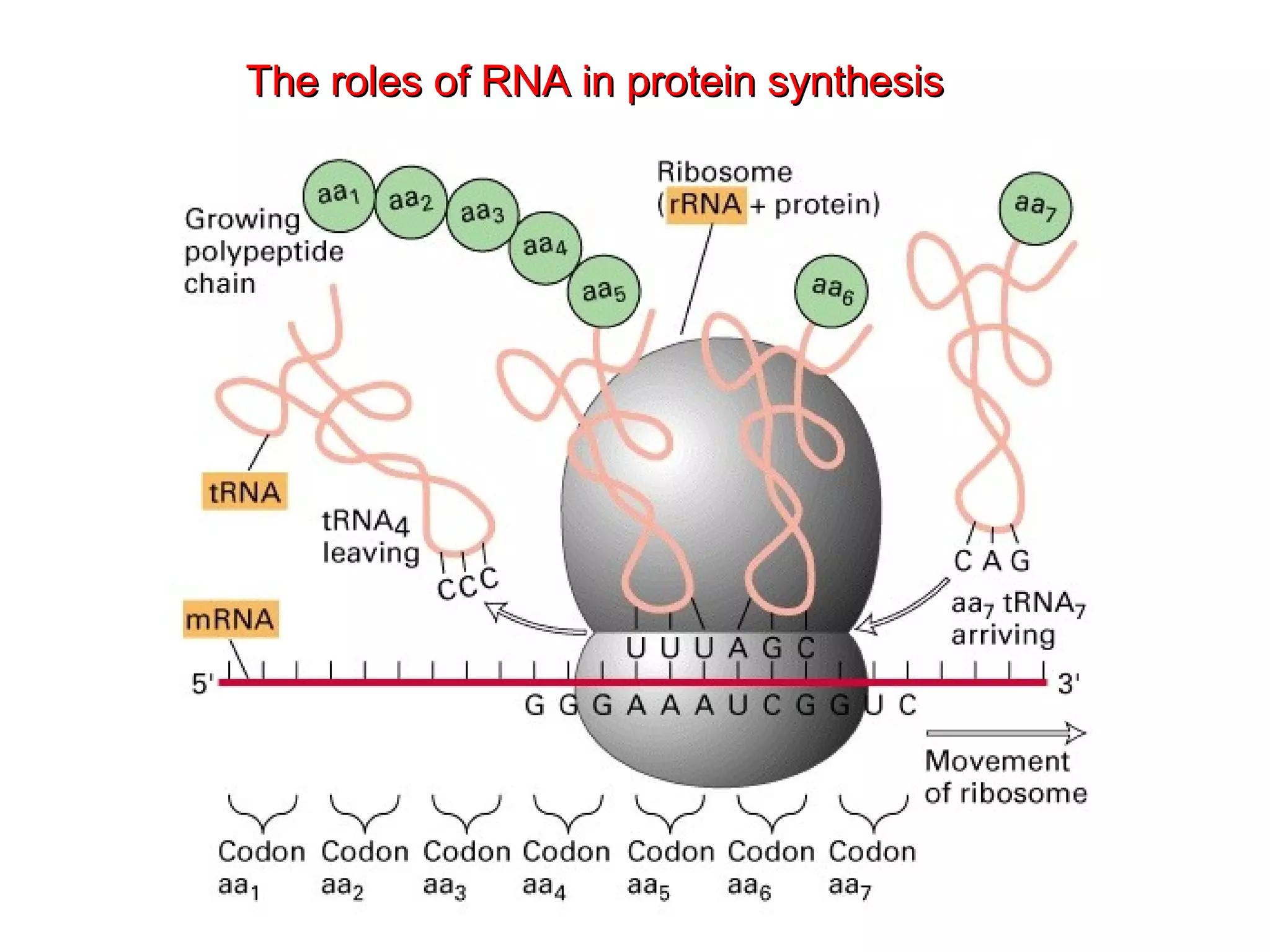
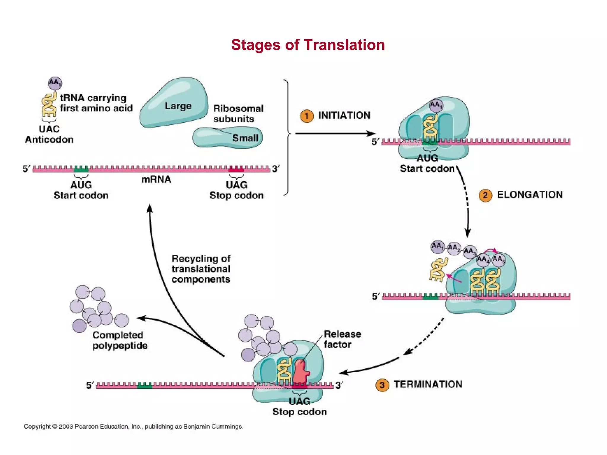
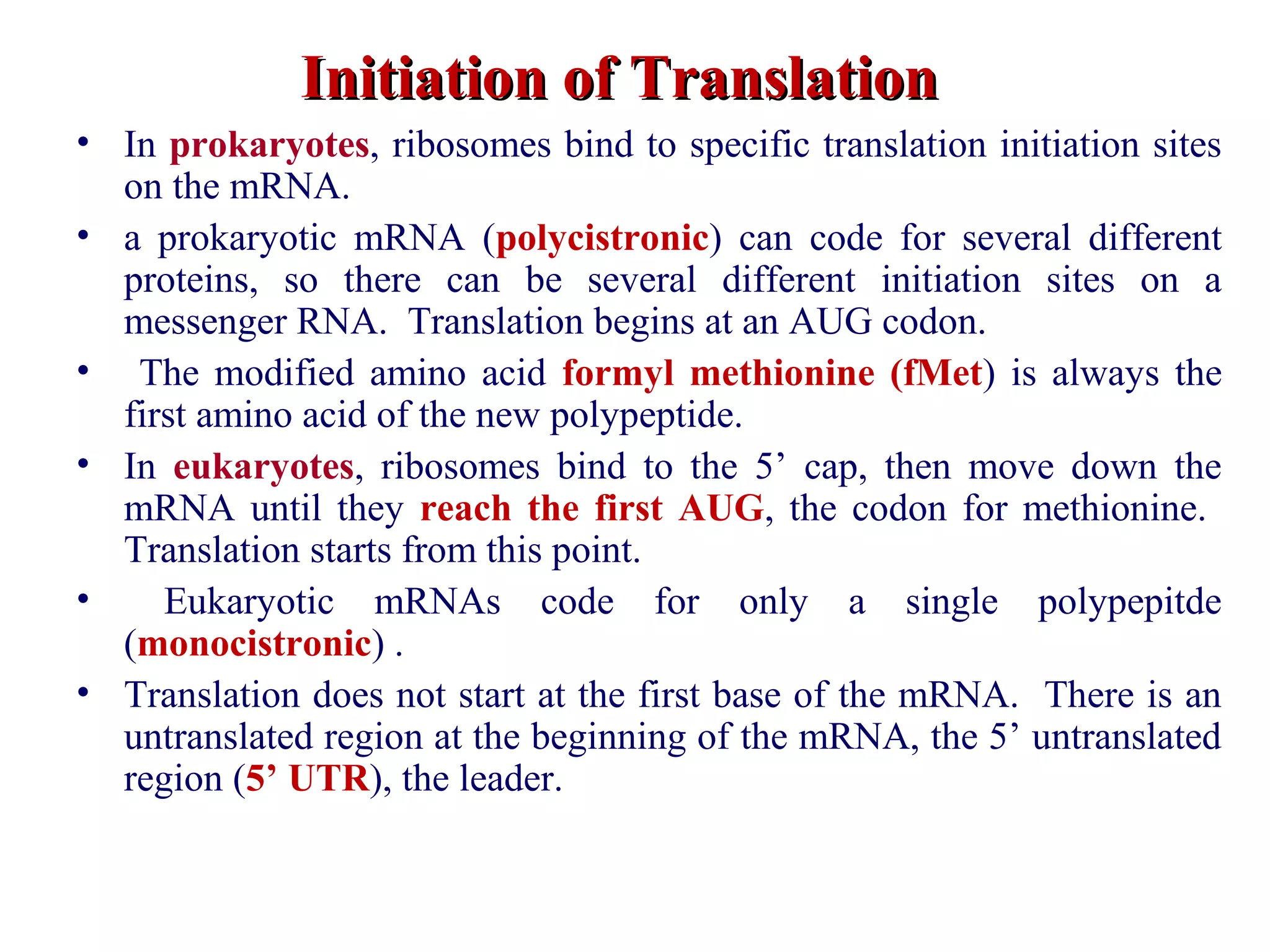
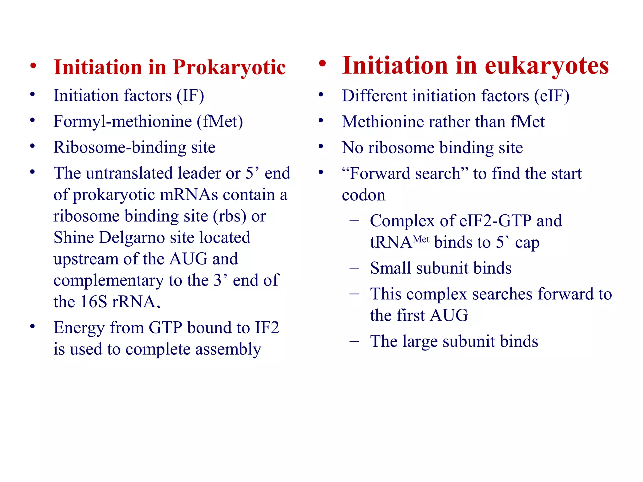
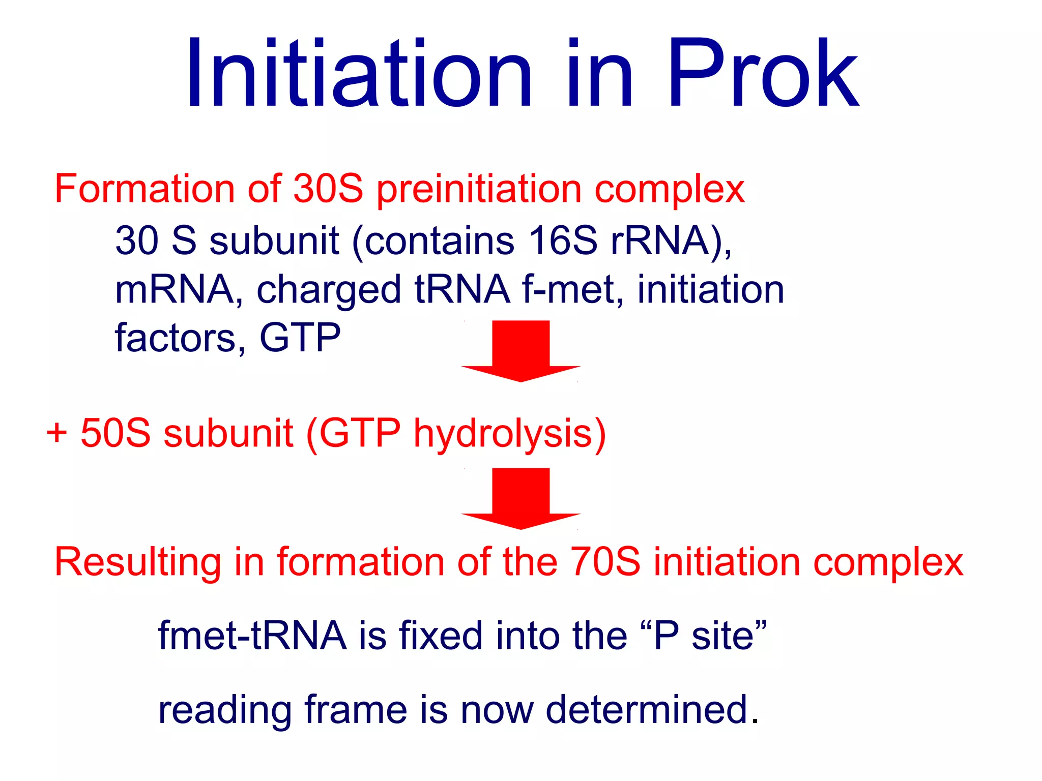
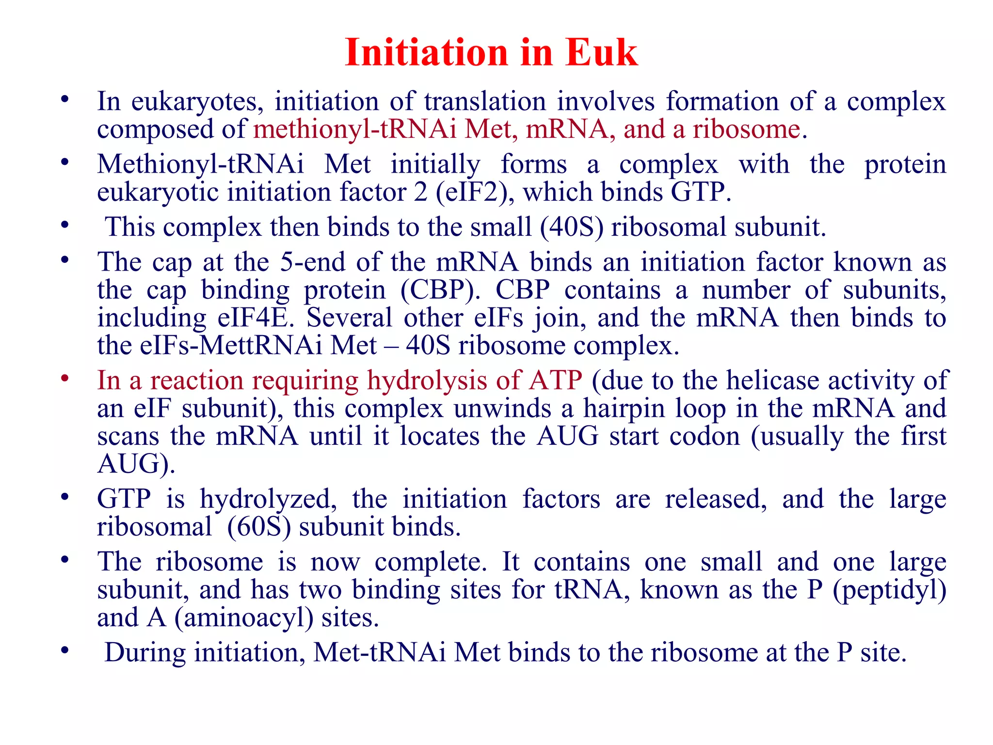
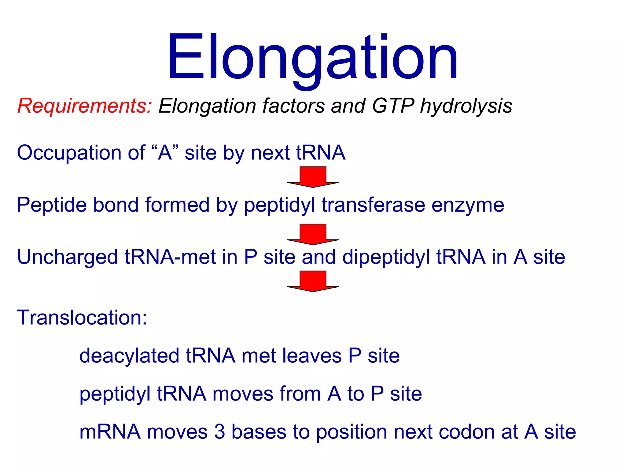
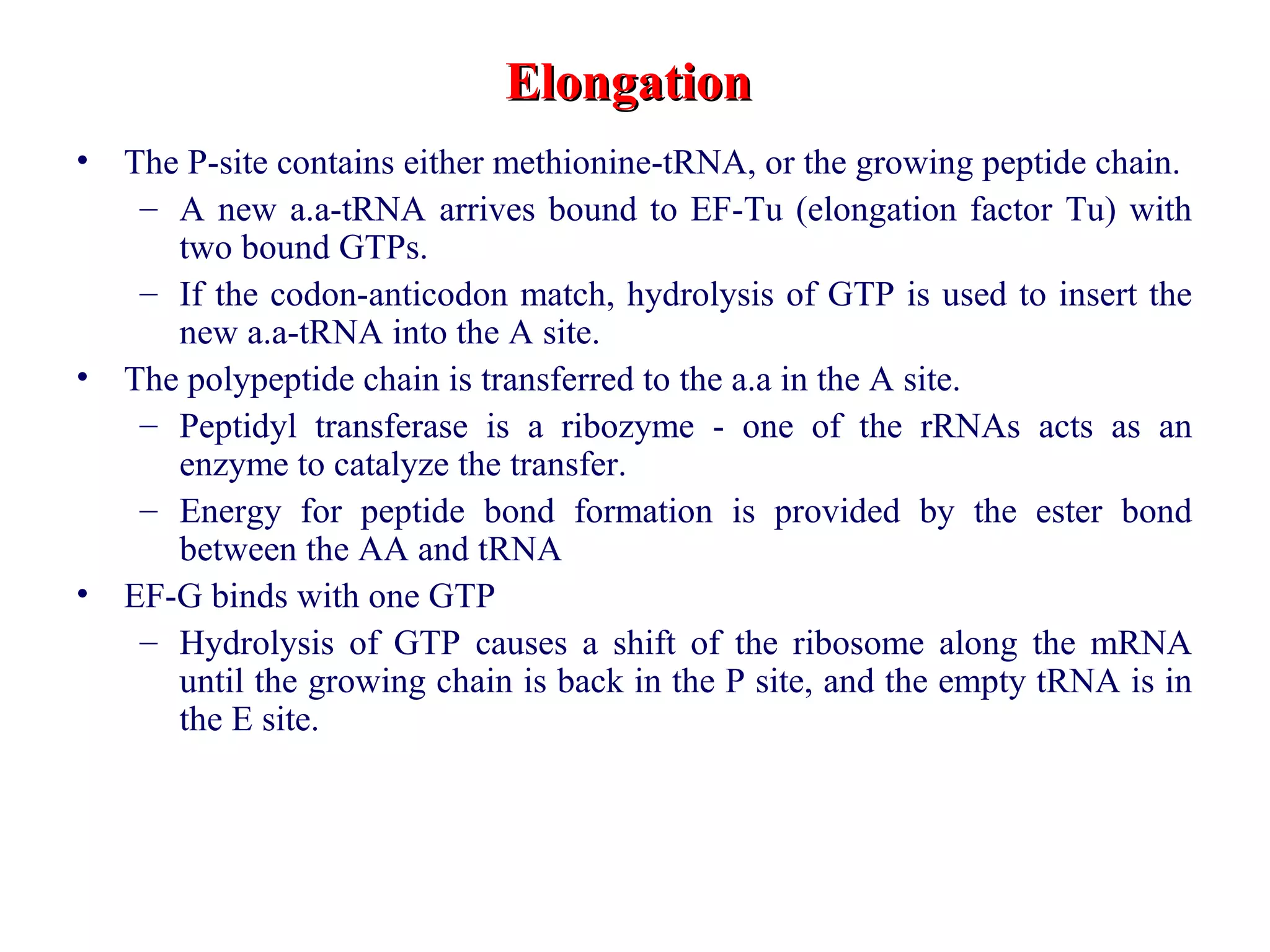
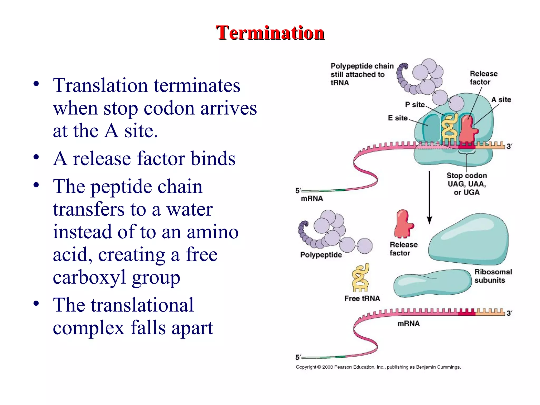
Translation: Synthesis of Proteins
• Translation is the transfer of the genetic message from the nucleotide
language of nucleic acids to the amino acid language of proteins.
• Scientists set out to determine the specific codons for each amino acid.
In 1961, Marshall Nirenberg produced the first crack in the genetic
code (the collection of codons that specify all the amino acids found in
proteins).
• The genetic code consists of triplets of nucleotides called codons. Each
codon codes for specific amino acid
• Amino acids cannot bind directly to the sets of three nucleotides that
form their codons, “adapters” are required (tRNA molecules).
• Each charged tRNA molecule contains an anticodon and covalently
binds a specific amino acid at its 3-end.
• The anticodon of a tRNA molecule is a set of three nucleotides that
must be complementary (base pairs in an antiparallel orientation)
to interact with a codon on mRNA.
• Each codon present within mRNA must correspond to a specific amino acid.
Trinucleotides (codon on the mRNA) of known base sequence could bind to
ribosomes and induce the binding of specific aminoacyl-tRNAs (i.e., tRNAs
with amino acids covalently attached).
• Three of the 64 possible codons (UGA, UAG, and UAA) terminate protein
synthesis and are known as “stop” or nonsense codons.
• The remaining 61 codons specify amino acids. Two amino acids each have
only one codon (AUG methionine; UGG tryptophan).
• The remaining amino acids have multiple codons.
• The genetic code is almost universal. It is used in both prokaryotes and
eukaryotes. However, some variants exist, mostly in mitochondria which
have very few genes.
• Redundancy : an amino acid can be coded for by more than one codon
• no ambiguity : each condon indicates a single, specific amino acid
• Non-overlapping: when translated, the "reading frame" is advanced 3 bases
at a time.
• Codons specify amino acids, while positioning on ribosome sets reading
frame
The Genetic Code
Codons and Reading Frame
Codons specify amino acids, while positioning on ribosome sets Reading Frame
• Inspection of a codon table shows that in most
instances of multiple codons for a single amino
acid, the variation occurs in the third base of the
codon
• the pairing between the 3’-base of the codon and
the 5’-base of the anticodon does not always
follow the strict base-pairing rules that had
previously discovered (i.e., A pairs with U, and G
with C). This observation resulted in the
“wobble” hypothesis.
• At the third base of the codon (the 3’-position of
the codon and the 5’-position of the anticodon),
the base pairs can “wobble”, e.g.: G can pair with
U; A, C, or U can pair with the unusual base
inosine (I) found in tRNA.
• Thus, three of the four codons for alanine (GCU,
GCC, and GCA) can pair with a single tRNA that
contains the anticodon 3’-CGI-5’.
• There are NOT 61 tRNAs
• Wobble: Base-pairing in the 3rd
position of the
anticodon is not strict or firm, this lets multiple
codons be recognized by one tRNA
WobbleWobble
Base inBase in
AnticodonAnticodon
Bases in 3Bases in 3rdrd
position ofposition of
CodonCodon
AA UU
CC GG
UU A or GA or G
GG C or UC or U
I (inosine)I (inosine) U, C or AU, C or A
N N
N
N
OH
N N
N
N
N
Adenine
Inosine
t-RNA
Aminoacyl-tRNA synthetasesAminoacyl-tRNA synthetases
• Aminoacyl-tRNA synthetases link amino acids to
tRNAs to form activated (charged) tRNA
• Each tRNA molecule is recognized by a specific
aminoacyl-tRNA synthetase
• One aminoacyl-tRNA synthetase per amino acid
• Uses ATP to complete the ester bond
• The free energy of this ester bond is used to form the
peptide bond during translation.
• Double sieve mechanism for error correction which
lies in specificity of synthetase and tRNA.
• No mechanism exists for error correction once tRNA is
mischarged and separated from synthetase
• Synthetases have 2 sites: active site, hydrolytic site.
• Amino acids larger than the correct amino acid are
never activated because they are too large to fit into the
active site.
• Smaller amino acids (than the correct one) fit into the
hydrolytic site (which excludes the correct amino acid)
and are hydrolyzed.
Reliability of protein synthesis determined by:Reliability of protein synthesis determined by:
• Correct aminoacylation of tRNACorrect aminoacylation of tRNA
• Codon-anticodon pairingCodon-anticodon pairing
LeaderLeader: Everything before the start codon: Everything before the start codon
TrailerTrailer: Everything after the stop codon: Everything after the stop codon
Coding sequenceCoding sequence: Everything from and including the start codon up until the: Everything from and including the start codon up until the
stop codonstop codon
Ribosomes: the macromolecular site for protein synthesisRibosomes: the macromolecular site for protein synthesis
S (Svedberg)S (Svedberg)
S (Svedberg) values are the sedimentation
coefficient: a measure of the rate at
which the particles are spun down in
the ultracentrifuge.
S values are not additive.
Prokaryotic ribosome
50S and 30S subunits in bacteria
Four sites:
mRNA binding site
A site (incoming tRNA with a.a )
P site (growing polypeptide)
E site (discharged tRNA)
The roles of RNA in protein synthesisThe roles of RNA in protein synthesis
Stages of Translation
Initiation of TranslationInitiation of Translation
• In prokaryotes, ribosomes bind to specific translation initiation sites
on the mRNA.
• a prokaryotic mRNA (polycistronic) can code for several different
proteins, so there can be several different initiation sites on a
messenger RNA. Translation begins at an AUG codon.
• The modified amino acid formyl methionine (fMet) is always the
first amino acid of the new polypeptide.
• In eukaryotes, ribosomes bind to the 5’ cap, then move down the
mRNA until they reach the first AUG, the codon for methionine.
Translation starts from this point.
• Eukaryotic mRNAs code for only a single polypepitde
(monocistronic) .
• Translation does not start at the first base of the mRNA. There is an
untranslated region at the beginning of the mRNA, the 5’ untranslated
region (5’ UTR), the leader.
• Initiation in Prokaryotic
• Initiation factors (IF)
• Formyl-methionine (fMet)
• Ribosome-binding site
• The untranslated leader or 5’ end
of prokaryotic mRNAs contain a
ribosome binding site (rbs) or
Shine Delgarno site located
upstream of the AUG and
complementary to the 3’ end of
the 16S rRNA..
• Energy from GTP bound to IF2
is used to complete assembly
• Initiation in eukaryotes
• Different initiation factors (eIF)
• Methionine rather than fMet
• No ribosome binding site
• “Forward search” to find the start
codon
– Complex of eIF2-GTP and
tRNAMet
binds to 5` cap
– Small subunit binds
– This complex searches forward to
the first AUG
– The large subunit binds
Initiation in Prok
Formation of 30S preinitiation complex
30 S subunit (contains 16S rRNA),
mRNA, charged tRNA f-met, initiation
factors, GTP
+ 50S subunit (GTP hydrolysis)
Resulting in formation of the 70S initiation complex
fmet-tRNA is fixed into the “P site”
reading frame is now determined.
Initiation in Euk
• In eukaryotes, initiation of translation involves formation of a complex
composed of methionyl-tRNAi Met, mRNA, and a ribosome.
• Methionyl-tRNAi Met initially forms a complex with the protein
eukaryotic initiation factor 2 (eIF2), which binds GTP.
• This complex then binds to the small (40S) ribosomal subunit.
• The cap at the 5-end of the mRNA binds an initiation factor known as
the cap binding protein (CBP). CBP contains a number of subunits,
including eIF4E. Several other eIFs join, and the mRNA then binds to
the eIFs-MettRNAi Met – 40S ribosome complex.
• In a reaction requiring hydrolysis of ATP (due to the helicase activity of
an eIF subunit), this complex unwinds a hairpin loop in the mRNA and
scans the mRNA until it locates the AUG start codon (usually the first
AUG).
• GTP is hydrolyzed, the initiation factors are released, and the large
ribosomal (60S) subunit binds.
• The ribosome is now complete. It contains one small and one large
subunit, and has two binding sites for tRNA, known as the P (peptidyl)
and A (aminoacyl) sites.
• During initiation, Met-tRNAi Met binds to the ribosome at the P site.
Elongation
Occupation of “A” site by next tRNA
Peptide bond formed by peptidyl transferase enzyme
Uncharged tRNA-met in P site and dipeptidyl tRNA in A site
Translocation:
deacylated tRNA met leaves P site
peptidyl tRNA moves from A to P site
mRNA moves 3 bases to position next codon at A site
Requirements: Elongation factors and GTP hydrolysis
ElongationElongation
• The P-site contains either methionine-tRNA, or the growing peptide chain.
– A new a.a-tRNA arrives bound to EF-Tu (elongation factor Tu) with
two bound GTPs.
– If the codon-anticodon match, hydrolysis of GTP is used to insert the
new a.a-tRNA into the A site.
• The polypeptide chain is transferred to the a.a in the A site.
– Peptidyl transferase is a ribozyme - one of the rRNAs acts as an
enzyme to catalyze the transfer.
– Energy for peptide bond formation is provided by the ester bond
between the AA and tRNA
• EF-G binds with one GTP
– Hydrolysis of GTP causes a shift of the ribosome along the mRNA
until the growing chain is back in the P site, and the empty tRNA is in
the E site.
TerminationTermination
• Translation terminates
when stop codon arrives
at the A site.
• A release factor binds
• The peptide chain
transfers to a water
instead of to an amino
acid, creating a free
carboxyl group
• The translational
complex falls apart
• Folding proceeds as the peptide chain is synthesized.
• 1 ATP and 3 GTPs are used for each amino acid
added.
• Many ribosomes proceed along a single mRNA in a
chain - polyribosomes - to maximize efficiency.
• Polycistronic mRNA can contain multiple ribosome
binding sequences - one for each protein in the
operon - allowing simultaneous but separate
translation of all encoded proteins
• New polypeptides usually fold themselves spontaneously into
their active conformation. However, some proteins are helped
and guided in the folding process by chaperone proteins
• Many proteins have sugars, phosphate groups, fatty acids, and
other molecules covalently attached to certain amino acids.
Most of this is done in the endoplasmic reticulum.
• Many proteins are targeted to specific organelles within the
cell. Targeting is accomplished through “signal sequences”
on the polypeptide. In the case of proteins that go into the
endoplasmic reticulum, the signal sequence is a group of
amino acids at the N terminal of the polypeptide, which are
removed from the final protein after translation.
Post-Translational Modification

The flow of genetic information translation (1)

  • 1.
    Translation: Synthesis ofProteins • Translation is the transfer of the genetic message from the nucleotide language of nucleic acids to the amino acid language of proteins. • Scientists set out to determine the specific codons for each amino acid. In 1961, Marshall Nirenberg produced the first crack in the genetic code (the collection of codons that specify all the amino acids found in proteins). • The genetic code consists of triplets of nucleotides called codons. Each codon codes for specific amino acid • Amino acids cannot bind directly to the sets of three nucleotides that form their codons, “adapters” are required (tRNA molecules). • Each charged tRNA molecule contains an anticodon and covalently binds a specific amino acid at its 3-end. • The anticodon of a tRNA molecule is a set of three nucleotides that must be complementary (base pairs in an antiparallel orientation) to interact with a codon on mRNA.
  • 2.
    • Each codonpresent within mRNA must correspond to a specific amino acid. Trinucleotides (codon on the mRNA) of known base sequence could bind to ribosomes and induce the binding of specific aminoacyl-tRNAs (i.e., tRNAs with amino acids covalently attached). • Three of the 64 possible codons (UGA, UAG, and UAA) terminate protein synthesis and are known as “stop” or nonsense codons. • The remaining 61 codons specify amino acids. Two amino acids each have only one codon (AUG methionine; UGG tryptophan). • The remaining amino acids have multiple codons. • The genetic code is almost universal. It is used in both prokaryotes and eukaryotes. However, some variants exist, mostly in mitochondria which have very few genes. • Redundancy : an amino acid can be coded for by more than one codon • no ambiguity : each condon indicates a single, specific amino acid • Non-overlapping: when translated, the "reading frame" is advanced 3 bases at a time. • Codons specify amino acids, while positioning on ribosome sets reading frame The Genetic Code
  • 4.
    Codons and ReadingFrame Codons specify amino acids, while positioning on ribosome sets Reading Frame
  • 5.
    • Inspection ofa codon table shows that in most instances of multiple codons for a single amino acid, the variation occurs in the third base of the codon • the pairing between the 3’-base of the codon and the 5’-base of the anticodon does not always follow the strict base-pairing rules that had previously discovered (i.e., A pairs with U, and G with C). This observation resulted in the “wobble” hypothesis. • At the third base of the codon (the 3’-position of the codon and the 5’-position of the anticodon), the base pairs can “wobble”, e.g.: G can pair with U; A, C, or U can pair with the unusual base inosine (I) found in tRNA. • Thus, three of the four codons for alanine (GCU, GCC, and GCA) can pair with a single tRNA that contains the anticodon 3’-CGI-5’. • There are NOT 61 tRNAs • Wobble: Base-pairing in the 3rd position of the anticodon is not strict or firm, this lets multiple codons be recognized by one tRNA WobbleWobble Base inBase in AnticodonAnticodon Bases in 3Bases in 3rdrd position ofposition of CodonCodon AA UU CC GG UU A or GA or G GG C or UC or U I (inosine)I (inosine) U, C or AU, C or A
  • 6.
  • 7.
  • 9.
    Aminoacyl-tRNA synthetasesAminoacyl-tRNA synthetases •Aminoacyl-tRNA synthetases link amino acids to tRNAs to form activated (charged) tRNA • Each tRNA molecule is recognized by a specific aminoacyl-tRNA synthetase • One aminoacyl-tRNA synthetase per amino acid • Uses ATP to complete the ester bond • The free energy of this ester bond is used to form the peptide bond during translation. • Double sieve mechanism for error correction which lies in specificity of synthetase and tRNA. • No mechanism exists for error correction once tRNA is mischarged and separated from synthetase • Synthetases have 2 sites: active site, hydrolytic site. • Amino acids larger than the correct amino acid are never activated because they are too large to fit into the active site. • Smaller amino acids (than the correct one) fit into the hydrolytic site (which excludes the correct amino acid) and are hydrolyzed.
  • 10.
    Reliability of proteinsynthesis determined by:Reliability of protein synthesis determined by: • Correct aminoacylation of tRNACorrect aminoacylation of tRNA • Codon-anticodon pairingCodon-anticodon pairing
  • 11.
    LeaderLeader: Everything beforethe start codon: Everything before the start codon TrailerTrailer: Everything after the stop codon: Everything after the stop codon Coding sequenceCoding sequence: Everything from and including the start codon up until the: Everything from and including the start codon up until the stop codonstop codon
  • 12.
    Ribosomes: the macromolecularsite for protein synthesisRibosomes: the macromolecular site for protein synthesis
  • 13.
    S (Svedberg)S (Svedberg) S(Svedberg) values are the sedimentation coefficient: a measure of the rate at which the particles are spun down in the ultracentrifuge. S values are not additive. Prokaryotic ribosome 50S and 30S subunits in bacteria Four sites: mRNA binding site A site (incoming tRNA with a.a ) P site (growing polypeptide) E site (discharged tRNA)
  • 14.
    The roles ofRNA in protein synthesisThe roles of RNA in protein synthesis
  • 16.
  • 17.
    Initiation of TranslationInitiationof Translation • In prokaryotes, ribosomes bind to specific translation initiation sites on the mRNA. • a prokaryotic mRNA (polycistronic) can code for several different proteins, so there can be several different initiation sites on a messenger RNA. Translation begins at an AUG codon. • The modified amino acid formyl methionine (fMet) is always the first amino acid of the new polypeptide. • In eukaryotes, ribosomes bind to the 5’ cap, then move down the mRNA until they reach the first AUG, the codon for methionine. Translation starts from this point. • Eukaryotic mRNAs code for only a single polypepitde (monocistronic) . • Translation does not start at the first base of the mRNA. There is an untranslated region at the beginning of the mRNA, the 5’ untranslated region (5’ UTR), the leader.
  • 18.
    • Initiation inProkaryotic • Initiation factors (IF) • Formyl-methionine (fMet) • Ribosome-binding site • The untranslated leader or 5’ end of prokaryotic mRNAs contain a ribosome binding site (rbs) or Shine Delgarno site located upstream of the AUG and complementary to the 3’ end of the 16S rRNA.. • Energy from GTP bound to IF2 is used to complete assembly • Initiation in eukaryotes • Different initiation factors (eIF) • Methionine rather than fMet • No ribosome binding site • “Forward search” to find the start codon – Complex of eIF2-GTP and tRNAMet binds to 5` cap – Small subunit binds – This complex searches forward to the first AUG – The large subunit binds
  • 19.
    Initiation in Prok Formationof 30S preinitiation complex 30 S subunit (contains 16S rRNA), mRNA, charged tRNA f-met, initiation factors, GTP + 50S subunit (GTP hydrolysis) Resulting in formation of the 70S initiation complex fmet-tRNA is fixed into the “P site” reading frame is now determined.
  • 21.
    Initiation in Euk •In eukaryotes, initiation of translation involves formation of a complex composed of methionyl-tRNAi Met, mRNA, and a ribosome. • Methionyl-tRNAi Met initially forms a complex with the protein eukaryotic initiation factor 2 (eIF2), which binds GTP. • This complex then binds to the small (40S) ribosomal subunit. • The cap at the 5-end of the mRNA binds an initiation factor known as the cap binding protein (CBP). CBP contains a number of subunits, including eIF4E. Several other eIFs join, and the mRNA then binds to the eIFs-MettRNAi Met – 40S ribosome complex. • In a reaction requiring hydrolysis of ATP (due to the helicase activity of an eIF subunit), this complex unwinds a hairpin loop in the mRNA and scans the mRNA until it locates the AUG start codon (usually the first AUG). • GTP is hydrolyzed, the initiation factors are released, and the large ribosomal (60S) subunit binds. • The ribosome is now complete. It contains one small and one large subunit, and has two binding sites for tRNA, known as the P (peptidyl) and A (aminoacyl) sites. • During initiation, Met-tRNAi Met binds to the ribosome at the P site.
  • 27.
    Elongation Occupation of “A”site by next tRNA Peptide bond formed by peptidyl transferase enzyme Uncharged tRNA-met in P site and dipeptidyl tRNA in A site Translocation: deacylated tRNA met leaves P site peptidyl tRNA moves from A to P site mRNA moves 3 bases to position next codon at A site Requirements: Elongation factors and GTP hydrolysis
  • 34.
    ElongationElongation • The P-sitecontains either methionine-tRNA, or the growing peptide chain. – A new a.a-tRNA arrives bound to EF-Tu (elongation factor Tu) with two bound GTPs. – If the codon-anticodon match, hydrolysis of GTP is used to insert the new a.a-tRNA into the A site. • The polypeptide chain is transferred to the a.a in the A site. – Peptidyl transferase is a ribozyme - one of the rRNAs acts as an enzyme to catalyze the transfer. – Energy for peptide bond formation is provided by the ester bond between the AA and tRNA • EF-G binds with one GTP – Hydrolysis of GTP causes a shift of the ribosome along the mRNA until the growing chain is back in the P site, and the empty tRNA is in the E site.
  • 35.
    TerminationTermination • Translation terminates whenstop codon arrives at the A site. • A release factor binds • The peptide chain transfers to a water instead of to an amino acid, creating a free carboxyl group • The translational complex falls apart
  • 36.
    • Folding proceedsas the peptide chain is synthesized. • 1 ATP and 3 GTPs are used for each amino acid added. • Many ribosomes proceed along a single mRNA in a chain - polyribosomes - to maximize efficiency. • Polycistronic mRNA can contain multiple ribosome binding sequences - one for each protein in the operon - allowing simultaneous but separate translation of all encoded proteins
  • 37.
    • New polypeptidesusually fold themselves spontaneously into their active conformation. However, some proteins are helped and guided in the folding process by chaperone proteins • Many proteins have sugars, phosphate groups, fatty acids, and other molecules covalently attached to certain amino acids. Most of this is done in the endoplasmic reticulum. • Many proteins are targeted to specific organelles within the cell. Targeting is accomplished through “signal sequences” on the polypeptide. In the case of proteins that go into the endoplasmic reticulum, the signal sequence is a group of amino acids at the N terminal of the polypeptide, which are removed from the final protein after translation. Post-Translational Modification