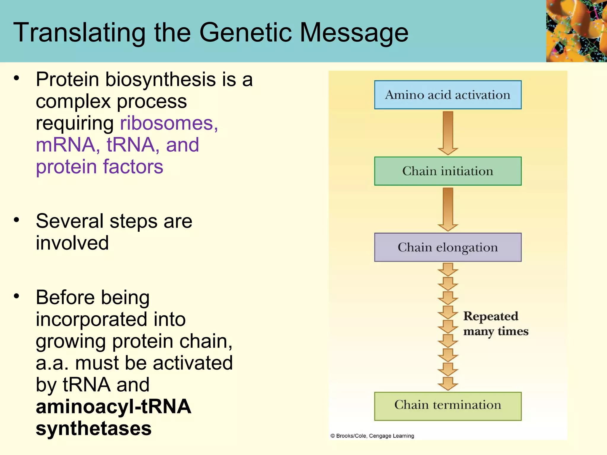
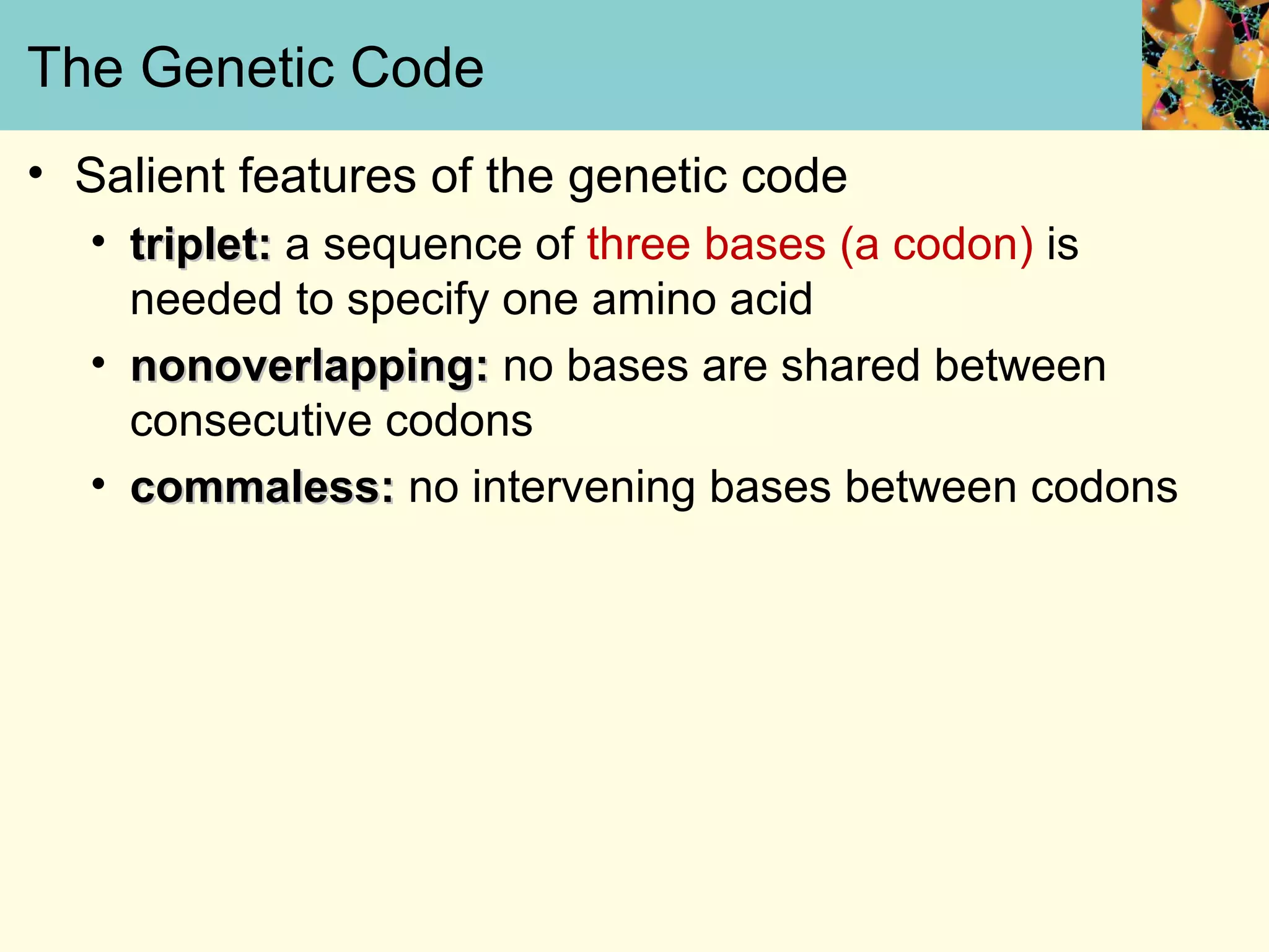
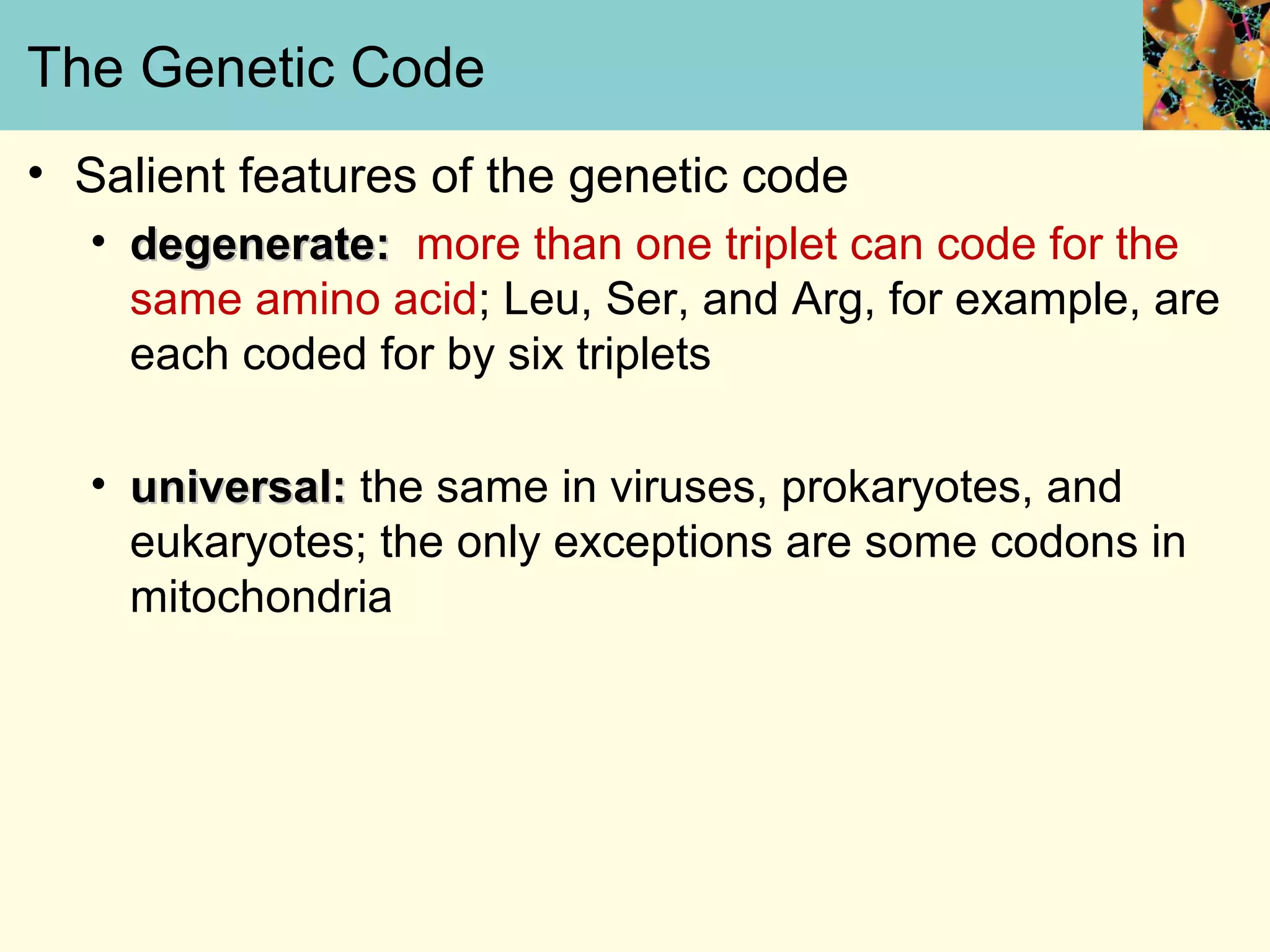
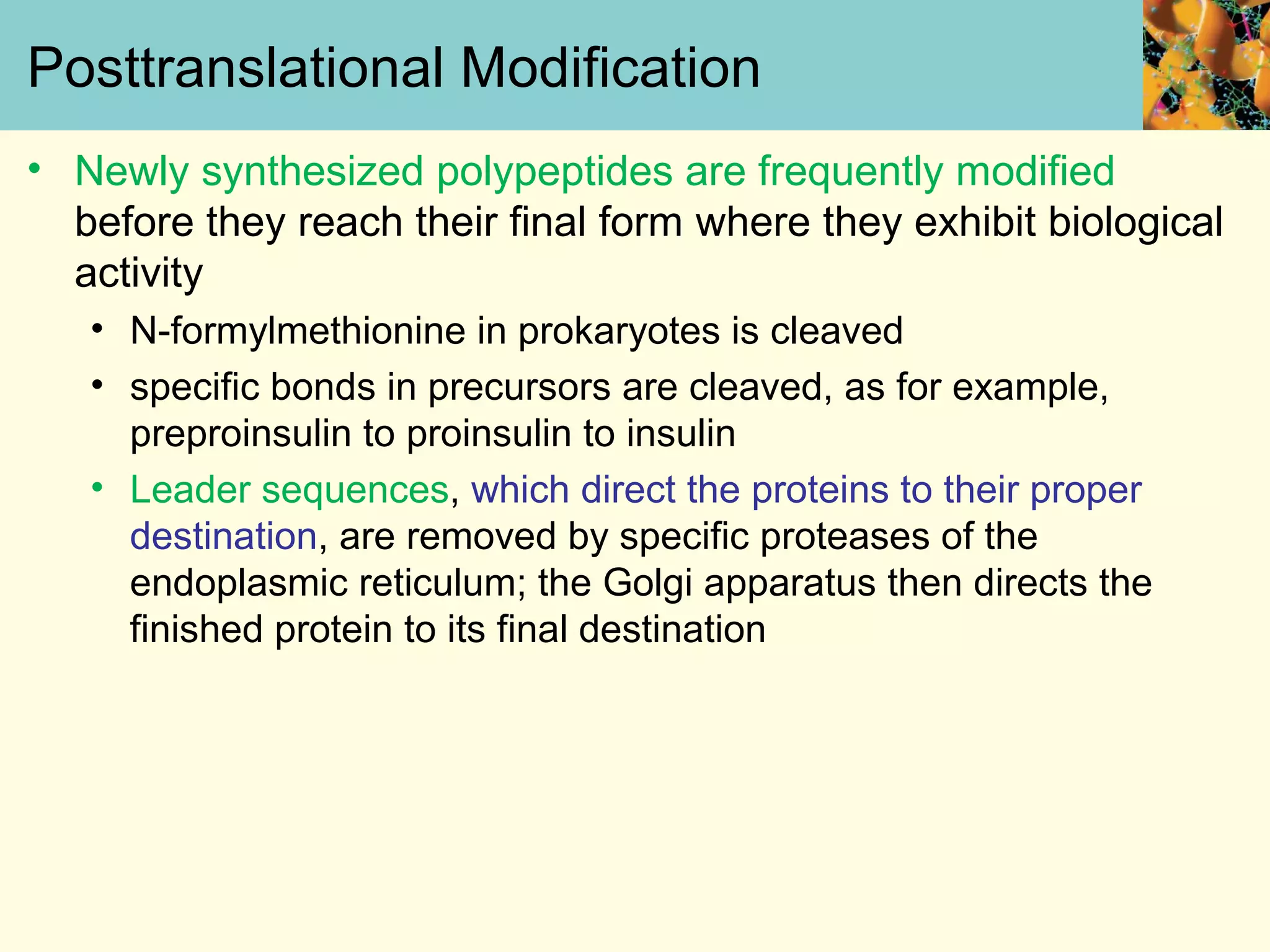
Paul D. Adams • University of Arkansas
Mary K. Campbell
Shawn O. Farrell
http://academic.cengage.com/chemistry/campbell
Chapter Twelve
Protein Synthesis: Translation of
the Genetic Message
Translating the Genetic Message
• Protein biosynthesis is a
complex process
requiring ribosomes,
mRNA, tRNA, and
protein factors
• Several steps are
involved
• Before being
incorporated into
growing protein chain,
a.a. must be activated
by tRNA and
aminoacyl-tRNA
synthetases
The Genetic Code
• Salient features of the genetic code
• triplet:triplet: a sequence of three bases (a codon) is
needed to specify one amino acid
• nonoverlapping:nonoverlapping: no bases are shared between
consecutive codons
• commaless:commaless: no intervening bases between codons
The Genetic Code
• Salient features of the genetic code
• degenerate:degenerate: more than one triplet can code for the
same amino acid; Leu, Ser, and Arg, for example, are
each coded for by six triplets
• universal:universal: the same in viruses, prokaryotes, and
eukaryotes; the only exceptions are some codons in
mitochondria
The Genetic Code (Cont’d)
• The ribosome moves
along the mRNA three
bases at a time rather
than one or two at a
time
• Theoretically possible
genetic codes are
shown in figure 12.2
The Genetic Code (Cont’d)
• All 64 codons have assigned meanings
• 61 code for amino acids
• 3 (UAA, UAG, and UGA) serve as termination signals
• only Trp and Met have one codon each
• the second base is important for the type of amino
acid; for example, if the second base is U, the amino
acids coded for are hydrophobic
The Genetic Code (Cont’d)
• Assignments of triplets in genetic code based on
several different experiments
• synthetic mRNA:synthetic mRNA: if mRNA is polyU, polyPhe is
formed; if mRNA is poly
---ACACACACACACACACACACA---, poly(Thr-His)
is formed
The Genetic Code (Cont’d)
• binding assay:binding assay: aminoacyl-tRNAs bind to ribosomes
in the presence of trinucleotides
•synthesize trinucleotides by chemical means
•carry out a binding assay for each type of
trinucleotide
•aminoacyl-tRNAs are tested for their ability to
bind in the presence of a given trinucleotide
The Filter-Binding Assay Helps Elucidate
the Genetic Code
The Filter-Binding Assay Helps Elucidate
the Genetic Code
• Various tRNA molecules, one of which is
radioactively labeled with carbon-14, are mixed with
ribosomes and synthetic trinucleotides that are
bound to a filter.
• If the radioactive label is detected on the filter, then
it is known that the particular tRNA did bind.
• This technique depends on the fact that aminoacyl-
tRNAs bind strongly to ribosomes in the presence of
the correct trinucleotide.
Wobble Base Pairing
• Some tRNAs bond to one codon exclusively, but
many tRNAs can recognize more than one codon
because of variations in allowed patterns of
hydrogen bonding
• the variation is called “wobble”
• wobble is in the first base of the anticodon
Base Pairing Combination in the Wobble
Scheme
Wobble Base Pairing
Amino Acid Activation
• Amino acid activation and formation of the
aminoacyl-tRNA take place in two separate steps
• Both catalyzed by amionacyl-tRNA synthetase
• Free energy of hydrolysis of ATP provides energy for
bond formation
Amino Acid Activation
Amino Acid Activation (Cont’d)
• This two-stage reaction allows selectivity at two
levels
• the amino acid:the amino acid: the aminoacyl-AMP remains bound
to the enzyme and binding of the correct amino acid is
verified by an editing site in the tRNA synthetase
• tRNA:tRNA: there are specific binding sites on tRNAs that
are recognized by aminoacyl-tRNA synthetases.
• Only 3’-esters are substrates for protein
synthesis.
tRNA Tertiary Structure
• There are several recognition sites for various amino
acids on the tRNA
Chain Initiation
• In all organisms, synthesis of polypeptide chain
starts at the N-terminal end, and grows from N-
terminus to C-terminus
• Initiation requires:
• tRNAfmet
• initiation codon (AUG) of mRNA
• 30S ribosomal subunit
• 50S ribosomal subunit
• initiation factors IF-1, IF-2, and IF-3
• GTP, Mg2+
• Forms the initiation complex
The Initiation Complex
Chain Initiation
• tRNAmet
and tRNAfmet
contain the triplet 3’-UAC-5’
• Triplet base pairs with 5’-AUG-3’ in mRNA
• 3’-UAC-5’ triplet on tRNAfmet
recognizes the AUG triplet
(the start signal) when it occurs at the beginning of the
mRNA sequence that directs polypeptide synthesis
Chain Initiation
• 3’-UAC-5’ triplet on tRNAmet
recognizes the AUG triplet
when it is found in an internal position in the mRNA
sequence
• Start signal is preceded by a Shine-Dalgarno purine-
rich leader segment, 5’-GGAGGU-3’, which usually
lies about 10 nucleotides upstream of the AUG start
signal and acts as a ribosomal binding site
Chain Elongation
• Uses three binding sites for tRNA present on the
50S subunit of the 70S ribosome:
P (peptidyl) site, A (aminoacyl) site, E (exit) site.
• Requires
• 70S ribosome
• codons of mRNA
• aminoacyl-tRNAs
• elongation factors EF-Tu (Elongation factor
temperature-unstable), EF-Ts (Elongation factor
temperature-stable), and EF-G (Elongation factor-
GTP)
• GTP, and Mg2+
Shine-Dalgarno Sequence Recognized by
E. Coli Ribosomes
Shine-Dalgarno Sequence
• The start signal is preceded by a purine-rich leader
segment of mRNA, called the Shine-Dalgarno
sequence (5’-GGAGGU-3’), which usually lies about
10 nucleotides upstream of the AUG start signal and
acts as a ribosomal binding site.
Elongation Steps
• Step 1
• an aminoacyl-tRNA is bound to the A site
• the P site is already occupied
• 2nd amino acid bound to 70S initiation complex. Defined by the
mRNA
• Step 2
• EF-Tu is released in a reaction requiring EF-Ts
• Step 3
• the peptide bond is formed, the P site is uncharged
• Step 4
• the uncharged tRNA is released
• the peptidyl-tRNA is translocated to the P site
• EF-G and GTP are required
• the next aminoacyl-tRNA occupies the empty A site
Why is EF-Tu so important in E.coli?
• If the tRNA and amino acid are mismatched, then
either the EF-Tu does not bind the activated tRNA
very well, in which case it does not deliver it well to
the ribosome.
Chain Elongation
Chain Termination
• Chain termination requires
• stop codons (UAA, UAG, or UGA) of mRNA
• RF-1 (Release factor-1) which binds to UAA and
UAG or RF-2 (Release factor-2) which binds to UAA
and UGA
• RF-3 which does not bind to any termination codon,
but facilitates the binding of RF-1 and RF-2
• GTP which is bound to RF-3
• The entire complex dissociates setting free the
completed polypeptide, the release factors, tRNA,
mRNA, and the 30S and 50S ribosomal subunits
Chain Termination
Components of Protein Synthesis
Protein Synthesis
• In prokaryotes, translation begins very soon after
mRNA transcription.
• It is possible to have several molecules of RNA
polymerase bound to a single DNA gene, each in a
different stage of transcription.
• It is also possible to have several ribosomes bound to
a single mRNA, each in a different stage of translation
Protein Synthesis
• Polysome:Polysome: mRNA bound to several ribosomes
• Coupled translation:Coupled translation: the process in which a
prokaryotic gene is being simultaneously transcribed
and translated.
Simultaneous Protein Synthesis on
Polysomes
• A single mRNA molecule is translated by several
ribosomes simultaneously.
• Each ribosome produces a copy of the polypeptide
chain specified by the mRNA.
• When protein has been completed, the ribosome
dissociates into subunits that are used again in
protein synthesis.
Simultaneous Protein Synthesis on
Polysomes (Cont’d)
Eukaryotic Translation
• Chain Initiation:
• the most different from process in prokaryotes
• 13 more initiation factors are given the designation eIF
(eukaryotic initiation factor) (Table 12.4)
Eukaryotic Translation (Cont’d)
• Chain elongation
• uses the same mechanism of peptidyl transferase and
ribosome translocation as prokaryotes
• there is no E site on eukaryotic ribosomes, only A and
P sites
• there are two elongation factors, eEF-1 and eEF-2
• eEF2 is the counterpart to EF-G, which causes
translocation
• Chain termination
• stop codons are the same: UAG, UAA, and UGA
• only one release factor that binds to all three stop
codons
Posttranslational Modification
• Newly synthesized polypeptides are frequently modified
before they reach their final form where they exhibit biological
activity
• N-formylmethionine in prokaryotes is cleaved
• specific bonds in precursors are cleaved, as for example,
preproinsulin to proinsulin to insulin
• Leader sequences, which direct the proteins to their proper
destination, are removed by specific proteases of the
endoplasmic reticulum; the Golgi apparatus then directs the
finished protein to its final destination
Posttranslational Modification
• Newly synthesized polypeptides are frequently modified
before they reach their final form where they exhibit biological
activity
• factors such as heme groups (funtion; electron transfer)may be
attached
• disulfide bonds may be formed
• amino acids may be modified, as for example, conversion of
proline to hydroxyproline
• other covalent modifications; e.g., addition of carbohydrates
Examples of Posttranslational Modification
Insulin
• Insulin is a hormone that has extensive effects on
metabolism and other body functions
• Insulin causes cells in the liver, muscle, and
fat tissue to take up glucose from the blood, storing it
as glycogen in the liver and muscle, and stopping
use of fat as an energy source.
• When insulin is absent (or low), glucose is not taken
up by body cells, and the body begins to use fat as
an energy source.
Insulin
Computer-generated image of six insulin molecules
assembled in a hexamer, highlighting the threefold
symmetry, the zinc ions holding it together, and the
histidine residues involved in zinc binding. Insulin is
stored in the body as a hexamer, while the active form
is the monomer
Examples of Posttranslational Modification
• After a precursor of preproinsulin is formed, it is
transformed into preproinsulin by formation of three
disulfide bonds.
• Specific cleavage that removes an end segment
converts preproinsulin to proinsulin.
• Finally, two further specific cleavages remove a
central segment, with insulin as the end result.
Protein Degradation
• Proteins are in a dynamic state and are often turned
over
• Degradative pathways are restricted to
• subcellular organelles such as lysosomes
• macromolecular structures called proteosomes
Lysosome
• Lysosomes are organelles containing digestive
enzymes.
Proteasomes
• Proteasomes are large protein complexes inside all
eukaryotes and archaea, as well as in some
bacteria.
• In eukaryotes, they are located in the nucleus and
the cytoplasm.
• The main function of the proteasome is to degrade
unneeded or damaged proteins by proteolysis, a
chemical reaction that breaks peptide bonds.
• Enzymes that carry out such reactions are called
proteases
Proteasome
-Its active sites are sheltered
inside the tube (blue).
-The caps (red; in this case,
11S regulatory particles) on
the ends regulate entry into
the destruction chamber,
where the protein is
degraded.
Protein Degradation
• In eukaryotes, ubiquitinylation (becoming bonded
to ubiquitin) targets a protein for destruction
• protein must have an N-terminus
• those with an N-terminus of Met, Ser, Ala, Thr, Val,
Gly, and Cys are resistant
• those with an N-terminus of Arg, Lys, His, Phe, Tyr,
Trp, Leu, Asn, Gln, Asp, Glu have short half-lives,
between 2 and 30 minutes.
Mechanism of Ubiquitinylation
• Three enzymes are involved ubiquitin-activating
enzyme (E1), ubiquitin-carrier protein(E2), and
ubiquitin-protein ligase(E3).
• The ligase transfers the ubiquitin to free amino group
on the targeted protein, either the N-termius or lysine
side chains.
• The tRNA for arginine, Arg-tRNA, is used to transfer
arginine to the N-terminus, making the protein much
more susceptible to the ubiquitin ligase.
Ubiquitin-Proteosome Degradation
Ubiquitin
- Ubiquitin (originally, ubiquitous immunopoietic polypeptide) was
first identified in 1975 as an 8.5-kDa protein of unknown function
expressed universally in living cells.
-The most prominent function of ubiquitin is labeling proteins for
proteasomal degradation
Acidic N-termini Induced Protein
Degradation

Campbell6e lecture ch12

  • 1.
    Paul D. Adams• University of Arkansas Mary K. Campbell Shawn O. Farrell http://academic.cengage.com/chemistry/campbell Chapter Twelve Protein Synthesis: Translation of the Genetic Message
  • 2.
    Translating the GeneticMessage • Protein biosynthesis is a complex process requiring ribosomes, mRNA, tRNA, and protein factors • Several steps are involved • Before being incorporated into growing protein chain, a.a. must be activated by tRNA and aminoacyl-tRNA synthetases
  • 3.
    The Genetic Code •Salient features of the genetic code • triplet:triplet: a sequence of three bases (a codon) is needed to specify one amino acid • nonoverlapping:nonoverlapping: no bases are shared between consecutive codons • commaless:commaless: no intervening bases between codons
  • 4.
    The Genetic Code •Salient features of the genetic code • degenerate:degenerate: more than one triplet can code for the same amino acid; Leu, Ser, and Arg, for example, are each coded for by six triplets • universal:universal: the same in viruses, prokaryotes, and eukaryotes; the only exceptions are some codons in mitochondria
  • 5.
    The Genetic Code(Cont’d) • The ribosome moves along the mRNA three bases at a time rather than one or two at a time • Theoretically possible genetic codes are shown in figure 12.2
  • 6.
    The Genetic Code(Cont’d) • All 64 codons have assigned meanings • 61 code for amino acids • 3 (UAA, UAG, and UGA) serve as termination signals • only Trp and Met have one codon each • the second base is important for the type of amino acid; for example, if the second base is U, the amino acids coded for are hydrophobic
  • 7.
    The Genetic Code(Cont’d)
  • 8.
    The Genetic Code(Cont’d) • Assignments of triplets in genetic code based on several different experiments • synthetic mRNA:synthetic mRNA: if mRNA is polyU, polyPhe is formed; if mRNA is poly ---ACACACACACACACACACACA---, poly(Thr-His) is formed
  • 9.
    The Genetic Code(Cont’d) • binding assay:binding assay: aminoacyl-tRNAs bind to ribosomes in the presence of trinucleotides •synthesize trinucleotides by chemical means •carry out a binding assay for each type of trinucleotide •aminoacyl-tRNAs are tested for their ability to bind in the presence of a given trinucleotide
  • 10.
    The Filter-Binding AssayHelps Elucidate the Genetic Code
  • 11.
    The Filter-Binding AssayHelps Elucidate the Genetic Code • Various tRNA molecules, one of which is radioactively labeled with carbon-14, are mixed with ribosomes and synthetic trinucleotides that are bound to a filter. • If the radioactive label is detected on the filter, then it is known that the particular tRNA did bind. • This technique depends on the fact that aminoacyl- tRNAs bind strongly to ribosomes in the presence of the correct trinucleotide.
  • 12.
    Wobble Base Pairing •Some tRNAs bond to one codon exclusively, but many tRNAs can recognize more than one codon because of variations in allowed patterns of hydrogen bonding • the variation is called “wobble” • wobble is in the first base of the anticodon
  • 13.
    Base Pairing Combinationin the Wobble Scheme
  • 14.
  • 15.
    Amino Acid Activation •Amino acid activation and formation of the aminoacyl-tRNA take place in two separate steps • Both catalyzed by amionacyl-tRNA synthetase • Free energy of hydrolysis of ATP provides energy for bond formation
  • 16.
  • 17.
    Amino Acid Activation(Cont’d) • This two-stage reaction allows selectivity at two levels • the amino acid:the amino acid: the aminoacyl-AMP remains bound to the enzyme and binding of the correct amino acid is verified by an editing site in the tRNA synthetase • tRNA:tRNA: there are specific binding sites on tRNAs that are recognized by aminoacyl-tRNA synthetases. • Only 3’-esters are substrates for protein synthesis.
  • 18.
    tRNA Tertiary Structure •There are several recognition sites for various amino acids on the tRNA
  • 19.
    Chain Initiation • Inall organisms, synthesis of polypeptide chain starts at the N-terminal end, and grows from N- terminus to C-terminus • Initiation requires: • tRNAfmet • initiation codon (AUG) of mRNA • 30S ribosomal subunit • 50S ribosomal subunit • initiation factors IF-1, IF-2, and IF-3 • GTP, Mg2+ • Forms the initiation complex
  • 20.
  • 21.
    Chain Initiation • tRNAmet andtRNAfmet contain the triplet 3’-UAC-5’ • Triplet base pairs with 5’-AUG-3’ in mRNA • 3’-UAC-5’ triplet on tRNAfmet recognizes the AUG triplet (the start signal) when it occurs at the beginning of the mRNA sequence that directs polypeptide synthesis
  • 22.
    Chain Initiation • 3’-UAC-5’triplet on tRNAmet recognizes the AUG triplet when it is found in an internal position in the mRNA sequence • Start signal is preceded by a Shine-Dalgarno purine- rich leader segment, 5’-GGAGGU-3’, which usually lies about 10 nucleotides upstream of the AUG start signal and acts as a ribosomal binding site
  • 23.
    Chain Elongation • Usesthree binding sites for tRNA present on the 50S subunit of the 70S ribosome: P (peptidyl) site, A (aminoacyl) site, E (exit) site. • Requires • 70S ribosome • codons of mRNA • aminoacyl-tRNAs • elongation factors EF-Tu (Elongation factor temperature-unstable), EF-Ts (Elongation factor temperature-stable), and EF-G (Elongation factor- GTP) • GTP, and Mg2+
  • 24.
  • 25.
    Shine-Dalgarno Sequence • Thestart signal is preceded by a purine-rich leader segment of mRNA, called the Shine-Dalgarno sequence (5’-GGAGGU-3’), which usually lies about 10 nucleotides upstream of the AUG start signal and acts as a ribosomal binding site.
  • 26.
    Elongation Steps • Step1 • an aminoacyl-tRNA is bound to the A site • the P site is already occupied • 2nd amino acid bound to 70S initiation complex. Defined by the mRNA • Step 2 • EF-Tu is released in a reaction requiring EF-Ts • Step 3 • the peptide bond is formed, the P site is uncharged • Step 4 • the uncharged tRNA is released • the peptidyl-tRNA is translocated to the P site • EF-G and GTP are required • the next aminoacyl-tRNA occupies the empty A site
  • 27.
    Why is EF-Tuso important in E.coli? • If the tRNA and amino acid are mismatched, then either the EF-Tu does not bind the activated tRNA very well, in which case it does not deliver it well to the ribosome.
  • 28.
  • 29.
    Chain Termination • Chaintermination requires • stop codons (UAA, UAG, or UGA) of mRNA • RF-1 (Release factor-1) which binds to UAA and UAG or RF-2 (Release factor-2) which binds to UAA and UGA • RF-3 which does not bind to any termination codon, but facilitates the binding of RF-1 and RF-2 • GTP which is bound to RF-3 • The entire complex dissociates setting free the completed polypeptide, the release factors, tRNA, mRNA, and the 30S and 50S ribosomal subunits
  • 30.
  • 31.
  • 32.
    Protein Synthesis • Inprokaryotes, translation begins very soon after mRNA transcription. • It is possible to have several molecules of RNA polymerase bound to a single DNA gene, each in a different stage of transcription. • It is also possible to have several ribosomes bound to a single mRNA, each in a different stage of translation
  • 33.
    Protein Synthesis • Polysome:Polysome:mRNA bound to several ribosomes • Coupled translation:Coupled translation: the process in which a prokaryotic gene is being simultaneously transcribed and translated.
  • 34.
    Simultaneous Protein Synthesison Polysomes • A single mRNA molecule is translated by several ribosomes simultaneously. • Each ribosome produces a copy of the polypeptide chain specified by the mRNA. • When protein has been completed, the ribosome dissociates into subunits that are used again in protein synthesis.
  • 35.
    Simultaneous Protein Synthesison Polysomes (Cont’d)
  • 36.
    Eukaryotic Translation • ChainInitiation: • the most different from process in prokaryotes • 13 more initiation factors are given the designation eIF (eukaryotic initiation factor) (Table 12.4)
  • 37.
    Eukaryotic Translation (Cont’d) •Chain elongation • uses the same mechanism of peptidyl transferase and ribosome translocation as prokaryotes • there is no E site on eukaryotic ribosomes, only A and P sites • there are two elongation factors, eEF-1 and eEF-2 • eEF2 is the counterpart to EF-G, which causes translocation • Chain termination • stop codons are the same: UAG, UAA, and UGA • only one release factor that binds to all three stop codons
  • 38.
    Posttranslational Modification • Newlysynthesized polypeptides are frequently modified before they reach their final form where they exhibit biological activity • N-formylmethionine in prokaryotes is cleaved • specific bonds in precursors are cleaved, as for example, preproinsulin to proinsulin to insulin • Leader sequences, which direct the proteins to their proper destination, are removed by specific proteases of the endoplasmic reticulum; the Golgi apparatus then directs the finished protein to its final destination
  • 39.
    Posttranslational Modification • Newlysynthesized polypeptides are frequently modified before they reach their final form where they exhibit biological activity • factors such as heme groups (funtion; electron transfer)may be attached • disulfide bonds may be formed • amino acids may be modified, as for example, conversion of proline to hydroxyproline • other covalent modifications; e.g., addition of carbohydrates
  • 40.
  • 41.
    Insulin • Insulin isa hormone that has extensive effects on metabolism and other body functions • Insulin causes cells in the liver, muscle, and fat tissue to take up glucose from the blood, storing it as glycogen in the liver and muscle, and stopping use of fat as an energy source. • When insulin is absent (or low), glucose is not taken up by body cells, and the body begins to use fat as an energy source.
  • 42.
    Insulin Computer-generated image ofsix insulin molecules assembled in a hexamer, highlighting the threefold symmetry, the zinc ions holding it together, and the histidine residues involved in zinc binding. Insulin is stored in the body as a hexamer, while the active form is the monomer
  • 43.
    Examples of PosttranslationalModification • After a precursor of preproinsulin is formed, it is transformed into preproinsulin by formation of three disulfide bonds. • Specific cleavage that removes an end segment converts preproinsulin to proinsulin. • Finally, two further specific cleavages remove a central segment, with insulin as the end result.
  • 44.
    Protein Degradation • Proteinsare in a dynamic state and are often turned over • Degradative pathways are restricted to • subcellular organelles such as lysosomes • macromolecular structures called proteosomes
  • 45.
    Lysosome • Lysosomes areorganelles containing digestive enzymes.
  • 46.
    Proteasomes • Proteasomes arelarge protein complexes inside all eukaryotes and archaea, as well as in some bacteria. • In eukaryotes, they are located in the nucleus and the cytoplasm. • The main function of the proteasome is to degrade unneeded or damaged proteins by proteolysis, a chemical reaction that breaks peptide bonds. • Enzymes that carry out such reactions are called proteases
  • 47.
    Proteasome -Its active sitesare sheltered inside the tube (blue). -The caps (red; in this case, 11S regulatory particles) on the ends regulate entry into the destruction chamber, where the protein is degraded.
  • 48.
    Protein Degradation • Ineukaryotes, ubiquitinylation (becoming bonded to ubiquitin) targets a protein for destruction • protein must have an N-terminus • those with an N-terminus of Met, Ser, Ala, Thr, Val, Gly, and Cys are resistant • those with an N-terminus of Arg, Lys, His, Phe, Tyr, Trp, Leu, Asn, Gln, Asp, Glu have short half-lives, between 2 and 30 minutes.
  • 49.
    Mechanism of Ubiquitinylation •Three enzymes are involved ubiquitin-activating enzyme (E1), ubiquitin-carrier protein(E2), and ubiquitin-protein ligase(E3). • The ligase transfers the ubiquitin to free amino group on the targeted protein, either the N-termius or lysine side chains. • The tRNA for arginine, Arg-tRNA, is used to transfer arginine to the N-terminus, making the protein much more susceptible to the ubiquitin ligase.
  • 50.
  • 51.
    Ubiquitin - Ubiquitin (originally,ubiquitous immunopoietic polypeptide) was first identified in 1975 as an 8.5-kDa protein of unknown function expressed universally in living cells. -The most prominent function of ubiquitin is labeling proteins for proteasomal degradation
  • 52.
    Acidic N-termini InducedProtein Degradation

Editor's Notes

  • #2 http://highered.mcgraw-hill.com/sites/0072437316/student_view0/chapter15/animations.html# http://www.biostudio.com/demo_freeman_protein_synthesis.htm
  • #4 Salient; 눈에 띄는 Consecutive : 연속적인 , 계속되는 Commaless ;
  • #5 Salient; 눈에 띄는 Consecutive : 연속적인 , 계속되는 Commaless ;
  • #45 Lysosome가수 분해 효소를 많이 지니고 있어서, 세균 등의 이물질을 소화하는 역할을 한다. 세포질 중에서 볼 수 있다. Proteosome
  • #52 Immunopoietic ; poietic; 만들어내는