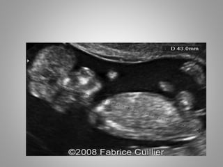
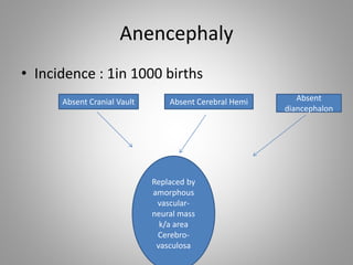
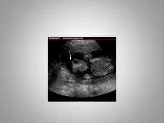
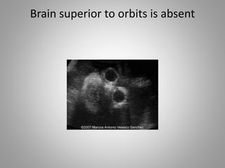
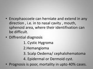
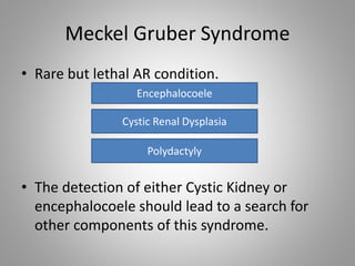
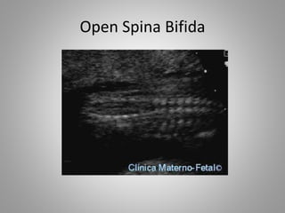
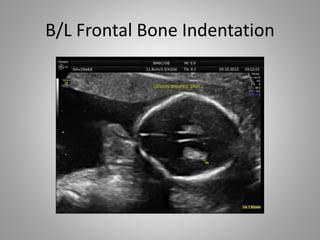
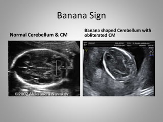
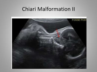
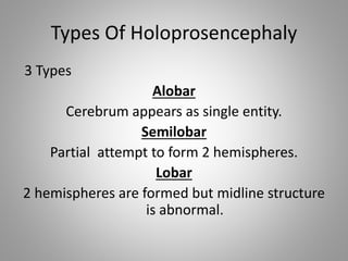
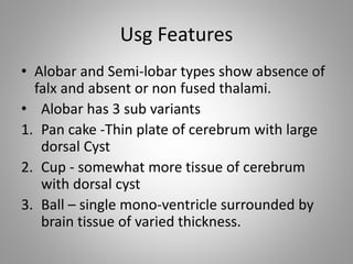
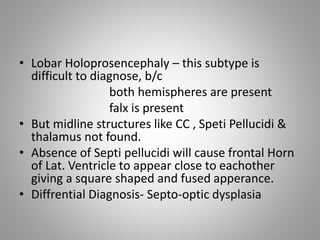
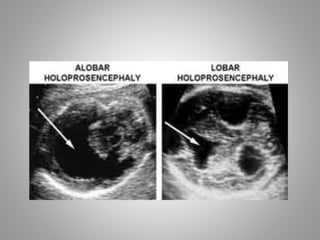
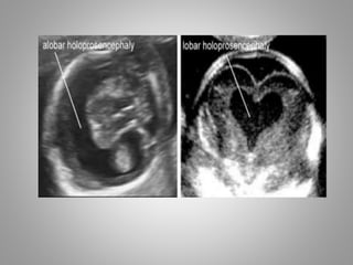
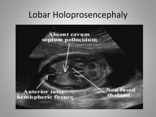
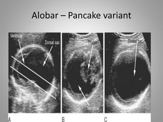
Fetal brain Usg.
Presented by: Dr Vrishit Saraswat
Topics for today
• Errors of Dorsal Induction
Acrania
Anencephaly
Cephalocoele
Encephalocoele
Spina Bifida
• Errors of Ventral Induction
mainly includes various
types of Holoprosencephaly ;i.e
1. Alobar
2. Semi-Lobar
3. Lobar
Error of Dorsal Induction
• Results in defect of closure of neural tube
which leads to various anomalies like
anencephaly, encephalocoele, spinal
dysraphism and chiari malformations.
Acrania
• Absence of cranial vault bones.
• Acrania can also occur with normal underlying
brain, so this term should not be used when
diagnosis is anencephaly or execncephaly.
Anencephaly
• Incidence : 1in 1000 births
Absent Cranial Vault Absent Cerebral Hemi Absent
diancephalon
Replaced by
amorphous
vascular-
neural mass
k/a area
Cerebro-
vasculosa
Brain superior to orbits is absent
Facial structures & orbits are present
• Anencephaly is always associated with
polyhydroamnios
• Outcome is fatal
• Termination of pregnancy is adviced at any
gestational age.
Cephalocoele & Encephalocoele
• Cephalocoele is herniation of intracranial
structures through a defect in cranial vault.
• Meningocoele – when meninges filled with
CSF fluid herniatethrough cranium & when
this meningocoele contains brain tissue, then
it is known as Encephalocoele.
• Encephalocoele herniate mostly through
midline and that too mostly in occipital
region(75%)
• Encephaocoele can herniate and extend in any
direction , i.e. in to nasal cavity , mouth,
sphenoid area, where their identifivation can
be difficult.
• Diffrential diagnosis
1. Cystic Hygroma
2.Hemangioma
3. Scalp Oedema/ cephalohematoma
4. Epidermal or Dermoid cyst.
• Prognosis is poor, mortality in upto 40% cases.
Meckel Gruber Syndrome
• Rare but lethal AR condition.
• The detection of either Cystic Kidney or
encephalocoele should lead to a search for
other components of this syndrome.
Encephalocoele
Cystic Renal Dysplasia
Polydactyly
Spina Bifida & Chiari II Malformation
• Spina bifida is classified as either
Open Closed
Covered with Skin Not covered with skin
• Virtually all fetuses with open variant have
cerebral changes typically of Chiari II
malformation .
• 80% of all diagnosed spina bifida are open
types
• Open spina bifida causes various changes in
head while closed variants, no matter how
big, will not cause any changes in fetal head.
• Head changes in Open Spina Bifida
1. Ventriculomegaly……….........
2. Lemon Sign……………………….
3. Banana Sign………………………
Closed Spina Bifida
Open Spina Bifida
B/L Frontal Bone Indentation
Banana Sign
Normal Cerebellum & CM
Banana shaped Cerebellum with
obliterated CM
Chiari Malformation II
MRI Chiari Malformation II
• The banana sign and effacement of cisterna
magna are due to fluid leakage allowing
cerebellum to compress into lower posterior
fossa.
• Cerebellum hemispheres wraps around lateral
aspect of brain stem and so asumes C-shape
{Banana Shape} in transcerebellar plane.
• These sonographic findings constitute Chiari II
malformation.
Errors of Ventral Induction
Holoprosencephaly
• Errors in induction of ventral portions of brain
and midline facial structures.
• Holoprosencephaly is a complex brain
malformation resulting from various degree of
incomplete clevage of Prosencephalon
• Most common anomaly of brain development
1 in 250 births.
• Prechordial Mesoderm (i.e mesoderm anterior to
notochord) which participates in midface
development ,which later induces forebrain
development.
• Due to any environmental or genetic insult this
above process fails causing Holoprosencephaly.
• From above pathogenesis , it is also clear that
severity of facial dysmorphism corelates with
cerebral anomaly
• Holoprosencephaly is a part of many different
syndromes, chromosomal & gene abnormality.
• Associations
1. DM type I
2. Vit A
3. Anti-convulsants
4. Alcohol
5. Velocardiofacial Syndrome
6. Smith Cemle-Opitz Syndrome
7. Tri 13 ***(70%)
8. Tri 18……………..
Types Of Holoprosencephaly
3 Types
Alobar
Cerebrum appears as single entity.
Semilobar
Partial attempt to form 2 hemispheres.
Lobar
2 hemispheres are formed but midline structure
is abnormal.
Usg Features
• Alobar and Semi-lobar types show absence of
falx and absent or non fused thalami.
• Alobar has 3 sub variants
1. Pan cake -Thin plate of cerebrum with large
dorsal Cyst
2. Cup - somewhat more tissue of cerebrum
with dorsal cyst
3. Ball – single mono-ventricle surrounded by
brain tissue of varied thickness.
• Lobar Holoprosencephaly – this subtype is
difficult to diagnose, b/c
both hemispheres are present
falx is present
• But midline structures like CC , Speti Pellucidi &
thalamus not found.
• Absence of Septi pellucidi will cause frontal Horn
of Lat. Ventricle to appear close to eachother
giving a square shaped and fused apperance.
• Diffrential Diagnosis- Septo-optic dysplasia
Lobar Holoprosencephaly
Alobar – Pancake variant
Alobar-Cup Variant
Alobar – Ball variant
• A new variant , also called as 4th type .
• Also known as middle interhemispheric form/
syntelemcephaly , in which seperation of
anterior and posterior parts of hemisphere
with partial formation of corpus callosum

Fetal brain usg 2

  • 1.
    Fetal brain Usg. Presentedby: Dr Vrishit Saraswat
  • 2.
    Topics for today •Errors of Dorsal Induction Acrania Anencephaly Cephalocoele Encephalocoele Spina Bifida
  • 3.
    • Errors ofVentral Induction mainly includes various types of Holoprosencephaly ;i.e 1. Alobar 2. Semi-Lobar 3. Lobar
  • 4.
    Error of DorsalInduction • Results in defect of closure of neural tube which leads to various anomalies like anencephaly, encephalocoele, spinal dysraphism and chiari malformations.
  • 5.
    Acrania • Absence ofcranial vault bones. • Acrania can also occur with normal underlying brain, so this term should not be used when diagnosis is anencephaly or execncephaly.
  • 7.
    Anencephaly • Incidence :1in 1000 births Absent Cranial Vault Absent Cerebral Hemi Absent diancephalon Replaced by amorphous vascular- neural mass k/a area Cerebro- vasculosa
  • 9.
    Brain superior toorbits is absent
  • 10.
    Facial structures &orbits are present
  • 11.
    • Anencephaly isalways associated with polyhydroamnios • Outcome is fatal • Termination of pregnancy is adviced at any gestational age.
  • 12.
    Cephalocoele & Encephalocoele •Cephalocoele is herniation of intracranial structures through a defect in cranial vault. • Meningocoele – when meninges filled with CSF fluid herniatethrough cranium & when this meningocoele contains brain tissue, then it is known as Encephalocoele. • Encephalocoele herniate mostly through midline and that too mostly in occipital region(75%)
  • 17.
    • Encephaocoele canherniate and extend in any direction , i.e. in to nasal cavity , mouth, sphenoid area, where their identifivation can be difficult. • Diffrential diagnosis 1. Cystic Hygroma 2.Hemangioma 3. Scalp Oedema/ cephalohematoma 4. Epidermal or Dermoid cyst. • Prognosis is poor, mortality in upto 40% cases.
  • 18.
    Meckel Gruber Syndrome •Rare but lethal AR condition. • The detection of either Cystic Kidney or encephalocoele should lead to a search for other components of this syndrome. Encephalocoele Cystic Renal Dysplasia Polydactyly
  • 19.
    Spina Bifida &Chiari II Malformation • Spina bifida is classified as either Open Closed Covered with Skin Not covered with skin
  • 20.
    • Virtually allfetuses with open variant have cerebral changes typically of Chiari II malformation . • 80% of all diagnosed spina bifida are open types • Open spina bifida causes various changes in head while closed variants, no matter how big, will not cause any changes in fetal head.
  • 21.
    • Head changesin Open Spina Bifida 1. Ventriculomegaly………......... 2. Lemon Sign………………………. 3. Banana Sign………………………
  • 22.
  • 23.
  • 24.
    B/L Frontal BoneIndentation
  • 25.
    Banana Sign Normal Cerebellum& CM Banana shaped Cerebellum with obliterated CM
  • 26.
  • 27.
  • 28.
    • The bananasign and effacement of cisterna magna are due to fluid leakage allowing cerebellum to compress into lower posterior fossa. • Cerebellum hemispheres wraps around lateral aspect of brain stem and so asumes C-shape {Banana Shape} in transcerebellar plane. • These sonographic findings constitute Chiari II malformation.
  • 29.
    Errors of VentralInduction Holoprosencephaly • Errors in induction of ventral portions of brain and midline facial structures. • Holoprosencephaly is a complex brain malformation resulting from various degree of incomplete clevage of Prosencephalon • Most common anomaly of brain development 1 in 250 births.
  • 30.
    • Prechordial Mesoderm(i.e mesoderm anterior to notochord) which participates in midface development ,which later induces forebrain development. • Due to any environmental or genetic insult this above process fails causing Holoprosencephaly. • From above pathogenesis , it is also clear that severity of facial dysmorphism corelates with cerebral anomaly
  • 31.
    • Holoprosencephaly isa part of many different syndromes, chromosomal & gene abnormality. • Associations 1. DM type I 2. Vit A 3. Anti-convulsants 4. Alcohol 5. Velocardiofacial Syndrome 6. Smith Cemle-Opitz Syndrome 7. Tri 13 ***(70%) 8. Tri 18……………..
  • 32.
    Types Of Holoprosencephaly 3Types Alobar Cerebrum appears as single entity. Semilobar Partial attempt to form 2 hemispheres. Lobar 2 hemispheres are formed but midline structure is abnormal.
  • 33.
    Usg Features • Alobarand Semi-lobar types show absence of falx and absent or non fused thalami. • Alobar has 3 sub variants 1. Pan cake -Thin plate of cerebrum with large dorsal Cyst 2. Cup - somewhat more tissue of cerebrum with dorsal cyst 3. Ball – single mono-ventricle surrounded by brain tissue of varied thickness.
  • 34.
    • Lobar Holoprosencephaly– this subtype is difficult to diagnose, b/c both hemispheres are present falx is present • But midline structures like CC , Speti Pellucidi & thalamus not found. • Absence of Septi pellucidi will cause frontal Horn of Lat. Ventricle to appear close to eachother giving a square shaped and fused apperance. • Diffrential Diagnosis- Septo-optic dysplasia
  • 37.
  • 38.
  • 39.
  • 40.
  • 41.
    • A newvariant , also called as 4th type . • Also known as middle interhemispheric form/ syntelemcephaly , in which seperation of anterior and posterior parts of hemisphere with partial formation of corpus callosum