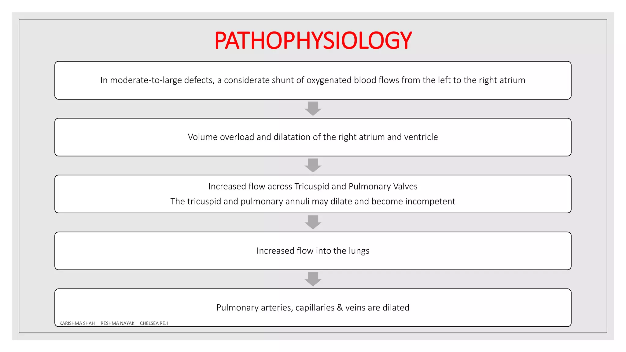
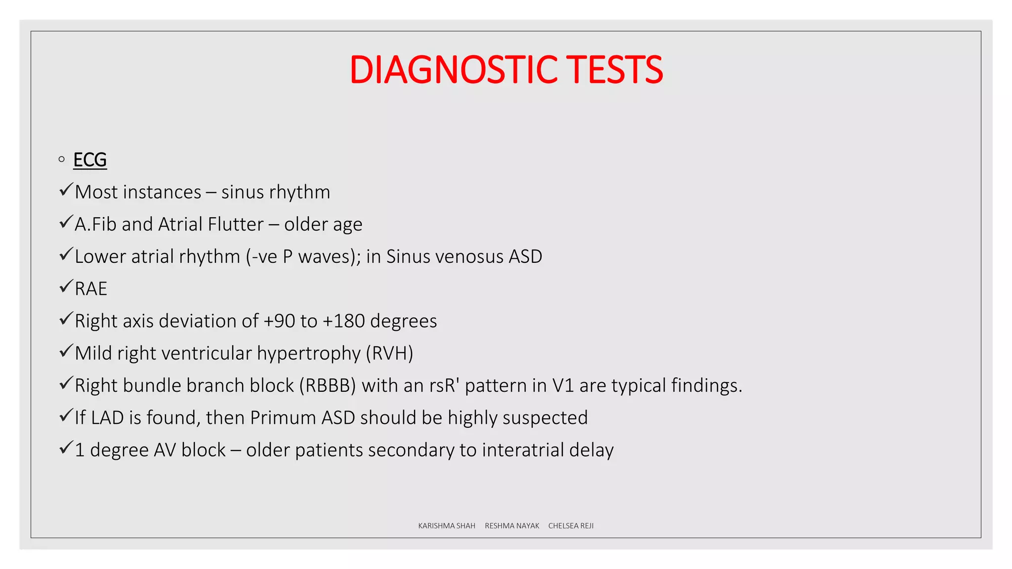
SlideShare a Scribd company logo
ATRIAL SEPTAL
DEFECT
BY GROUP 1: KARISHMA K SHAH, RESHMA
NAYAK & CHELSEA REJI
(MAHE, MCHP, Bsc.CVT)
CONTENTS
◦ DEFINITION
◦ PREVALENCE AND INCIDENCE
◦ GENETICS
◦ EMBRYOLOGY
◦ CLASSIFICATION
◦ PATHOPHYSIOLOGY
◦ RIMS
◦ CLINICAL MANIFESTATIONS
◦ DIAGNOSTIC TESTS
◦ MANAGEMENT
KARISHMA SHAH RESHMA NAYAK CHELSEA REJI
DEFINITION
◦ Atrial septal defect (ASD) is a heart defect that is present at birth (congenital).
◦ As a baby develops in the womb, a wall (called the interatrial septum) forms that
divides the upper chamber into a left and right atrium.
◦ An abnormal formation of this wall can result in a hole that remains after birth.
◦ This defect allows oxygen-rich blood to leak into the oxygen-poor blood chambers in
the heart.
◦ ASD refers to an opening between the 2 atria other than the component of the
Foramen Ovale.
KARISHMA SHAH RESHMA NAYAK CHELSEA REJI
PREVALENCE
◦ 30-50% of children with CHD have an ASD as a part of Cardiac defect.
◦ ASD (ostium secundum defect) occurs as an isolated anomaly in 5 % to 10% of all congenital heart
defects.
◦ It is more common in females than in males (male/female ratio of 1:2).
INCIDENCE
◦ The true incidence of interatrial defects is underestimated due to the high rate of spontaneous closure
and the delay in the presentation into adulthood due to subtle signs and symptoms.
KARISHMA SHAH RESHMA NAYAK CHELSEA REJI
GENETICS
◦ The genetic basis of the ASD is not completely understood.
◦ In the majority of cases this is a sporadic lesion, yet some gene defects have been found to explain some
of the well-known familial cases with ASDs, such as NKX2- chromosome 5, which has an autosomal
dominant inheritance and AV conduction defect.
◦ Other genetic syndromes with skeletal abnormality such as Holt-Oram syndrome, which is caused by
mutations in the transcription factor TBX5, essential in the development of both the heart and upper
limbs.
◦ Atrial septal defects can be part of many other syndromes and more complex congenital heart diseases.
Down syndrome is associated with both primum and secundum ASD, while Noonan syndrome most
commonly is associated with secundum ASD and pulmonary valve stenosis
KARISHMA SHAH RESHMA NAYAK CHELSEA REJI
EMBRYOLOGY
◦ Heart begins to develop in the middle of the 3rd week from the aggregation of the
splanchnic mesoderm cells in the cardiogenic area.
◦ 2 endocardial tubes are developed
◦ The 2 tubes fuse to form a single tube
◦ The heart tube differentiates into:
1. Truncus Arteriosus
2. Bulbus cordis
3. Primitive ventricle
4. Primitive atria
5. Sinus venosus
KARISHMA SHAH RESHMA NAYAK CHELSEA REJI
EMBRYOLOGY
◦ The heart tube then undergoes looping from day 23 to day 28 where the :
1. Cephalic portion bends ventrally, caudally and to the right
2. Caudal portion bends dorsocranially and to the left
KARISHMA SHAH RESHMA NAYAK CHELSEA REJI
EMBRYOLOGY
◦ The development of the normal atrial septum occurs following the initial looping of the
heart that is after 28 days of gestation.
◦ The SEPTUM PRIMUM arises from the roof of the atrium to the left of the septum
spurium. It grows downwards towards the atrioventricular canal and ultimately fuses
with the septum intermedium.
KARISHMA SHAH RESHMA NAYAK CHELSEA REJI
EMBRYOLOGY
◦ Throughout the fetal life oxygenated blood reaches the RA from the placenta. This
blood has to reach the LA. For this purpose, a communication between the 2 atria is
essential.
◦ Before the septum primum fuses with the septum intermedium, blood flows through
the gap between them. This gap is referred to as the FORAMEN PRIMUM.
◦ Before foramen primum can be closed it is essential that another path for the flow of
blood is created.
◦ This is achieved by breaking down of the upper part of the septum primum.
◦ New gap is referred to as FORAMEN SECUNDUM.
KARISHMA SHAH RESHMA NAYAK CHELSEA REJI
EMBRYOLOGY
◦ The septum primum now has a free upper edge.
◦ The SEPTUM SECUNDUM grows down from the roof of the
atrial chamber to the right of the septum primum.
◦ As it grows, it comes to overlap and the blood has to flow
through the interval between the septa. This gap is referred
to as FORAMEN OVALE.
◦ After birth the foramen ovale is obliterated by the fusion of
the septum primum and septum secundum.
KARISHMA SHAH RESHMA NAYAK CHELSEA REJI
EMBRYOLOGY
◦ Any abnormality in the formation of the IAS results in an ASD:
◦ SECUNDUM ASD occurs secondary to deficiency of septum primum in majority of the
cases and rarely due to deficient septum secundum. Occurs in the fossa ovalis position.
◦ PRIMUM ASD occurs as a result of abnormal formation of the endocardial cushion. Are
usually anteroinferior in location relative to the fossa ovalis.
◦ SINUS VENOSUS and CORONARY SINUS defects occur as a consequence of abnormal
development of embryologic venous tributaries.
KARISHMA SHAH RESHMA NAYAK CHELSEA REJI
CLASSIFICATION
◦ OSTIUM SECUNDUM ASD:
Most common type; 50-70% of all ASDs
This defect is present at the site of Fossa Ovalis, allowing L R shunting of blood from
LA to RA
PAPVC is present in about 10% of the cases
◦ OSTIUM PRIMUM ASD:
Occurs in 30% of all ASDs, if those that occur as part of complete endocardial cushion
defect.
Isolated ostium primum ASD occurs in about 15% of all ASDs.
KARISHMA SHAH RESHMA NAYAK CHELSEA REJI
CLASSIFICATION
◦ SINUS VENOSUS DEFECT:
Occurs in about 10% of all ASDs.
There are 2 types; SVC and IVC type
The defect is most commonly located at the entry of the SVC into the RA and rarely at
the entry of the IVC into the RA.
Commonly associated with PAPVC (RUPV RA)
Is often associated with anomalous Pulmonary venous drainage of the Right lung into
the IVC (SCIMITAR SYNDROME)
KARISHMA SHAH RESHMA NAYAK CHELSEA REJI
CLASSIFICATION
◦ CORONARY SINUS DEFECT:
There is a defect in the roof of the coronary sinus and the LA blood shunts through the
defect and empties into RA through the CS Ostium, producing clinical picture similar to
other types of ASDs.
This is usually seen in dilated coronary sinus opening into RA and is often associated
with a persistent left SVC.
NB: Mitral valve prolapse (MVP) occurs in 20% of patients with either ostium
secundum or sinus venosus defects
KARISHMA SHAH RESHMA NAYAK CHELSEA REJI
CLASSIFICATION
KARISHMA SHAH RESHMA NAYAK CHELSEA REJI
CLASSIFICATION
BASED ON SIZE:
1. McMohan et al (2000):
Small; >3mm - <6mm
Moderate; ≥6 - <12mm
Large; ≥12mm
2. Anita Saxena (2005):
Small; 4-5mm
Medium; 6-8mm
Large; > 8mm KARISHMA SHAH RESHMA NAYAK CHELSEA REJI
PATHOPHYSIOLOGY
◦ The degree of the left to right shunting is dependent on:
The size of the defect
The relative compliance of the RV and LV
The relative vascular resistance in the pulmonary and systemic circulations.
KARISHMA SHAH RESHMA NAYAK CHELSEA REJI
PATHOPHYSIOLOGY
In moderate-to-large defects, a considerate shunt of oxygenated blood flows from the left to the right atrium
Volume overload and dilatation of the right atrium and ventricle
Increased flow across Tricuspid and Pulmonary Valves
The tricuspid and pulmonary annuli may dilate and become incompetent
Increased flow into the lungs
Pulmonary arteries, capillaries & veins are dilated
KARISHMA SHAH RESHMA NAYAK CHELSEA REJI
PATHOPHYSIOLOGY
Flow- related pulmonary artery hypertension
Medial hypertrophy of pulmonary arteries and muscularization of arterioles resulting in PVOD
Reversal of the shunt
Eisenmenger syndrome
KARISHMA SHAH RESHMA NAYAK CHELSEA REJI
RIMS
◦ AORTIC RIM – The superior/anterior rim between the ASD and aortic annulus and
aortic root.
◦ AV VALVE RIM – The inferior/anterior rim between the ASD and the AV valves.
◦ SVC RIM – The superior/posterior rim between the ASD and the SVC.
◦ IVC RIM – The inferior/posterior rim between the ASD and the IVC.
◦ POSTERIOR RIM – The posterior rim between the ASD and the posterior atrial walls.
◦ RIGHT UPPER PV RIM – The posterior rim between the ASD and RUPV.
KARISHMA SHAH RESHMA NAYAK CHELSEA REJI
CLINICAL MANIFESTATIONS
◦ HISTORY
Infants and children are usually asymptomatic.
Most of the patients present symptoms at the 4-5th decade.
◦ SYMPTOMS
Fatigue
Dyspnoea on exertion
Peripheral edema
Other symptoms of heart failure
KARISHMA SHAH RESHMA NAYAK CHELSEA REJI
CLINICAL MANIFESTATIONS
◦ PHYSICAL EXAMINATION
A relatively slender body build is typical.
A widely split and fixed S2 due to a delay in the closure of the pulmonary valve as a reflection of the
dilated pulmonary artery and prolonged RV emptying.
Grade 2 to 3/6 systolic ejection murmur are characteristic findings of ASD in older infants and children.
With a large left-to-right shunt, a mid-diastolic rumble resulting from relative tricuspid stenosis may be
audible at the lower left sternal border
KARISHMA SHAH RESHMA NAYAK CHELSEA REJI
CLINICAL MANIFESTATIONS
◦ NATURAL HISTORY
Earlier reports have indicated that spontaneous closure of the secundum defect occurs in about 40% of patients in
the first 4 years of life. In patients with an ASD less than 3 mm in size diagnosed before 3 months of age,
spontaneous closure occurs in 100% of patients at 1½ years of age. Spontaneous closure occurs more than 80% of
the time in patients with defects between 3 and 8 mm before 1½ years of age. An ASD with a diameter greater than
8 mm rarely closes spontaneously.
Most children with an ASD remain active and asymptomatic. Rarely, congestive heart failure (CHF) can develop in
infancy.
If a large defect is untreated, CHF and pulmonary hypertension develop in adults who are in their 20s and 30s.
With or without surgery, atrial arrhythmias (flutter or fibrillation) may occur in adults.
Infective endocarditis does not occur in patients with isolated ASDs.
Cerebrovascular accident, resulting from paradoxical embolization through an ASD, is a rare complication.
KARISHMA SHAH RESHMA NAYAK CHELSEA REJI
DIAGNOSTIC TESTS
◦ CHEST X-RAY
Cardiomegaly with enlargement of the RA and
RV may be present.
A prominent pulmonary artery (PA) segment
and increased pulmonary vascular markings are
seen when the shunt is significant.
KARISHMA SHAH RESHMA NAYAK CHELSEA REJI
DIAGNOSTIC TESTS
KARISHMA SHAH RESHMA NAYAK CHELSEA REJI
DIAGNOSTIC TESTS
◦ ECG
Most instances – sinus rhythm
A.Fib and Atrial Flutter – older age
Lower atrial rhythm (-ve P waves); in Sinus venosus ASD
RAE
Right axis deviation of +90 to +180 degrees
Mild right ventricular hypertrophy (RVH)
Right bundle branch block (RBBB) with an rsR' pattern in V1 are typical findings.
If LAD is found, then Primum ASD should be highly suspected
1 degree AV block – older patients secondary to interatrial delay
KARISHMA SHAH RESHMA NAYAK CHELSEA REJI
DIAGNOSTIC TESTS
KARISHMA SHAH RESHMA NAYAK CHELSEA REJI
DIAGNOSTIC TESTS
KARISHMA SHAH RESHMA NAYAK CHELSEA REJI
DIAGNOSTIC TESTS
KARISHMA SHAH RESHMA NAYAK CHELSEA REJI
DIAGNOSTIC TESTS
◦ ECHOCARDIOGRAPHY
Goals :
◦ To identify and confirm ASD
◦ To identify associated anomaly
◦ To diagnose complications of ASD
◦ For therapeutic purpose
◦ 2D echo shows location, size of the defect
◦ If the dropout is seen at the mid-atrial level then it is SECUNDUM ASD
◦ If dropout at the lower atrial septum-PRIMUM ASD
◦ Defect in the posterosuperior atrial septum-SINUS VENOSUS ASD
◦ Defect in the wall that separates coronary sinus from LA- CORONARY SINUS ASD
KARISHMA SHAH RESHMA NAYAK CHELSEA REJI
DIAGNOSTIC TESTS
Apical 4 chamber
◦ Hemodynamic consequences like
RA/RV dilatation
◦ RV pressure
◦ Evaluation of R->L shunt
KARISHMA SHAH RESHMA NAYAK CHELSEA REJI
DIAGNOSTIC TESTS
Parasternal short axis (PSAX)
◦ Ideal to identify aortic rim
◦ Highlights posterior rim in sinus
venosus and posteroinferior
secundum ASD
◦ Shows dilatation of RV
◦ Volume overload of RV flattens IVS in
diastole
KARISHMA SHAH RESHMA NAYAK CHELSEA REJI
DIAGNOSTIC TESTS
Subcostal 4C view
◦ Viewed with breath held in
inspiration- index marker in 3 O clock
position
◦ Distinguishes OS, OP, SV ASD
◦ This view is ideal for SVC ASD
◦ Measurements of septum can be
taken
◦ Anomalous drainage of PVs
◦ Useful in patients with COPD and
ventilated patients
KARISHMA SHAH RESHMA NAYAK CHELSEA REJI
DIAGNOSTIC TESTS
Suprasternal short axis
◦ To visualize PV drainage into LA
Hemodynamics
◦ Indirect signs of L->R atrial shunt include RA/RV/PA enlargement which often accompanies increase in
flow velocity across PV
◦ Pulse doppler reveals characteristic flow pattern with max L-> R shunt occupying in diastole
1. L->R shunt begins in ventricular late systole.
2. Diminishes through mid diastole.
3. Enhanced by atrial contraction.
4. Reverses in early systole.
KARISHMA SHAH RESHMA NAYAK CHELSEA REJI
DIAGNOSTIC TESTS
KARISHMA SHAH RESHMA NAYAK CHELSEA REJI
DIAGNOSTIC TESTS
KARISHMA SHAH RESHMA NAYAK CHELSEA REJI
DIAGNOSTIC TESTS
◦ Qp/Qs
◦ Qp/Qs= 1 in normal
◦ Qp/Qs>1.5 significant shunt
◦ Qp=VTI,RVOT diameter
◦ Qs=VTI, LVOT diameter
𝑄𝑝
𝑄𝑠
=
𝑅𝑉𝑂𝑇 𝑑2 ∗ 0.785 ∗ 𝑅𝑉𝑂𝑇 𝑉𝑇𝐼
𝐿𝑉𝑂𝑇 𝑑2 ∗ 0.785 ∗ 𝐿𝑉𝑂𝑇 𝑉𝑇𝐼
ASD in Eisenmenger syndrome
◦ Pulmonary hypertension
◦ Reversal of flow
◦ Cyanosis KARISHMA SHAH RESHMA NAYAK CHELSEA REJI
DIAGNOSTIC TESTS
◦ CONTRAST
ECHOCARDIOGRAPHY
Apical 4 chamber view is
used
Agitated saline used – 5ml
in each 10ml syringe, o.5ml
of air taken in the syringe
and agitated to create
microbubbles
KARISHMA SHAH RESHMA NAYAK CHELSEA REJI
DIAGNOSTIC TESTS
TRANSESOPHAGIAL ECHO
◦ Gold standard
◦ Evaluation of number and location of defect, dimensions and adequacy of the rims
◦ It is used in individuals whom TTE acoustic window is suboptimal
◦ Guide trans catheter device closure
3D ECHO
◦ Clear image with spatial relationship to surrounding landmarks
◦ Used when attempting to close the multiple defects percutaneously
KARISHMA SHAH RESHMA NAYAK CHELSEA REJI
DIAGNOSTIC TESTS
CATH
◦ Pulmonary artery blood oxygen saturation is more than 80%, the possibility of left to
right intracardiac shunt should be considered.
◦ Oxygen saturation in PA, RA, RV, VC can be measured.
◦ Step up oxygen level in RA.
◦ In case of right to left shunt there will be step down oxygen level in LA.
KARISHMA SHAH RESHMA NAYAK CHELSEA REJI
MANAGEMENT
1. Medical
◦ Exercise restriction is unnecessary.
◦ In infants with CHF, medical management (with a diuretic) is
recommended because of its high success rate and the possibility of
spontaneous closure of the defect
KARISHMA SHAH RESHMA NAYAK CHELSEA REJI
MANAGEMENT
2. Nonsurgical Closure
◦ Nonsurgical closure using a catheter-delivered device closure has become a preferred method, provided
the indications are met.
◦ Several closure devices that can be delivered through cardiac catheters have been shown to be safe and
efficacious for secundum ASD closure.
Indication for device closure :
◦ Secundum ASD measuring 5mm or more in diameter (but less than 32 mm for Amplatzer device and
less than 18 mm for Helex device)
◦ A hemodynamically significant L-R shunt with clinical evidence of RV volume overload (i.e., Qp/Qs ratio
of 1.5:1 or greater or RV enlargement)
◦ There must be enough rim (4 mm) of septal tissue around the defect for appropriate placement of the
device
KARISHMA SHAH RESHMA NAYAK CHELSEA REJI
MANAGEMENT
◦ The rim size is estimated in four directions:
anterosuperior, anteroinferior, posterosuperior, and
posteroinferior.
◦ Two-dimensional echocardiographic estimates of the
ASD rim size: The posterosuperior (PS) and
posteroinferior (PI) rims are estimated in the bi–-vena
cava view from the subcostal transducer position, the
anteroinferior (AI) rim from the apical four-chamber
view, and the anterosuperior (AS) (or retro-aortic) rim
from the parasternal short-axis view
KARISHMA SHAH RESHMA NAYAK CHELSEA REJI
MANAGEMENT
◦ Indication for infants:
Considering the possibility of spontaneous closure, it is wise not to use the device in infancy unless the
patient is symptomatic with heart failure.
ASD closure devices can be implanted successfully in children younger than 2 years of age.
◦ Advantages of nonsurgical closure
Complete avoidance of cardiopulmonary bypass with its attendant risk
Avoidance of pain and residual thoracotomy scars
 A less than 24-hour hospital stay
 Rapid recovery
KARISHMA SHAH RESHMA NAYAK CHELSEA REJI
MANAGEMENT
◦ Complications / Disadvantages
Early ECG abnormalities are common within the first 24 hours after implant
Early or late erosion of the device into the aortic root (Amplatzer device)
Thrombus formation in the right and left atrium occurs (Rarely)
Higher rate of small residual leak
◦ Postdevice Closure Follow-up
The patients are prescribed aspirin- 81 mg per day for 6 months.
 Post-procedure echocardiographic studies check for a residual atrial shunt and unobstructed flow of
pulmonary veins, coronary sinus, and venae cavae, and proper function of the mitral and tricuspid
valves.
KARISHMA SHAH RESHMA NAYAK CHELSEA REJI
MANAGEMENT
3. Surgical Closure
◦ Indications and Timing
Surgical closure is indicated only when device closure is not considered appropriate.
Therefore, most patients with secundum ASD are not candidates for surgical closure of the defect.
A left-to-right shunt with a pulmonary-to-systemic blood flow ratio (Qp/Qs ratio of 1.5:1 or greater) is a
surgical indication.
Surgery is usually delayed until 2 to 4 years of age because the possibility of spontaneous closure exists.
If CHF does not respond to medical management, surgery is performed during infancy, again if device
closure is considered inappropriate.
High pulmonary vascular resistance (PVR) (i.e., >10 units/m2, >7 units/m2 with vasodilators) may be a
contraindication for surgery (or device closure).
KARISHMA SHAH RESHMA NAYAK CHELSEA REJI
MANAGEMENT
◦ Procedure
For secundum ASD, the defect is traditionally repaired through a midsternal incision under
cardiopulmonary bypass by either a simple suture or a pericardial or Teflon patch.
For a sinus venosus defect without associated anomalous pulmonary venous return, the defect is
closed using an autologous pericardial patch.
When it is associated with pulmonary venous anomaly, a tunnel is created between the anomalous
pulmonary vein and the ASD by using a Teflon or pericardial patch.
For coronary sinus ASD, the ostium of the coronary sinus is closed with an autologous pericardium with
care to avoid conduction tissues, provided it is not associated with persistent left SVC. This will result in
drainage of coronary sinus blood into the left atrium.
KARISHMA SHAH RESHMA NAYAK CHELSEA REJI
MANAGEMENT
PERICARDIAL PATCH NORMAL PATCH
KARISHMA SHAH RESHMA NAYAK CHELSEA REJI
MANAGEMENT
◦ Complications
Cerebrovascular accident and postoperative arrhythmias may develop in the immediate
postoperative period.
◦ Mortality
Fewer than 0.5% of patients die
however, there is a greater risk for small infants and those with increased PVR.
◦ Postoperative Follow-up
Cardiomegaly on chest radiographs and enlarged RV dimension on echo as well as the wide splitting of the S2 may
persist for 1 or 2 years after surgery.
 The ECG typically demonstrates RBBB (or RV conduction disturbance).
 Atrial or nodal arrhythmias occur in 7% to 20% of postoperative patients. Occasionally, sick sinus syndrome, which
occurs especially after the repair of a sinus venosus defect, may require antiarrhythmic drugs, pacemaker therapy,
or both.
KARISHMA SHAH RESHMA NAYAK CHELSEA REJI

More Related Content

What's hot

aortic stenosis AHA guidlines 2014
aortic stenosis AHA guidlines 2014aortic stenosis AHA guidlines 2014
aortic stenosis AHA guidlines 2014
Basem Enany
 
Tissue doppler Echocardiography (TDE)
Tissue doppler Echocardiography (TDE)Tissue doppler Echocardiography (TDE)
Tissue doppler Echocardiography (TDE)
sruthiMeenaxshiSR
 

What's hot (20)

Aorto pulmonary window may 2021
Aorto   pulmonary window  may 2021Aorto   pulmonary window  may 2021
Aorto pulmonary window may 2021
 
Echo assessment of mitral regurgitation
Echo assessment of mitral regurgitationEcho assessment of mitral regurgitation
Echo assessment of mitral regurgitation
 
Constrictive pericarditis
Constrictive pericarditis Constrictive pericarditis
Constrictive pericarditis
 
Tricuspid and pulmonic valves 2011
Tricuspid and pulmonic valves 2011Tricuspid and pulmonic valves 2011
Tricuspid and pulmonic valves 2011
 
ECHOCARDIOGRAPHIC EVALUATION OF AORTIC REGURGITATION
ECHOCARDIOGRAPHIC EVALUATION OF AORTIC REGURGITATIONECHOCARDIOGRAPHIC EVALUATION OF AORTIC REGURGITATION
ECHOCARDIOGRAPHIC EVALUATION OF AORTIC REGURGITATION
 
Echo Mitral Stenosis
Echo Mitral StenosisEcho Mitral Stenosis
Echo Mitral Stenosis
 
aortic stenosis AHA guidlines 2014
aortic stenosis AHA guidlines 2014aortic stenosis AHA guidlines 2014
aortic stenosis AHA guidlines 2014
 
Echo assessment of aortic valve disease
Echo assessment of aortic valve diseaseEcho assessment of aortic valve disease
Echo assessment of aortic valve disease
 
Atrioventricular canal defect, Firas Aljanadi,MD
Atrioventricular canal defect, Firas Aljanadi,MDAtrioventricular canal defect, Firas Aljanadi,MD
Atrioventricular canal defect, Firas Aljanadi,MD
 
Tissue doppler Echocardiography (TDE)
Tissue doppler Echocardiography (TDE)Tissue doppler Echocardiography (TDE)
Tissue doppler Echocardiography (TDE)
 
Echo in pericardial diseases
Echo in pericardial diseasesEcho in pericardial diseases
Echo in pericardial diseases
 
Congenital Heart Disease An Approach for Simple and Complex Anomalies
Congenital Heart Disease An Approach for Simple and Complex AnomaliesCongenital Heart Disease An Approach for Simple and Complex Anomalies
Congenital Heart Disease An Approach for Simple and Complex Anomalies
 
Aortic stenosis
Aortic stenosis Aortic stenosis
Aortic stenosis
 
CONTRAST ECHOCARDIOGRAPHY
CONTRAST ECHOCARDIOGRAPHYCONTRAST ECHOCARDIOGRAPHY
CONTRAST ECHOCARDIOGRAPHY
 
Atrial septal defect Echocardiography
Atrial septal defect EchocardiographyAtrial septal defect Echocardiography
Atrial septal defect Echocardiography
 
Atrioventricular canal defect
Atrioventricular canal defectAtrioventricular canal defect
Atrioventricular canal defect
 
MVP Mitral Valve Prolapse - Echocardiographic Evaluation
 MVP Mitral Valve  Prolapse - Echocardiographic Evaluation MVP Mitral Valve  Prolapse - Echocardiographic Evaluation
MVP Mitral Valve Prolapse - Echocardiographic Evaluation
 
Echocardiography assessment of Aortic Regurgitation severity
Echocardiography assessment of Aortic Regurgitation severityEchocardiography assessment of Aortic Regurgitation severity
Echocardiography assessment of Aortic Regurgitation severity
 
Echocardiographic evaluation of Aortic stenosis
Echocardiographic evaluation of Aortic stenosisEchocardiographic evaluation of Aortic stenosis
Echocardiographic evaluation of Aortic stenosis
 
Tricuspid valve disease
Tricuspid valve diseaseTricuspid valve disease
Tricuspid valve disease
 

Similar to Atrial septal defect (ASD)

Similar to Atrial septal defect (ASD) (20)

Congenital heart disease
Congenital heart diseaseCongenital heart disease
Congenital heart disease
 
Sami asd work
Sami asd workSami asd work
Sami asd work
 
seminar ASD
seminar ASDseminar ASD
seminar ASD
 
Atrial Septal Defect
Atrial Septal DefectAtrial Septal Defect
Atrial Septal Defect
 
Asd may 2021
Asd  may 2021Asd  may 2021
Asd may 2021
 
Asd new
Asd newAsd new
Asd new
 
Acyanotic congenital heart defects
Acyanotic congenital heart defectsAcyanotic congenital heart defects
Acyanotic congenital heart defects
 
Acyanoticcongenitalheartdisease 150417031927-conversion-gate01
Acyanoticcongenitalheartdisease 150417031927-conversion-gate01Acyanoticcongenitalheartdisease 150417031927-conversion-gate01
Acyanoticcongenitalheartdisease 150417031927-conversion-gate01
 
Atrial septal defects 16 3-15
Atrial septal defects 16 3-15Atrial septal defects 16 3-15
Atrial septal defects 16 3-15
 
CARDIOVASCULAR PATHOLOGY.pptx
CARDIOVASCULAR PATHOLOGY.pptxCARDIOVASCULAR PATHOLOGY.pptx
CARDIOVASCULAR PATHOLOGY.pptx
 
An approach to a patient with Atrial septal defect (ASD)
An approach to a patient with Atrial  septal defect (ASD)An approach to a patient with Atrial  septal defect (ASD)
An approach to a patient with Atrial septal defect (ASD)
 
Atrial septal defect
Atrial septal defect Atrial septal defect
Atrial septal defect
 
Atrial septal defect .by shanta
Atrial septal defect .by shantaAtrial septal defect .by shanta
Atrial septal defect .by shanta
 
Av canal defect
Av canal defectAv canal defect
Av canal defect
 
Endocardial Cushion Defect / AVSD
Endocardial Cushion Defect / AVSDEndocardial Cushion Defect / AVSD
Endocardial Cushion Defect / AVSD
 
Congenital_Heart_Disaese.pptx
Congenital_Heart_Disaese.pptxCongenital_Heart_Disaese.pptx
Congenital_Heart_Disaese.pptx
 
Cyanotic &amp; acyanotic heart disease
Cyanotic &amp; acyanotic heart diseaseCyanotic &amp; acyanotic heart disease
Cyanotic &amp; acyanotic heart disease
 
Congenital heart disease
Congenital heart diseaseCongenital heart disease
Congenital heart disease
 
Atrial Septal Defects.pptx
Atrial Septal Defects.pptxAtrial Septal Defects.pptx
Atrial Septal Defects.pptx
 
Acynotic heart defects
Acynotic heart defectsAcynotic heart defects
Acynotic heart defects
 

Recently uploaded

Benefits of Dentulu's Salivary Testing.pptx
Benefits of Dentulu's Salivary Testing.pptxBenefits of Dentulu's Salivary Testing.pptx
Benefits of Dentulu's Salivary Testing.pptx
Dentulu Inc
 
Immunity to Veterinary parasitic infections power point presentation
Immunity to Veterinary parasitic infections power point presentationImmunity to Veterinary parasitic infections power point presentation
Immunity to Veterinary parasitic infections power point presentation
BeshedaWedajo
 
Urinary Elimination BY ANUSHRI SRIVASTAVA.pptx
Urinary Elimination BY ANUSHRI SRIVASTAVA.pptxUrinary Elimination BY ANUSHRI SRIVASTAVA.pptx
Urinary Elimination BY ANUSHRI SRIVASTAVA.pptx
AnushriSrivastav
 
GENERAL PHARMACOLOGY - INTRODUCTION DENTAL.ppt
GENERAL PHARMACOLOGY - INTRODUCTION DENTAL.pptGENERAL PHARMACOLOGY - INTRODUCTION DENTAL.ppt
GENERAL PHARMACOLOGY - INTRODUCTION DENTAL.ppt
Mangaiarkkarasi
 
BOWEL ELIMINATION BY ANUSHRI SRIVASTAVA.pptx
BOWEL ELIMINATION BY ANUSHRI SRIVASTAVA.pptxBOWEL ELIMINATION BY ANUSHRI SRIVASTAVA.pptx
BOWEL ELIMINATION BY ANUSHRI SRIVASTAVA.pptx
AnushriSrivastav
 

Recently uploaded (20)

Global launch of the Healthy Ageing and Prevention Index 2nd wave – alongside...
Global launch of the Healthy Ageing and Prevention Index 2nd wave – alongside...Global launch of the Healthy Ageing and Prevention Index 2nd wave – alongside...
Global launch of the Healthy Ageing and Prevention Index 2nd wave – alongside...
 
Benefits of Dentulu's Salivary Testing.pptx
Benefits of Dentulu's Salivary Testing.pptxBenefits of Dentulu's Salivary Testing.pptx
Benefits of Dentulu's Salivary Testing.pptx
 
Jesse Jhaj: Building Relationships with Patients as a Doctor or Healthcare Wo...
Jesse Jhaj: Building Relationships with Patients as a Doctor or Healthcare Wo...Jesse Jhaj: Building Relationships with Patients as a Doctor or Healthcare Wo...
Jesse Jhaj: Building Relationships with Patients as a Doctor or Healthcare Wo...
 
VVIP Dehradun Girls 9719300533 Heat-bake { Dehradun } Genteel ℂall Serviℂe By...
VVIP Dehradun Girls 9719300533 Heat-bake { Dehradun } Genteel ℂall Serviℂe By...VVIP Dehradun Girls 9719300533 Heat-bake { Dehradun } Genteel ℂall Serviℂe By...
VVIP Dehradun Girls 9719300533 Heat-bake { Dehradun } Genteel ℂall Serviℂe By...
 
Immunity to Veterinary parasitic infections power point presentation
Immunity to Veterinary parasitic infections power point presentationImmunity to Veterinary parasitic infections power point presentation
Immunity to Veterinary parasitic infections power point presentation
 
Roti bank chennai PPT [Autosaved].pptx1
Roti bank  chennai PPT [Autosaved].pptx1Roti bank  chennai PPT [Autosaved].pptx1
Roti bank chennai PPT [Autosaved].pptx1
 
Navigating Women's Health: Understanding Prenatal Care and Beyond
Navigating Women's Health: Understanding Prenatal Care and BeyondNavigating Women's Health: Understanding Prenatal Care and Beyond
Navigating Women's Health: Understanding Prenatal Care and Beyond
 
Nose-Nasal Cavity & Paranasal Sinuses BY Dr.Rabia Inam Gandapore.pptx
Nose-Nasal Cavity & Paranasal Sinuses BY Dr.Rabia Inam Gandapore.pptxNose-Nasal Cavity & Paranasal Sinuses BY Dr.Rabia Inam Gandapore.pptx
Nose-Nasal Cavity & Paranasal Sinuses BY Dr.Rabia Inam Gandapore.pptx
 
Storage_of _Bariquin_Components_in_Storage_Boxes.pptx
Storage_of _Bariquin_Components_in_Storage_Boxes.pptxStorage_of _Bariquin_Components_in_Storage_Boxes.pptx
Storage_of _Bariquin_Components_in_Storage_Boxes.pptx
 
Chris Shade BS MEd MS LPC-Associate "Presume" (What Do I Do?)
Chris Shade BS MEd MS LPC-Associate "Presume" (What Do I Do?)Chris Shade BS MEd MS LPC-Associate "Presume" (What Do I Do?)
Chris Shade BS MEd MS LPC-Associate "Presume" (What Do I Do?)
 
A Community health , health for prisoners
A Community health  , health for prisonersA Community health  , health for prisoners
A Community health , health for prisoners
 
Artificial Intelligence to Optimize Cardiovascular Therapy
Artificial Intelligence to Optimize Cardiovascular TherapyArtificial Intelligence to Optimize Cardiovascular Therapy
Artificial Intelligence to Optimize Cardiovascular Therapy
 
Unlocking the Benefits of Cognitive Behavioural Therapy (CBT) with Renewed Edge
Unlocking the Benefits of Cognitive Behavioural Therapy (CBT) with Renewed EdgeUnlocking the Benefits of Cognitive Behavioural Therapy (CBT) with Renewed Edge
Unlocking the Benefits of Cognitive Behavioural Therapy (CBT) with Renewed Edge
 
Contact Now 89011**83002 Dehradun ℂall Girls By Full Service ℂall Girl In De...
Contact Now  89011**83002 Dehradun ℂall Girls By Full Service ℂall Girl In De...Contact Now  89011**83002 Dehradun ℂall Girls By Full Service ℂall Girl In De...
Contact Now 89011**83002 Dehradun ℂall Girls By Full Service ℂall Girl In De...
 
HEAT WAVE presented by priya bhojwani..pptx
HEAT WAVE presented by priya bhojwani..pptxHEAT WAVE presented by priya bhojwani..pptx
HEAT WAVE presented by priya bhojwani..pptx
 
Management of psoriasis.pptx (Recent advances)
Management of psoriasis.pptx (Recent advances)Management of psoriasis.pptx (Recent advances)
Management of psoriasis.pptx (Recent advances)
 
Urinary Elimination BY ANUSHRI SRIVASTAVA.pptx
Urinary Elimination BY ANUSHRI SRIVASTAVA.pptxUrinary Elimination BY ANUSHRI SRIVASTAVA.pptx
Urinary Elimination BY ANUSHRI SRIVASTAVA.pptx
 
GENERAL PHARMACOLOGY - INTRODUCTION DENTAL.ppt
GENERAL PHARMACOLOGY - INTRODUCTION DENTAL.pptGENERAL PHARMACOLOGY - INTRODUCTION DENTAL.ppt
GENERAL PHARMACOLOGY - INTRODUCTION DENTAL.ppt
 
BOWEL ELIMINATION BY ANUSHRI SRIVASTAVA.pptx
BOWEL ELIMINATION BY ANUSHRI SRIVASTAVA.pptxBOWEL ELIMINATION BY ANUSHRI SRIVASTAVA.pptx
BOWEL ELIMINATION BY ANUSHRI SRIVASTAVA.pptx
 
Myopia Management & Control Strategies.pptx
Myopia Management & Control Strategies.pptxMyopia Management & Control Strategies.pptx
Myopia Management & Control Strategies.pptx
 

Atrial septal defect (ASD)

  • 1. ATRIAL SEPTAL DEFECT BY GROUP 1: KARISHMA K SHAH, RESHMA NAYAK & CHELSEA REJI (MAHE, MCHP, Bsc.CVT)
  • 2. CONTENTS ◦ DEFINITION ◦ PREVALENCE AND INCIDENCE ◦ GENETICS ◦ EMBRYOLOGY ◦ CLASSIFICATION ◦ PATHOPHYSIOLOGY ◦ RIMS ◦ CLINICAL MANIFESTATIONS ◦ DIAGNOSTIC TESTS ◦ MANAGEMENT KARISHMA SHAH RESHMA NAYAK CHELSEA REJI
  • 3. DEFINITION ◦ Atrial septal defect (ASD) is a heart defect that is present at birth (congenital). ◦ As a baby develops in the womb, a wall (called the interatrial septum) forms that divides the upper chamber into a left and right atrium. ◦ An abnormal formation of this wall can result in a hole that remains after birth. ◦ This defect allows oxygen-rich blood to leak into the oxygen-poor blood chambers in the heart. ◦ ASD refers to an opening between the 2 atria other than the component of the Foramen Ovale. KARISHMA SHAH RESHMA NAYAK CHELSEA REJI
  • 4. KARISHMA SHAH RESHMA NAYAK CHELSEA REJI
  • 5. PREVALENCE ◦ 30-50% of children with CHD have an ASD as a part of Cardiac defect. ◦ ASD (ostium secundum defect) occurs as an isolated anomaly in 5 % to 10% of all congenital heart defects. ◦ It is more common in females than in males (male/female ratio of 1:2). INCIDENCE ◦ The true incidence of interatrial defects is underestimated due to the high rate of spontaneous closure and the delay in the presentation into adulthood due to subtle signs and symptoms. KARISHMA SHAH RESHMA NAYAK CHELSEA REJI
  • 6. GENETICS ◦ The genetic basis of the ASD is not completely understood. ◦ In the majority of cases this is a sporadic lesion, yet some gene defects have been found to explain some of the well-known familial cases with ASDs, such as NKX2- chromosome 5, which has an autosomal dominant inheritance and AV conduction defect. ◦ Other genetic syndromes with skeletal abnormality such as Holt-Oram syndrome, which is caused by mutations in the transcription factor TBX5, essential in the development of both the heart and upper limbs. ◦ Atrial septal defects can be part of many other syndromes and more complex congenital heart diseases. Down syndrome is associated with both primum and secundum ASD, while Noonan syndrome most commonly is associated with secundum ASD and pulmonary valve stenosis KARISHMA SHAH RESHMA NAYAK CHELSEA REJI
  • 7. EMBRYOLOGY ◦ Heart begins to develop in the middle of the 3rd week from the aggregation of the splanchnic mesoderm cells in the cardiogenic area. ◦ 2 endocardial tubes are developed ◦ The 2 tubes fuse to form a single tube ◦ The heart tube differentiates into: 1. Truncus Arteriosus 2. Bulbus cordis 3. Primitive ventricle 4. Primitive atria 5. Sinus venosus KARISHMA SHAH RESHMA NAYAK CHELSEA REJI
  • 8. EMBRYOLOGY ◦ The heart tube then undergoes looping from day 23 to day 28 where the : 1. Cephalic portion bends ventrally, caudally and to the right 2. Caudal portion bends dorsocranially and to the left KARISHMA SHAH RESHMA NAYAK CHELSEA REJI
  • 9. EMBRYOLOGY ◦ The development of the normal atrial septum occurs following the initial looping of the heart that is after 28 days of gestation. ◦ The SEPTUM PRIMUM arises from the roof of the atrium to the left of the septum spurium. It grows downwards towards the atrioventricular canal and ultimately fuses with the septum intermedium. KARISHMA SHAH RESHMA NAYAK CHELSEA REJI
  • 10. EMBRYOLOGY ◦ Throughout the fetal life oxygenated blood reaches the RA from the placenta. This blood has to reach the LA. For this purpose, a communication between the 2 atria is essential. ◦ Before the septum primum fuses with the septum intermedium, blood flows through the gap between them. This gap is referred to as the FORAMEN PRIMUM. ◦ Before foramen primum can be closed it is essential that another path for the flow of blood is created. ◦ This is achieved by breaking down of the upper part of the septum primum. ◦ New gap is referred to as FORAMEN SECUNDUM. KARISHMA SHAH RESHMA NAYAK CHELSEA REJI
  • 11. EMBRYOLOGY ◦ The septum primum now has a free upper edge. ◦ The SEPTUM SECUNDUM grows down from the roof of the atrial chamber to the right of the septum primum. ◦ As it grows, it comes to overlap and the blood has to flow through the interval between the septa. This gap is referred to as FORAMEN OVALE. ◦ After birth the foramen ovale is obliterated by the fusion of the septum primum and septum secundum. KARISHMA SHAH RESHMA NAYAK CHELSEA REJI
  • 12. EMBRYOLOGY ◦ Any abnormality in the formation of the IAS results in an ASD: ◦ SECUNDUM ASD occurs secondary to deficiency of septum primum in majority of the cases and rarely due to deficient septum secundum. Occurs in the fossa ovalis position. ◦ PRIMUM ASD occurs as a result of abnormal formation of the endocardial cushion. Are usually anteroinferior in location relative to the fossa ovalis. ◦ SINUS VENOSUS and CORONARY SINUS defects occur as a consequence of abnormal development of embryologic venous tributaries. KARISHMA SHAH RESHMA NAYAK CHELSEA REJI
  • 13. CLASSIFICATION ◦ OSTIUM SECUNDUM ASD: Most common type; 50-70% of all ASDs This defect is present at the site of Fossa Ovalis, allowing L R shunting of blood from LA to RA PAPVC is present in about 10% of the cases ◦ OSTIUM PRIMUM ASD: Occurs in 30% of all ASDs, if those that occur as part of complete endocardial cushion defect. Isolated ostium primum ASD occurs in about 15% of all ASDs. KARISHMA SHAH RESHMA NAYAK CHELSEA REJI
  • 14. CLASSIFICATION ◦ SINUS VENOSUS DEFECT: Occurs in about 10% of all ASDs. There are 2 types; SVC and IVC type The defect is most commonly located at the entry of the SVC into the RA and rarely at the entry of the IVC into the RA. Commonly associated with PAPVC (RUPV RA) Is often associated with anomalous Pulmonary venous drainage of the Right lung into the IVC (SCIMITAR SYNDROME) KARISHMA SHAH RESHMA NAYAK CHELSEA REJI
  • 15. CLASSIFICATION ◦ CORONARY SINUS DEFECT: There is a defect in the roof of the coronary sinus and the LA blood shunts through the defect and empties into RA through the CS Ostium, producing clinical picture similar to other types of ASDs. This is usually seen in dilated coronary sinus opening into RA and is often associated with a persistent left SVC. NB: Mitral valve prolapse (MVP) occurs in 20% of patients with either ostium secundum or sinus venosus defects KARISHMA SHAH RESHMA NAYAK CHELSEA REJI
  • 17. CLASSIFICATION BASED ON SIZE: 1. McMohan et al (2000): Small; >3mm - <6mm Moderate; ≥6 - <12mm Large; ≥12mm 2. Anita Saxena (2005): Small; 4-5mm Medium; 6-8mm Large; > 8mm KARISHMA SHAH RESHMA NAYAK CHELSEA REJI
  • 18. PATHOPHYSIOLOGY ◦ The degree of the left to right shunting is dependent on: The size of the defect The relative compliance of the RV and LV The relative vascular resistance in the pulmonary and systemic circulations. KARISHMA SHAH RESHMA NAYAK CHELSEA REJI
  • 19. PATHOPHYSIOLOGY In moderate-to-large defects, a considerate shunt of oxygenated blood flows from the left to the right atrium Volume overload and dilatation of the right atrium and ventricle Increased flow across Tricuspid and Pulmonary Valves The tricuspid and pulmonary annuli may dilate and become incompetent Increased flow into the lungs Pulmonary arteries, capillaries & veins are dilated KARISHMA SHAH RESHMA NAYAK CHELSEA REJI
  • 20. PATHOPHYSIOLOGY Flow- related pulmonary artery hypertension Medial hypertrophy of pulmonary arteries and muscularization of arterioles resulting in PVOD Reversal of the shunt Eisenmenger syndrome KARISHMA SHAH RESHMA NAYAK CHELSEA REJI
  • 21. RIMS ◦ AORTIC RIM – The superior/anterior rim between the ASD and aortic annulus and aortic root. ◦ AV VALVE RIM – The inferior/anterior rim between the ASD and the AV valves. ◦ SVC RIM – The superior/posterior rim between the ASD and the SVC. ◦ IVC RIM – The inferior/posterior rim between the ASD and the IVC. ◦ POSTERIOR RIM – The posterior rim between the ASD and the posterior atrial walls. ◦ RIGHT UPPER PV RIM – The posterior rim between the ASD and RUPV. KARISHMA SHAH RESHMA NAYAK CHELSEA REJI
  • 22. CLINICAL MANIFESTATIONS ◦ HISTORY Infants and children are usually asymptomatic. Most of the patients present symptoms at the 4-5th decade. ◦ SYMPTOMS Fatigue Dyspnoea on exertion Peripheral edema Other symptoms of heart failure KARISHMA SHAH RESHMA NAYAK CHELSEA REJI
  • 23. CLINICAL MANIFESTATIONS ◦ PHYSICAL EXAMINATION A relatively slender body build is typical. A widely split and fixed S2 due to a delay in the closure of the pulmonary valve as a reflection of the dilated pulmonary artery and prolonged RV emptying. Grade 2 to 3/6 systolic ejection murmur are characteristic findings of ASD in older infants and children. With a large left-to-right shunt, a mid-diastolic rumble resulting from relative tricuspid stenosis may be audible at the lower left sternal border KARISHMA SHAH RESHMA NAYAK CHELSEA REJI
  • 24. CLINICAL MANIFESTATIONS ◦ NATURAL HISTORY Earlier reports have indicated that spontaneous closure of the secundum defect occurs in about 40% of patients in the first 4 years of life. In patients with an ASD less than 3 mm in size diagnosed before 3 months of age, spontaneous closure occurs in 100% of patients at 1½ years of age. Spontaneous closure occurs more than 80% of the time in patients with defects between 3 and 8 mm before 1½ years of age. An ASD with a diameter greater than 8 mm rarely closes spontaneously. Most children with an ASD remain active and asymptomatic. Rarely, congestive heart failure (CHF) can develop in infancy. If a large defect is untreated, CHF and pulmonary hypertension develop in adults who are in their 20s and 30s. With or without surgery, atrial arrhythmias (flutter or fibrillation) may occur in adults. Infective endocarditis does not occur in patients with isolated ASDs. Cerebrovascular accident, resulting from paradoxical embolization through an ASD, is a rare complication. KARISHMA SHAH RESHMA NAYAK CHELSEA REJI
  • 25. DIAGNOSTIC TESTS ◦ CHEST X-RAY Cardiomegaly with enlargement of the RA and RV may be present. A prominent pulmonary artery (PA) segment and increased pulmonary vascular markings are seen when the shunt is significant. KARISHMA SHAH RESHMA NAYAK CHELSEA REJI
  • 26. DIAGNOSTIC TESTS KARISHMA SHAH RESHMA NAYAK CHELSEA REJI
  • 27. DIAGNOSTIC TESTS ◦ ECG Most instances – sinus rhythm A.Fib and Atrial Flutter – older age Lower atrial rhythm (-ve P waves); in Sinus venosus ASD RAE Right axis deviation of +90 to +180 degrees Mild right ventricular hypertrophy (RVH) Right bundle branch block (RBBB) with an rsR' pattern in V1 are typical findings. If LAD is found, then Primum ASD should be highly suspected 1 degree AV block – older patients secondary to interatrial delay KARISHMA SHAH RESHMA NAYAK CHELSEA REJI
  • 28. DIAGNOSTIC TESTS KARISHMA SHAH RESHMA NAYAK CHELSEA REJI
  • 29. DIAGNOSTIC TESTS KARISHMA SHAH RESHMA NAYAK CHELSEA REJI
  • 30. DIAGNOSTIC TESTS KARISHMA SHAH RESHMA NAYAK CHELSEA REJI
  • 31. DIAGNOSTIC TESTS ◦ ECHOCARDIOGRAPHY Goals : ◦ To identify and confirm ASD ◦ To identify associated anomaly ◦ To diagnose complications of ASD ◦ For therapeutic purpose ◦ 2D echo shows location, size of the defect ◦ If the dropout is seen at the mid-atrial level then it is SECUNDUM ASD ◦ If dropout at the lower atrial septum-PRIMUM ASD ◦ Defect in the posterosuperior atrial septum-SINUS VENOSUS ASD ◦ Defect in the wall that separates coronary sinus from LA- CORONARY SINUS ASD KARISHMA SHAH RESHMA NAYAK CHELSEA REJI
  • 32. DIAGNOSTIC TESTS Apical 4 chamber ◦ Hemodynamic consequences like RA/RV dilatation ◦ RV pressure ◦ Evaluation of R->L shunt KARISHMA SHAH RESHMA NAYAK CHELSEA REJI
  • 33. DIAGNOSTIC TESTS Parasternal short axis (PSAX) ◦ Ideal to identify aortic rim ◦ Highlights posterior rim in sinus venosus and posteroinferior secundum ASD ◦ Shows dilatation of RV ◦ Volume overload of RV flattens IVS in diastole KARISHMA SHAH RESHMA NAYAK CHELSEA REJI
  • 34. DIAGNOSTIC TESTS Subcostal 4C view ◦ Viewed with breath held in inspiration- index marker in 3 O clock position ◦ Distinguishes OS, OP, SV ASD ◦ This view is ideal for SVC ASD ◦ Measurements of septum can be taken ◦ Anomalous drainage of PVs ◦ Useful in patients with COPD and ventilated patients KARISHMA SHAH RESHMA NAYAK CHELSEA REJI
  • 35. DIAGNOSTIC TESTS Suprasternal short axis ◦ To visualize PV drainage into LA Hemodynamics ◦ Indirect signs of L->R atrial shunt include RA/RV/PA enlargement which often accompanies increase in flow velocity across PV ◦ Pulse doppler reveals characteristic flow pattern with max L-> R shunt occupying in diastole 1. L->R shunt begins in ventricular late systole. 2. Diminishes through mid diastole. 3. Enhanced by atrial contraction. 4. Reverses in early systole. KARISHMA SHAH RESHMA NAYAK CHELSEA REJI
  • 36. DIAGNOSTIC TESTS KARISHMA SHAH RESHMA NAYAK CHELSEA REJI
  • 37. DIAGNOSTIC TESTS KARISHMA SHAH RESHMA NAYAK CHELSEA REJI
  • 38. KARISHMA SHAH RESHMA NAYAK CHELSEA REJI
  • 39. KARISHMA SHAH RESHMA NAYAK CHELSEA REJI
  • 40. KARISHMA SHAH RESHMA NAYAK CHELSEA REJI
  • 41. KARISHMA SHAH RESHMA NAYAK CHELSEA REJI
  • 42. KARISHMA SHAH RESHMA NAYAK CHELSEA REJI
  • 43. KARISHMA SHAH RESHMA NAYAK CHELSEA REJI
  • 44. DIAGNOSTIC TESTS ◦ Qp/Qs ◦ Qp/Qs= 1 in normal ◦ Qp/Qs>1.5 significant shunt ◦ Qp=VTI,RVOT diameter ◦ Qs=VTI, LVOT diameter 𝑄𝑝 𝑄𝑠 = 𝑅𝑉𝑂𝑇 𝑑2 ∗ 0.785 ∗ 𝑅𝑉𝑂𝑇 𝑉𝑇𝐼 𝐿𝑉𝑂𝑇 𝑑2 ∗ 0.785 ∗ 𝐿𝑉𝑂𝑇 𝑉𝑇𝐼 ASD in Eisenmenger syndrome ◦ Pulmonary hypertension ◦ Reversal of flow ◦ Cyanosis KARISHMA SHAH RESHMA NAYAK CHELSEA REJI
  • 45. DIAGNOSTIC TESTS ◦ CONTRAST ECHOCARDIOGRAPHY Apical 4 chamber view is used Agitated saline used – 5ml in each 10ml syringe, o.5ml of air taken in the syringe and agitated to create microbubbles KARISHMA SHAH RESHMA NAYAK CHELSEA REJI
  • 46. DIAGNOSTIC TESTS TRANSESOPHAGIAL ECHO ◦ Gold standard ◦ Evaluation of number and location of defect, dimensions and adequacy of the rims ◦ It is used in individuals whom TTE acoustic window is suboptimal ◦ Guide trans catheter device closure 3D ECHO ◦ Clear image with spatial relationship to surrounding landmarks ◦ Used when attempting to close the multiple defects percutaneously KARISHMA SHAH RESHMA NAYAK CHELSEA REJI
  • 47. DIAGNOSTIC TESTS CATH ◦ Pulmonary artery blood oxygen saturation is more than 80%, the possibility of left to right intracardiac shunt should be considered. ◦ Oxygen saturation in PA, RA, RV, VC can be measured. ◦ Step up oxygen level in RA. ◦ In case of right to left shunt there will be step down oxygen level in LA. KARISHMA SHAH RESHMA NAYAK CHELSEA REJI
  • 48. MANAGEMENT 1. Medical ◦ Exercise restriction is unnecessary. ◦ In infants with CHF, medical management (with a diuretic) is recommended because of its high success rate and the possibility of spontaneous closure of the defect KARISHMA SHAH RESHMA NAYAK CHELSEA REJI
  • 49. MANAGEMENT 2. Nonsurgical Closure ◦ Nonsurgical closure using a catheter-delivered device closure has become a preferred method, provided the indications are met. ◦ Several closure devices that can be delivered through cardiac catheters have been shown to be safe and efficacious for secundum ASD closure. Indication for device closure : ◦ Secundum ASD measuring 5mm or more in diameter (but less than 32 mm for Amplatzer device and less than 18 mm for Helex device) ◦ A hemodynamically significant L-R shunt with clinical evidence of RV volume overload (i.e., Qp/Qs ratio of 1.5:1 or greater or RV enlargement) ◦ There must be enough rim (4 mm) of septal tissue around the defect for appropriate placement of the device KARISHMA SHAH RESHMA NAYAK CHELSEA REJI
  • 50. MANAGEMENT ◦ The rim size is estimated in four directions: anterosuperior, anteroinferior, posterosuperior, and posteroinferior. ◦ Two-dimensional echocardiographic estimates of the ASD rim size: The posterosuperior (PS) and posteroinferior (PI) rims are estimated in the bi–-vena cava view from the subcostal transducer position, the anteroinferior (AI) rim from the apical four-chamber view, and the anterosuperior (AS) (or retro-aortic) rim from the parasternal short-axis view KARISHMA SHAH RESHMA NAYAK CHELSEA REJI
  • 51. MANAGEMENT ◦ Indication for infants: Considering the possibility of spontaneous closure, it is wise not to use the device in infancy unless the patient is symptomatic with heart failure. ASD closure devices can be implanted successfully in children younger than 2 years of age. ◦ Advantages of nonsurgical closure Complete avoidance of cardiopulmonary bypass with its attendant risk Avoidance of pain and residual thoracotomy scars  A less than 24-hour hospital stay  Rapid recovery KARISHMA SHAH RESHMA NAYAK CHELSEA REJI
  • 52. MANAGEMENT ◦ Complications / Disadvantages Early ECG abnormalities are common within the first 24 hours after implant Early or late erosion of the device into the aortic root (Amplatzer device) Thrombus formation in the right and left atrium occurs (Rarely) Higher rate of small residual leak ◦ Postdevice Closure Follow-up The patients are prescribed aspirin- 81 mg per day for 6 months.  Post-procedure echocardiographic studies check for a residual atrial shunt and unobstructed flow of pulmonary veins, coronary sinus, and venae cavae, and proper function of the mitral and tricuspid valves. KARISHMA SHAH RESHMA NAYAK CHELSEA REJI
  • 53. KARISHMA SHAH RESHMA NAYAK CHELSEA REJI
  • 54. MANAGEMENT 3. Surgical Closure ◦ Indications and Timing Surgical closure is indicated only when device closure is not considered appropriate. Therefore, most patients with secundum ASD are not candidates for surgical closure of the defect. A left-to-right shunt with a pulmonary-to-systemic blood flow ratio (Qp/Qs ratio of 1.5:1 or greater) is a surgical indication. Surgery is usually delayed until 2 to 4 years of age because the possibility of spontaneous closure exists. If CHF does not respond to medical management, surgery is performed during infancy, again if device closure is considered inappropriate. High pulmonary vascular resistance (PVR) (i.e., >10 units/m2, >7 units/m2 with vasodilators) may be a contraindication for surgery (or device closure). KARISHMA SHAH RESHMA NAYAK CHELSEA REJI
  • 55. MANAGEMENT ◦ Procedure For secundum ASD, the defect is traditionally repaired through a midsternal incision under cardiopulmonary bypass by either a simple suture or a pericardial or Teflon patch. For a sinus venosus defect without associated anomalous pulmonary venous return, the defect is closed using an autologous pericardial patch. When it is associated with pulmonary venous anomaly, a tunnel is created between the anomalous pulmonary vein and the ASD by using a Teflon or pericardial patch. For coronary sinus ASD, the ostium of the coronary sinus is closed with an autologous pericardium with care to avoid conduction tissues, provided it is not associated with persistent left SVC. This will result in drainage of coronary sinus blood into the left atrium. KARISHMA SHAH RESHMA NAYAK CHELSEA REJI
  • 56. MANAGEMENT PERICARDIAL PATCH NORMAL PATCH KARISHMA SHAH RESHMA NAYAK CHELSEA REJI
  • 57. MANAGEMENT ◦ Complications Cerebrovascular accident and postoperative arrhythmias may develop in the immediate postoperative period. ◦ Mortality Fewer than 0.5% of patients die however, there is a greater risk for small infants and those with increased PVR. ◦ Postoperative Follow-up Cardiomegaly on chest radiographs and enlarged RV dimension on echo as well as the wide splitting of the S2 may persist for 1 or 2 years after surgery.  The ECG typically demonstrates RBBB (or RV conduction disturbance).  Atrial or nodal arrhythmias occur in 7% to 20% of postoperative patients. Occasionally, sick sinus syndrome, which occurs especially after the repair of a sinus venosus defect, may require antiarrhythmic drugs, pacemaker therapy, or both. KARISHMA SHAH RESHMA NAYAK CHELSEA REJI
  • 58. KARISHMA SHAH RESHMA NAYAK CHELSEA REJI
  • 59. KARISHMA SHAH RESHMA NAYAK CHELSEA REJI

Editor's Notes

  1. Prevalence refers to proportion of persons who have a condition at or during a particular time period, whereas incidence refers to the proportion or rate of persons who develop a condition during a particular time period.
  2. HOLT ORAM syndrome – abnormal formed bones and heart problems.