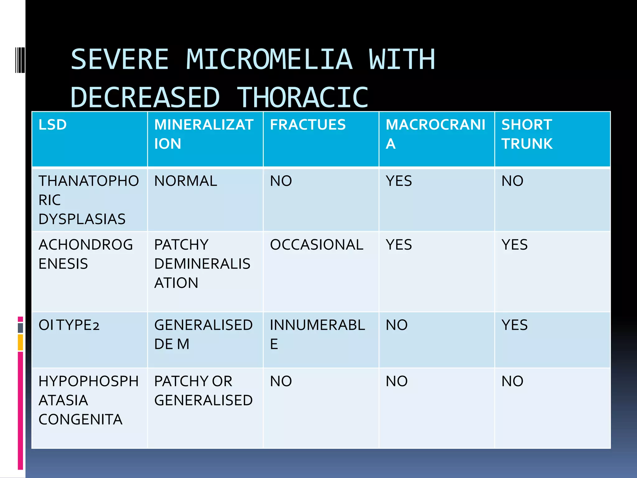
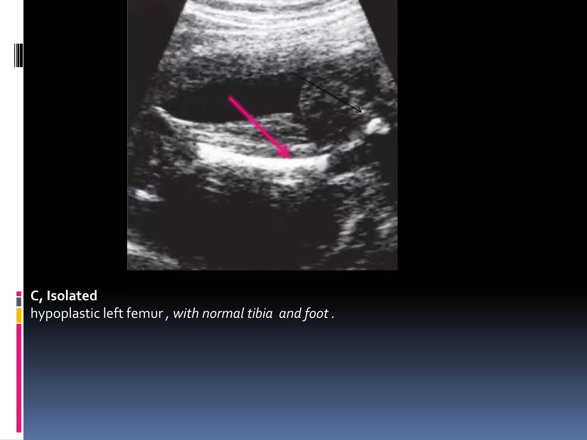
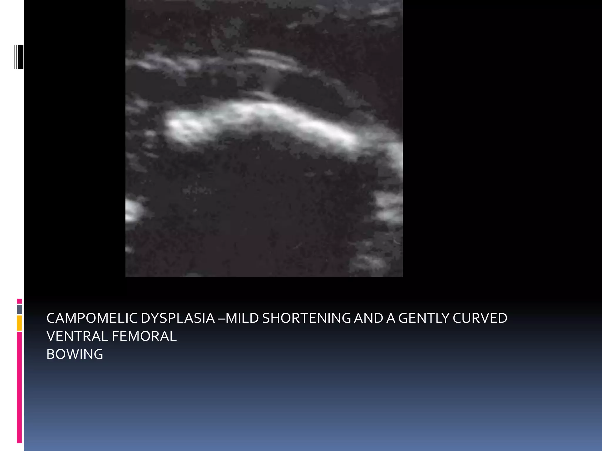
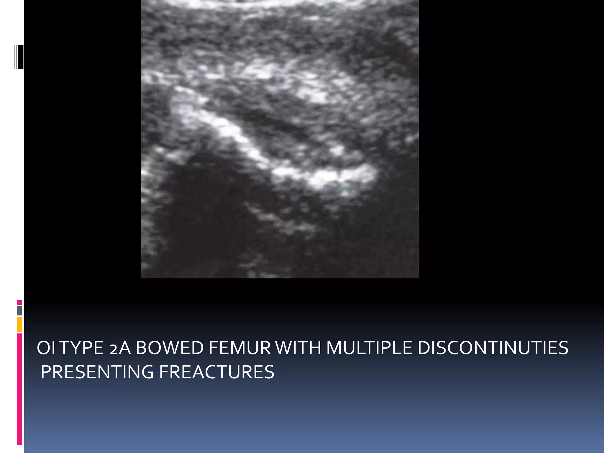
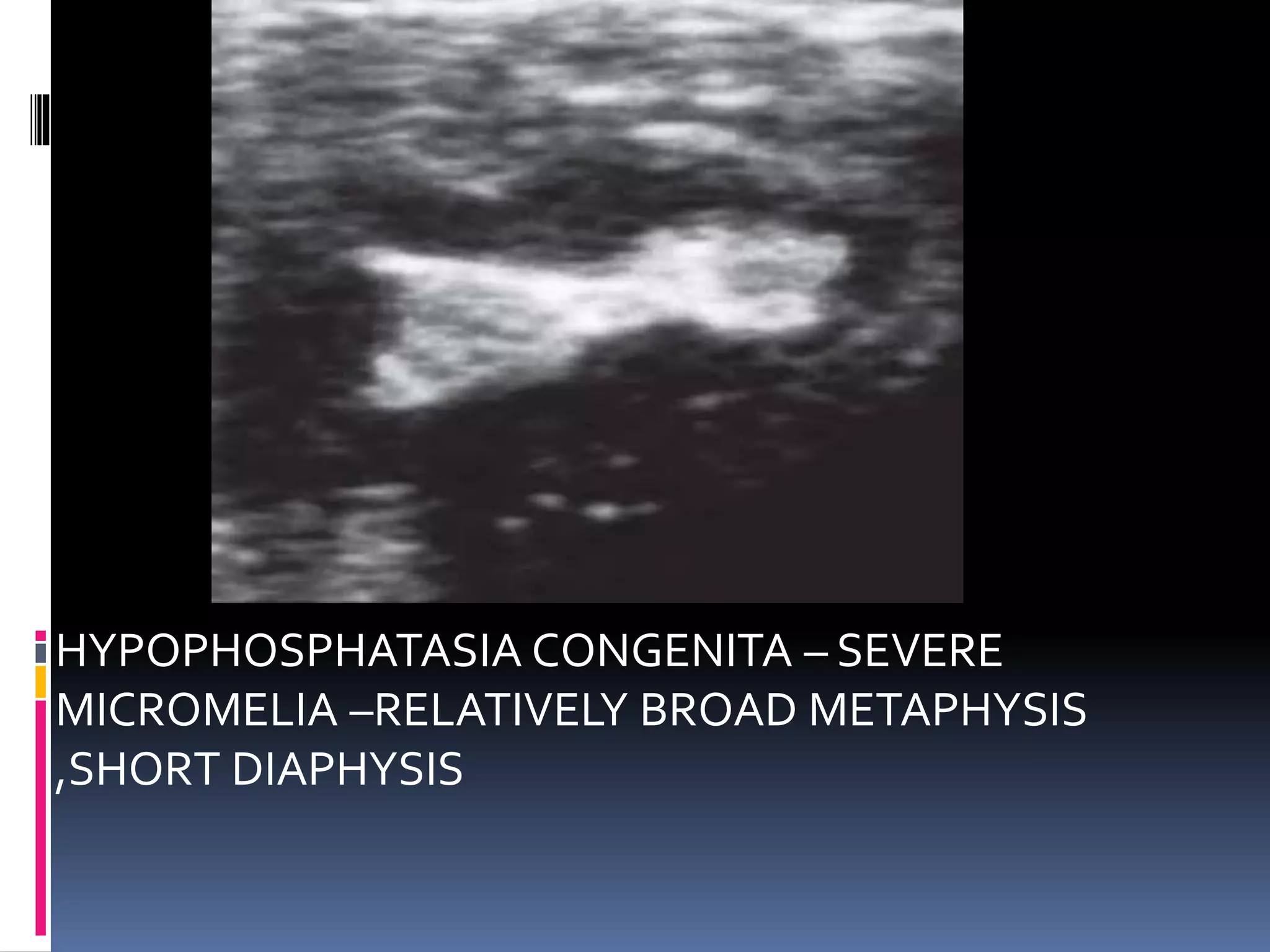
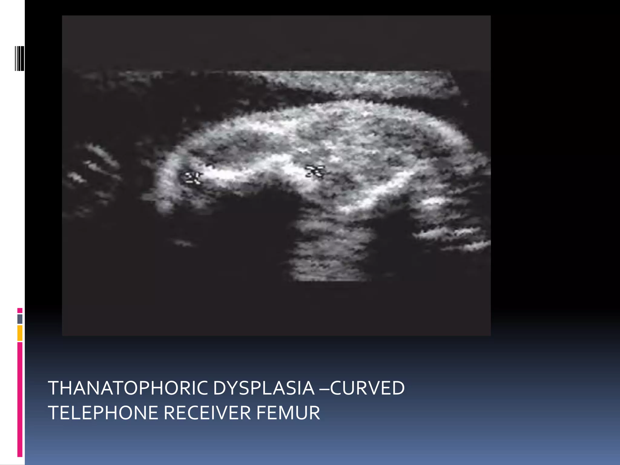
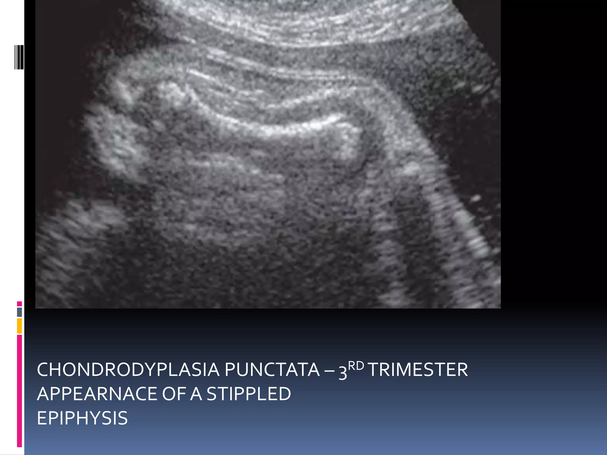
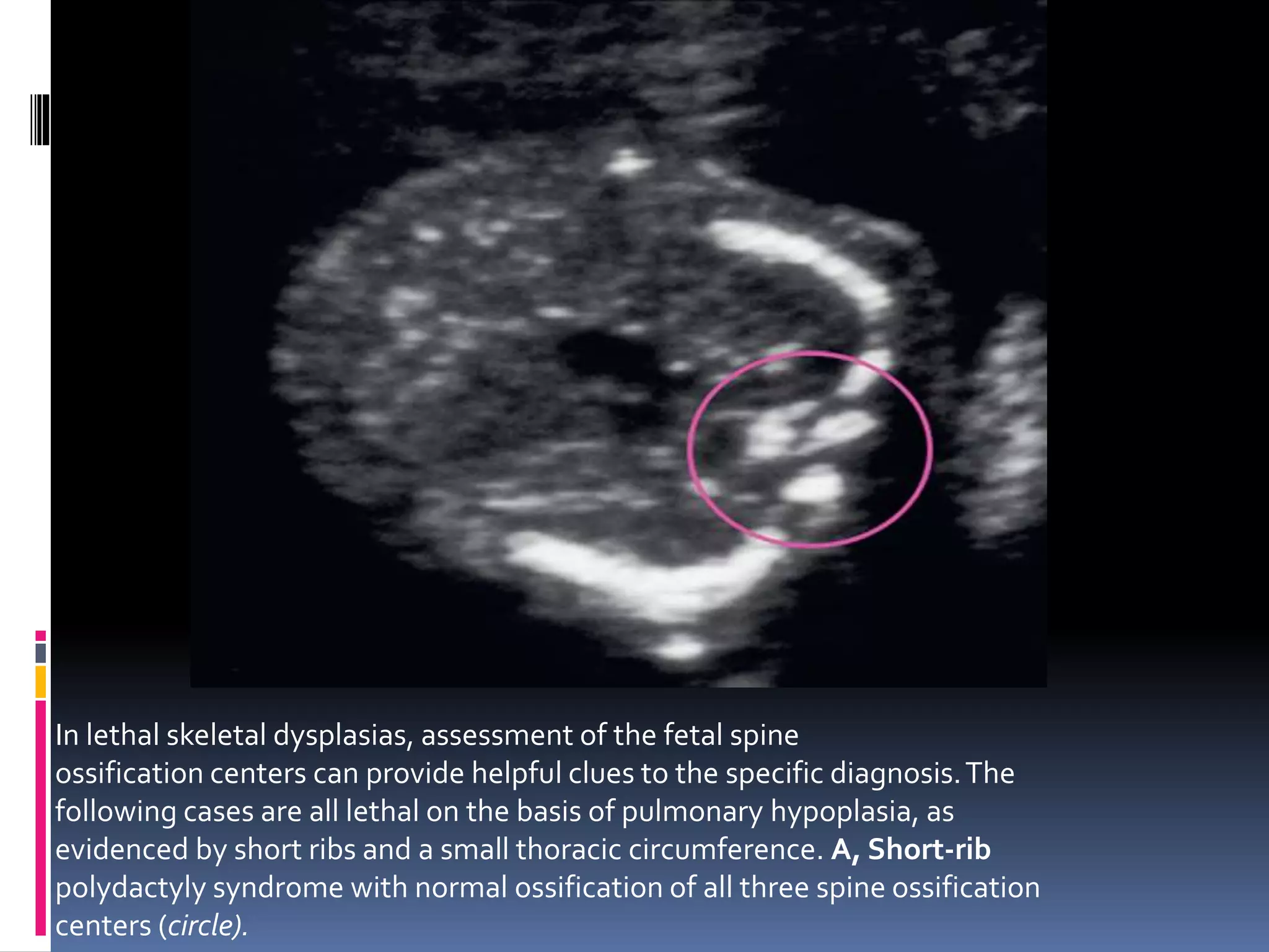
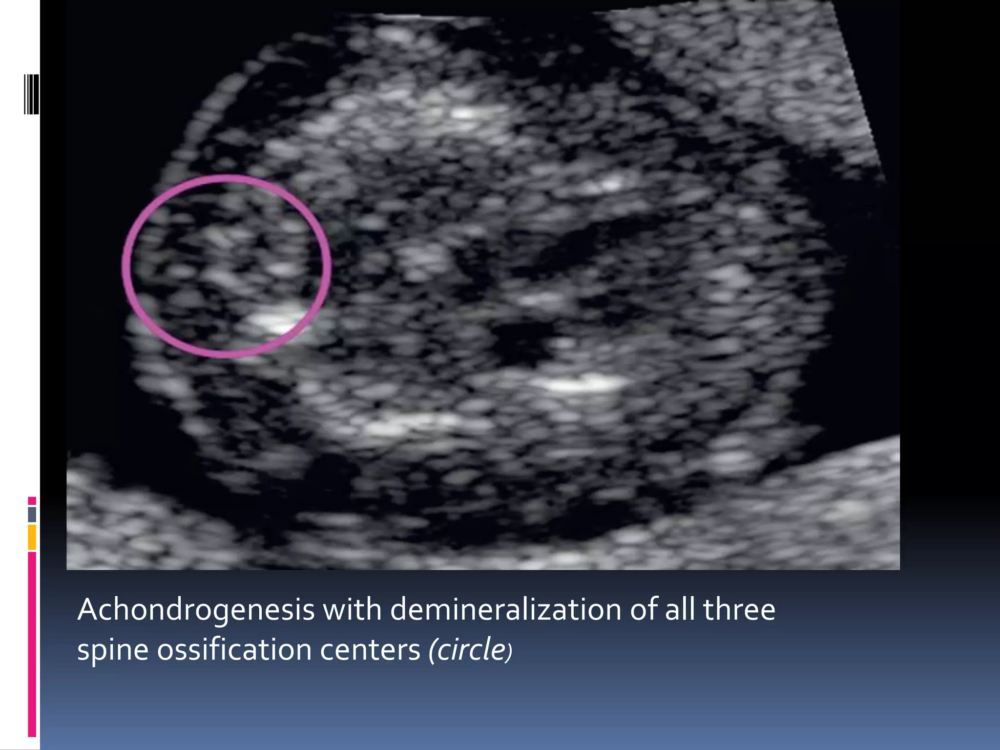
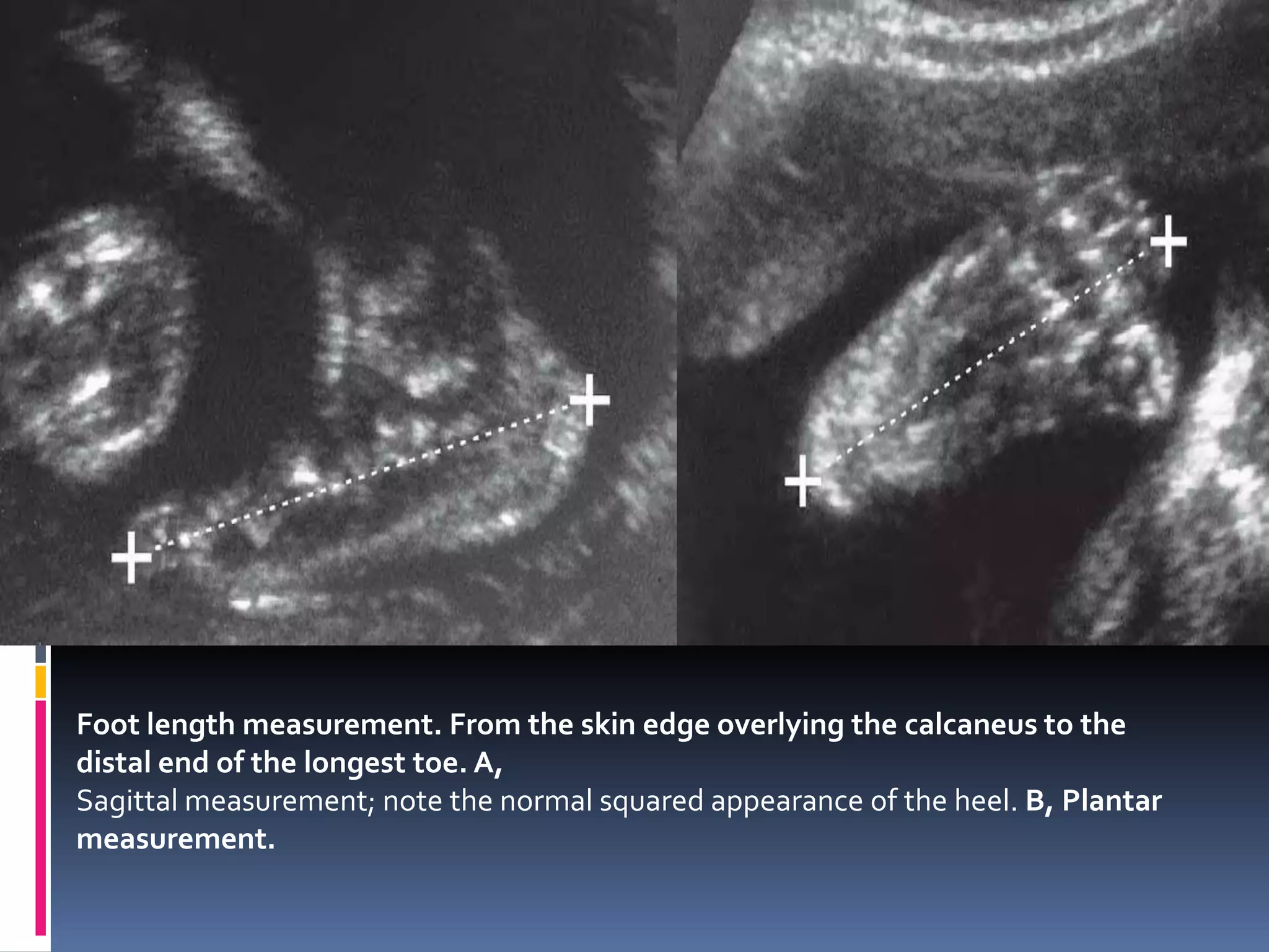
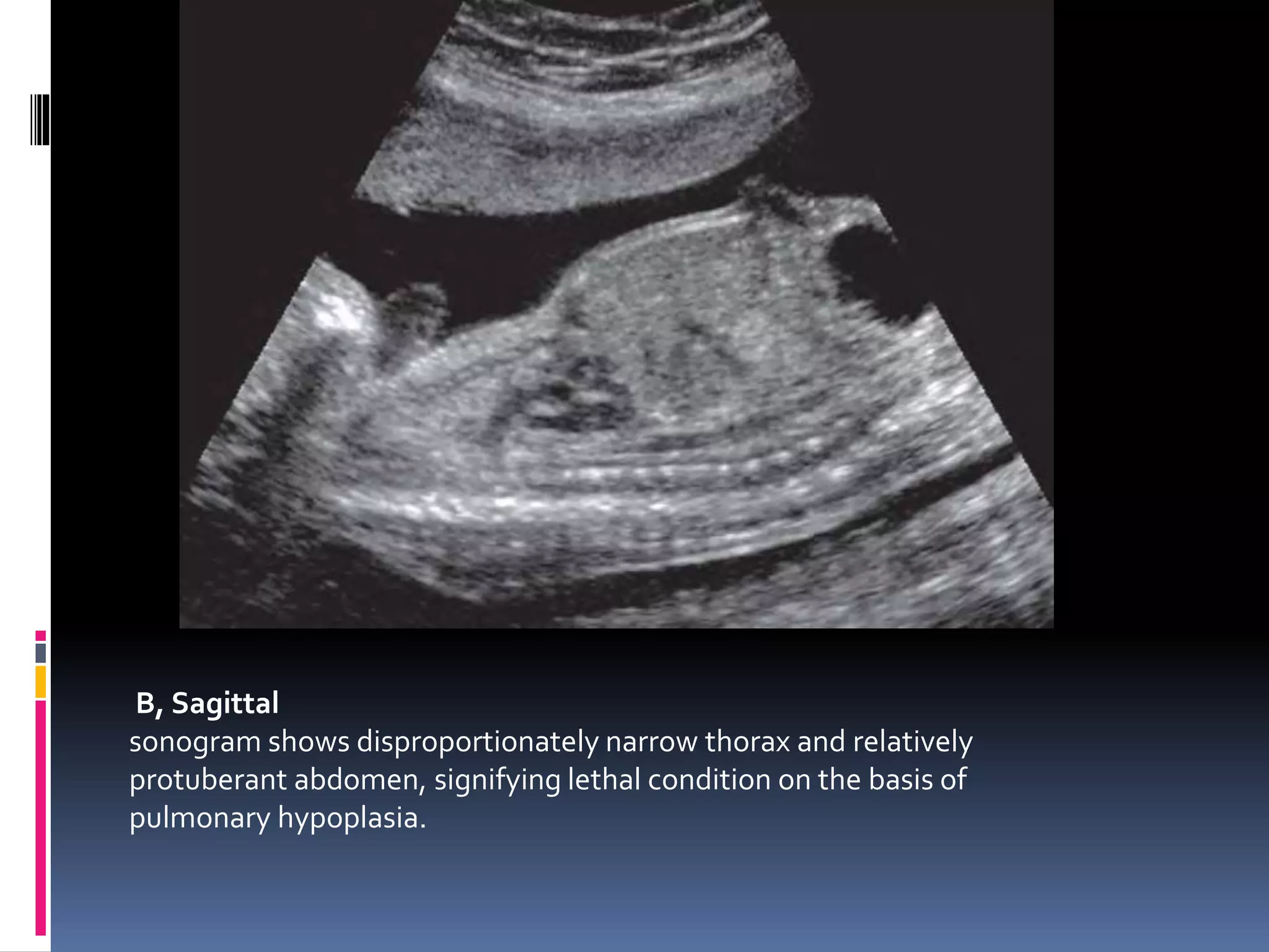
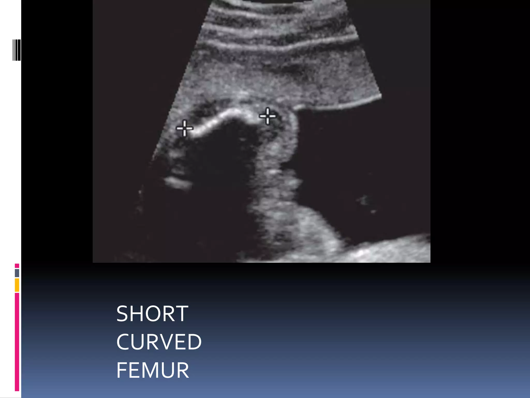
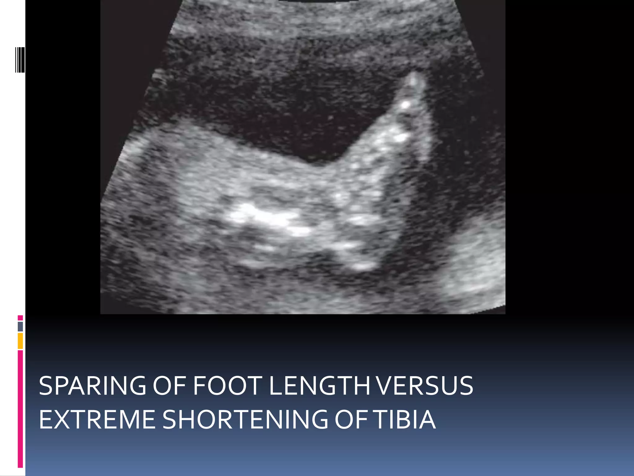
THE FETAL MUSCULOSKELETAL
SYSTEM
LETHAL SKELETAL DYSPLASIAS
 ISTHERE A LETHAL
SKELETAL DYSPLASIA?
CHARACTERISTIC FEATURES
 Severe micromelia
 Pulmonary hypoplasia-M/I DETERMINANT
DISTINGUISHING FEATURES
 Abnormal mineralization
 Fractures
 Presence or absence of macrocranium
 Thoracic length
 In many fetal skeletal dysplasias ,the skin and
s/c tissue continues to grow at a rate
proportionately greater than the long bones
resulting in relatively thickened skin folds (on
occasion mistaken for hydrops fetalis ) .
 Polyhydraminos –common .cause –variable
combination of the following –oesophageal
compression by the small chest ,GI
abnormalities ,micrognathia ,or hypotonia .
 3 M/C lethal skeletal dysplasias-
 THANATOPHORIC DYSPLASIA
 ACHONDROGENESIS
 OSTEOGENESIS IMPERFECTATYPE 2
SEVERE MICROMELIA WITH
DECREASED THORACIC
CIRCUMFERENCELSD MINERALIZAT
ION
FRACTUES MACROCRANI
A
SHORT
TRUNK
THANATOPHO
RIC
DYSPLASIAS
NORMAL NO YES NO
ACHONDROG
ENESIS
PATCHY
DEMINERALIS
ATION
OCCASIONAL YES YES
OITYPE2 GENERALISED
DE M
INNUMERABL
E
NO YES
HYPOPHOSPH
ATASIA
CONGENITA
PATCHY OR
GENERALISED
NO NO NO
SONOGRAPHIC ASSESSMENT OF BONES
Long Bones
 Degree of limb shortening
 Pattern of limb shortening
 Degree of mineralization
 Presence of fractures, bowing, or angulation
 Abnormal shape or contour
 Limb reduction anomalies
 Hypoplastic or aplastic bones
SPINE
 Degree and pattern of demineralization
 Platyspondyly
 Segmentation or curvature anomalies
 Caudal regression syndrome
 Myelodysplasia
THORAX
 Thoracic length and circumference
 Hypoplastic ribs
 Bell-shaped thorax of pulmonary hypoplasia
 Convex contour in cross section
HANDS AND FEET
 Postural deformities
 Abnormal number of digits
 Syndactyly
CALVARIUM
 Macrocranium
 Frontal bossing
 Craniosynostosis
 Compressibility/abnormal degree of
mineralization
FACIAL FEATURES
 Cleft lip and palate
 Hypertelorism and hypotelorism
 Midface hypoplasia/flat nasal bridge
DEGREE OF LIMB SHORTENING
 FL –AN abnormal FL –below -2SD for GA .
 Using this cutoff ,2.5% of all fetuses would be
classified as having short limbs .this exceeds the
expected frequency of skeletal dysplasias thus
additional investigations are required to confirm
the diagnosis .
 When 1 or all long bones measure < -2SD for
gestational age , a follow up USG should be done
in 3-4 weeks to evaluate the interval growth .if
interval growth is normal , high chances of being
normal .
 However, further deviation from the mean by
atleast 1 SD should suggest the diagnosis of
skeletal dysplasia or severe IUGR .
 If FL is 1 to 4mm below -2SD further serial
measurements are required to detemine if
skeletal dysplasia is present .
 If FL is > 5mm below the -2SD there is a high
likelihood of skeletal dysplasias .
 M/C etiology of short femur –either
inaccurate dating or constitutionally small
fetus or family history of less than average
stature.
 Isolated ,symmetrical short femurs identified
at mid 2nd trimester –group of fetuses at
increased risk of LBW, SMALL FOR GA ,OR
SEVERE IUGR .they will also have small
abdominal circumferences .
 Occasionally ,severe IUGR may present with
greatly shortened long bones ,associated
findings of normal or decreased skin fold
measurements ,oligohydraminos ,abnormal
placental morphology ,and abnormal doppler
waveforms .( whereas redundant thickened
skin folds and polyhydraminos typically
accompany short limb dysplasias )
 Nonlethal skeletal dysplasias like
heterozygous achondroplasia are generally
not evident before 20 weeks gestation .
 The findings of short long bones before 20
weeks indicate a more serious and usually
fatal skeletal dysplasia .
 The earlier the detection of shortening ,the
worse is the prognosis .
PATTERNS OF LIMB SHORTENING
 Rhizomelia: shortening of proximal segment
(femur, humerus).
 Mesomelia: shortening of middle segment
(radius,ulna/tibia, fibula).
 Acromelia: shortening of distal segment (hands,
feet).
 Micromelia: shortening of entire limb (mild, mild/
bowed, severe).
MESOMELIA
RHIZOMELIA
MILD
MICROMELIA
MILD AND
BOWED
MICROMELIA
SEVERE
MICROMELIA
 The shape ,contour ,and density should be
assessed for the presence of bowing ,angulations
,fractures and thickening .
 BOWING –nonspecific finding typically caused
by underlying osseous fragility .
 Diseases associated with bowed ,bent or
angulated femur -40 distinct types but majority
belong to 3 disorders –campomelic dysplasia
,thantophoric dysplasia ,and osteogenesis
imperfecta .
 Patients with OI types 1 and 4 can present with
apparent inutero bowing and shortening without
frank fractures of the long bones .
 Anterior bowing of tibia ,fibula and humerus may
–suggest the diagnosis of campomelic dysplasia
associated findings –hypoplastic scapula
,cervical kyphosis are typicallypresent.
 Bone fractures –may appear as angulations ,or
inteurrptions in the bone contour ,or as thick
,wrinkled contours corresponding tpo repititive
cycles of fracture and callous formation .
 ACOUSTIC SHADOWING –decreased or absent indicates
reduced mineralisation of the long bones but its presence
doesn’t confirm normal mineralisation .
 Spine – assess for segmentation anomalies ,kyphoscoliosis
,platyspondyly –M/C ( flattened vertebral bodies )
,demineralisation ,myelodysplasia , caudal regression
syndrome.
 Demineralisation of spine –appearance of ghost vertebrae
or nonvisualisation of one or all the 3 ossification centres .
 A progressively narrowed lumbar interpedicular distance is
associated with achondroplasia , a widened one is
associated with myelodysplasia .
 PULMONARY HYPOPLASIA – M/I determinant in prognosis of
lethality of a given skeletal dysplasia .USG specifity is 85-95%
accurate in the diagnosis in its presence for th4e diagnosis of a
lethal skeletal dysplasia .
 THORACIC CIRCUMFERENCE – measured at the level of the 4
chamberer heart and compared to normograms . A thoracic/
abdominal circumference ratio< 0.8 is considered abnormal .
 THORACIC LENGTH – from the neck to the diaphragm .
 Ribs are detemined if they are short.At the level of 4 chamber
cardiac view ,the ribs should normally encircle at least 70-80% of
the thoracic circumference .the ribs remain in a relatively
horizontal plane , as does the cardiac axis ,facilitating this
evaluation .
 In the sagital view ,a markedly narrow AP diameter of thorax ,in
coronal view , a concave or bell shaped contour associated with
pulmonary hypolplasia .
 Fetal cranium – assess for macrocranium ,frontal
bossing ,cloverleaf skull deformity underlying brain
abnormalities ,and facial abnormalities such as
saddle nose ,hypertelorism ,and cleftlip and palate.
 An abnormal cranial contour may be seen with
craniosynostosis or premature fusion of the sutures .
 The most reliable sonographic sign of reduced
mineralisation is increased compressibility of the
calvarium .typically seen in OI type 2,
achondrogenesis ,and hypophosphotasia .the falx
may appear abnormally bright or echogenic
compared to the demineralised calvarium .
View of the femur and
distal femoral ossification center
A, Normal femur: measure the longest length,
excluding the proximal and distal epiphysis and the specular reflection of the lateral aspect
of the distal femoral epiphysis (arrow).
B, Normal femur in the near field, with straight lateral border versus the curved medial
border in the far field of the transducer.
C, Isolated
hypoplastic left femur , with normal tibia and foot .
OI TYPE 1 ISOLATED FEMORAL FRACTURE
WITH ACUTE ANGULATION
CAMPOMELIC DYSPLASIA –MILD SHORTENINGAND A GENTLY CURVED
VENTRAL FEMORAL
BOWING
OITYPE 2A BOWED FEMUR WITH MULTIPLE DISCONTINUTIES
PRESENTING FREACTURES
HYPOPHOSPHATASIA CONGENITA – SEVERE
MICROMELIA –RELATIVELY BROAD METAPHYSIS
,SHORT DIAPHYSIS
THANATOPHORIC DYSPLASIA –CURVED
TELEPHONE RECEIVER FEMUR
CHONDRODYPLASIA PUNCTATA – 3RD TRIMESTER
APPEARNACE OF A STIPPLED
EPIPHYSIS
In lethal skeletal dysplasias, assessment of the fetal spine
ossification centers can provide helpful clues to the specific diagnosis.The
following cases are all lethal on the basis of pulmonary hypoplasia, as
evidenced by short ribs and a small thoracic circumference. A, Short-rib
polydactyly syndrome with normal ossification of all three spine ossification
centers (circle).
Achondrogenesis with demineralization of all three
spine ossification centers (circle)
C, Hypophosphatasia with demineralization
of the posterior ossification centers but mineralization of the vertebral
body (circle).
Triplet B affected with campomelic dysplasia and pulmonary hypoplasia. Coronal
ultrasound
images of the thorax in triplet pregnancy at 27 weeks’ gestation. A, Normal triplet A
shows normal convex contour of the thorax. Calipers
measure the scapula. B,Triplet B shows a bell-shaped thorax. C, Radiograph of
triplet B confirms a bell-shaped thorax consistent with
pulmonary hypoplasia.
Foot length measurement. From the skin edge overlying the calcaneus to the
distal end of the longest toe. A,
Sagittal measurement; note the normal squared appearance of the heel. B, Plantar
measurement.
 NORMAL FEMUR /FOOT LENGTH RATIO=1
 Ratio remains constant from 14 weeks of
gestation onwards .
 If fetus is constitutively small or has severe
IUGR this ratio remains 0.9 or greater
generally .
 In most skeletal dysplasias charaterised by
short limbs ,this ratio is generally <
0.9because of the relative sparing of the
hands and feet .
THANATOPHORIC DYSPLASIA
Thanatophoric dysplasia at 33 weeks. A, Anteroposterior (AP) radiograph
shows normal mineralization,
short curved extremity bones, severe platyspondyly with U-shaped vertebral
bodies, and narrow thorax with short ribs.
B, AP specimen
photograph shows severe micromelia with relative sparing of the feet,
telescoping of the redundant skin folds, and small, bell-shaped
thorax. C, Profile specimen photograph shows macrocranium, frontal
bossing, and flattened nasal bridge.
Thanatophoric dysplasia at 22 weeks.
A, Profile; midface hypoplasia with flat
nasal bridge.
B, Sagittal
sonogram shows disproportionately narrow thorax and relatively
protuberant abdomen, signifying lethal condition on the basis of
pulmonary hypoplasia.
SHORT
CURVED
FEMUR
SPARING OF FOOT LENGTHVERSUS
EXTREME SHORTENING OFTIBIA
Cloverleaf deformity of thanatophoric
dysplasia. A, Severe variant. B, Mild
variant
THANATOPHORIC DYSPLASIA
 M/C LETHAL SKELETAL DYSPLASIA
 Thanatos meaning death and foros meaning
bearing or carrying ,and dysplasia means
abnormal growth of a tissue or organ .
 Prevalence – 0.24 to 0.69 per 10,000 births .
 KEY FEATURES – SEVERE MICROMELIA
WITH RHIZOMELIC PREDOMINANCE,
MACROCRANIA ,DECREASEDTHORACIC
CIRCUMFERENCE BUT A NORMALTRUNK
LENGTH .
 MINERALISATION –NORMAL
 NO FRACTURES
 FORESHORTENEND EXTREMITIESTHATTHEY
PROTUDE AT RIGHT ANGLESTOTHE BODY .
 SKIN FOLDS –thickened and redundant
secondary to a relatively greater rate of growth
of the skin and s/c layers than the bones .
 Clinical presentation usually caused by large for
date measurements secondary to
polyhydraminos .
 2TYPES –
 1) type 1 –more common ,caused by the R248C and
Y373C mutations in the fibroblast growth factor
receptor 3 gene displays the typical telephone
receiver shape of the extremities .This bowed or
curved appearance is secondary to the broadened
metaphyses at the ends of the severely shortened
tubular bones .
 Associated with frontal bossing ,flattened nasal
bridge with midface hypoplasia .occasionally
,craniosynostosis results in a mild variant of
cloverleaf skull deformity .
 Platysondyly .
 TYPE 2- usually caused by the K650E mutation in
the FGFR3 gene ,the femurs are typically straight
with flared metaphses .
 Most specific feature –cloverleaf skull due to
premature craniosynostosis of the lambdoid and
coronal sutures resulting in a trilobed
appearance of skull in coronal plane .other
conditions with cloverleaf deformity –
homozygous achondroplasia ,campomelic
dysplasia and trisomy 13 .
 Both types are AD conditions .
 D/D -1) HOMOZYGOUSACONDROPLASIA –
positive family history
 2) CAMPOMELIC DYSPLASIA – moderate
and bowed form of micromelia typically
affecting the tibias with associated anomalies
.
 LIMBS – RHIZOMELIC MICROMELIA
-TELEPHONE RECEIVER CURVED FEMUR
AND HUMERUS
 CHEST – NARROW ,SHORT HORIZONTAL
RIBS,SMALL SCAPULAE
 SKULL AND FACE –RELATIVE MACROCEPHALY
,FRONTAL BOSSING ,NASAL BRIDGE
FLATTENING ,CLOVERLEAF SKULL
 SPINE –PLATYSPONDYLY ,NORMALTRUNK
LENGTH
 Associated CNS findings –holoprosencephaly
,agenesis of the corpus callosum ,
polymicrogyria ,heterotopia ,and
ventriculomegaly.
 Other anomalies- horseshoe kidneys
,hydronephrosis ,CHD ( ASD ,tricuspid
insufficiency ) ,radioulnar synostosis ,and
imperforate anus .
Homozygous achondroplasia at 34 weeks. A, Lateral profile is similar to
thanatophoric dysplasia with
macrocranium, frontal bossing, and flat nasal bridge. B, Axial image through the orbits
(calipers denote outer orbital diameter) and nasal
bones confirms a flat nasal bridge.
Thanatophoric dysplasia
at 33 weeks. A, Platyspondyly appears on ultrasound as a wafer-thin vertebral
body (arrows) with relatively larger hypoechoic intervertebral disc space on either side of
the vertebral body. B, Correlative lateral spine
radiograph. Note the short ribs with widecupped
metaphyseal ends.
CAMPOMELIC DYSPLASIA
 CAMPO means BENT and melic means limb .
 Bent limb dysplasia .
 Rare autosomal dominant condition that usually
results from a new dominant mutation in the
SOX9 gene ( sex- determining protein homeobox
9 )
 Most cases are lethal because of respiratory
insufficiency because of laryngotracheomalacia
in combination with a mildly narrowed thorax .
 Characteristic skeletal features –short and ventrally
bowed tibia and femur , a hypoplastic or absent
fibula , talipes equinovarus ,and hypoplastic scapulae
,bowing may also be seen in upper extremities.
 Additional skeletal features –scoliosis ,hypoplastic or
poorly ossified cervicothoracic vertebrae ,dislocated
hips ,11 rib pairs .
 Facial abnormalities – micrognathia ,cleft palate (
pierre robin sequence )
 Approx 33% fetuses have CHD and brain (
ventriculomegaly ) and renal ( pyelectasis )
abnormalities .
 Sex reversal found in about 75% of the
affected 46XY cases, with a gradation of the
defects ranging from ambiguous genitalia to
normal female genital phenotype .
 The gene responsible for campomelic
dysplasia is expressed in the fetal brain , the
testes , and the perichondrium and
chondrocytes of the long bones .
Campomelic dysplasia at 27 weeks.
A, Shortened femur and tibia with ventral bowing.
C, Short and curved dysplastic scapula.
ACHONDROGENESIS
 2nd M/C lethal skeletal dysplasia .
 PREVALENCE-0.09 to0.23 per 10,000 births
 Osteochondrodysplasia –deficiency of both bone and cartilage
development .
 2 types -1&2( .type 1 –a &b AUTOSOMAL RECESSSIVE ,20%)
 Genetics – type 1b- mutation diastrophic dysplasia sulfate
transporter gene
 Type 2 –LANGER SALDINO FORM new dominant mutation in
COL2A1 GENE .(80%) NORMAL CALVARIAL OSSIFICATION BUT
absent ossification in the vertebral column ,sacral and pubic
bones
 TYPE 1A – unknown pattern of inheritance , includes rib fractures
which is not seen in b type .
 Type 1 –partial or complete lack of ossification of the calvarium
,vertebral bodies ,sacral and pubic bones .
 MACROCRANIUM
 SEVERE MICROMELIA
 DECREASEDTHORACICCIRCUMFERENCEANDTRUNK
LENGTH
 DECREASED MINERALISATION – most marked in the
vertebral bodies ,ischium and pubic bones leading to a
greatly shortened trunk length , decresed thoracic
circumference and occasional fractures
 In hypophosphatasia congenita demineralisation
predominantly involves the post elements with only patchy
involvement of the vertebral bodies .
 POLYHYDRAMINOSANDTHICK,REDUNDANT SKIN
FOLDS .
Achondrogenesis at 18 weeks. A, Coronal sonogram shows small thorax, redundant
subcutaneous tissues,
absent spine ossification (arrows), and decreased calvarial ossification. B, Postmortem
radiograph demonstrates macrocranium, decreased
calvarial ossification, virtually absent spine ossification (only some posterior elements are
ossified in the cervical region).There is severe
micromelia with strikingly short wide bones with metaphyseal spurs.The ribs are short and
horizontal with splayed ends
OSTEOGENESIS IMPERFECTA
Osteogenesis imperfecta type IIA
at 32 weeks. Postmortem radiograph shows severe micromelia;
thickened bones with wavy contours caused by innumerable
fractures and exuberant callus formation; shortened ribs with multiple
fractures; and platyspondyly.

The fetal musculoskeletal system

  • 1.
  • 2.
    LETHAL SKELETAL DYSPLASIAS ISTHERE A LETHAL SKELETAL DYSPLASIA? CHARACTERISTIC FEATURES  Severe micromelia  Pulmonary hypoplasia-M/I DETERMINANT DISTINGUISHING FEATURES  Abnormal mineralization  Fractures  Presence or absence of macrocranium  Thoracic length
  • 3.
     In manyfetal skeletal dysplasias ,the skin and s/c tissue continues to grow at a rate proportionately greater than the long bones resulting in relatively thickened skin folds (on occasion mistaken for hydrops fetalis ) .  Polyhydraminos –common .cause –variable combination of the following –oesophageal compression by the small chest ,GI abnormalities ,micrognathia ,or hypotonia .
  • 4.
     3 M/Clethal skeletal dysplasias-  THANATOPHORIC DYSPLASIA  ACHONDROGENESIS  OSTEOGENESIS IMPERFECTATYPE 2
  • 5.
    SEVERE MICROMELIA WITH DECREASEDTHORACIC CIRCUMFERENCELSD MINERALIZAT ION FRACTUES MACROCRANI A SHORT TRUNK THANATOPHO RIC DYSPLASIAS NORMAL NO YES NO ACHONDROG ENESIS PATCHY DEMINERALIS ATION OCCASIONAL YES YES OITYPE2 GENERALISED DE M INNUMERABL E NO YES HYPOPHOSPH ATASIA CONGENITA PATCHY OR GENERALISED NO NO NO
  • 6.
    SONOGRAPHIC ASSESSMENT OFBONES Long Bones  Degree of limb shortening  Pattern of limb shortening  Degree of mineralization  Presence of fractures, bowing, or angulation  Abnormal shape or contour  Limb reduction anomalies  Hypoplastic or aplastic bones
  • 7.
    SPINE  Degree andpattern of demineralization  Platyspondyly  Segmentation or curvature anomalies  Caudal regression syndrome  Myelodysplasia
  • 8.
    THORAX  Thoracic lengthand circumference  Hypoplastic ribs  Bell-shaped thorax of pulmonary hypoplasia  Convex contour in cross section
  • 9.
    HANDS AND FEET Postural deformities  Abnormal number of digits  Syndactyly
  • 10.
    CALVARIUM  Macrocranium  Frontalbossing  Craniosynostosis  Compressibility/abnormal degree of mineralization
  • 11.
    FACIAL FEATURES  Cleftlip and palate  Hypertelorism and hypotelorism  Midface hypoplasia/flat nasal bridge
  • 12.
    DEGREE OF LIMBSHORTENING  FL –AN abnormal FL –below -2SD for GA .  Using this cutoff ,2.5% of all fetuses would be classified as having short limbs .this exceeds the expected frequency of skeletal dysplasias thus additional investigations are required to confirm the diagnosis .  When 1 or all long bones measure < -2SD for gestational age , a follow up USG should be done in 3-4 weeks to evaluate the interval growth .if interval growth is normal , high chances of being normal .
  • 13.
     However, furtherdeviation from the mean by atleast 1 SD should suggest the diagnosis of skeletal dysplasia or severe IUGR .  If FL is 1 to 4mm below -2SD further serial measurements are required to detemine if skeletal dysplasia is present .  If FL is > 5mm below the -2SD there is a high likelihood of skeletal dysplasias .
  • 14.
     M/C etiologyof short femur –either inaccurate dating or constitutionally small fetus or family history of less than average stature.  Isolated ,symmetrical short femurs identified at mid 2nd trimester –group of fetuses at increased risk of LBW, SMALL FOR GA ,OR SEVERE IUGR .they will also have small abdominal circumferences .
  • 15.
     Occasionally ,severeIUGR may present with greatly shortened long bones ,associated findings of normal or decreased skin fold measurements ,oligohydraminos ,abnormal placental morphology ,and abnormal doppler waveforms .( whereas redundant thickened skin folds and polyhydraminos typically accompany short limb dysplasias )
  • 16.
     Nonlethal skeletaldysplasias like heterozygous achondroplasia are generally not evident before 20 weeks gestation .  The findings of short long bones before 20 weeks indicate a more serious and usually fatal skeletal dysplasia .  The earlier the detection of shortening ,the worse is the prognosis .
  • 17.
    PATTERNS OF LIMBSHORTENING  Rhizomelia: shortening of proximal segment (femur, humerus).  Mesomelia: shortening of middle segment (radius,ulna/tibia, fibula).  Acromelia: shortening of distal segment (hands, feet).  Micromelia: shortening of entire limb (mild, mild/ bowed, severe).
  • 18.
  • 19.
     The shape,contour ,and density should be assessed for the presence of bowing ,angulations ,fractures and thickening .  BOWING –nonspecific finding typically caused by underlying osseous fragility .  Diseases associated with bowed ,bent or angulated femur -40 distinct types but majority belong to 3 disorders –campomelic dysplasia ,thantophoric dysplasia ,and osteogenesis imperfecta .
  • 20.
     Patients withOI types 1 and 4 can present with apparent inutero bowing and shortening without frank fractures of the long bones .  Anterior bowing of tibia ,fibula and humerus may –suggest the diagnosis of campomelic dysplasia associated findings –hypoplastic scapula ,cervical kyphosis are typicallypresent.  Bone fractures –may appear as angulations ,or inteurrptions in the bone contour ,or as thick ,wrinkled contours corresponding tpo repititive cycles of fracture and callous formation .
  • 21.
     ACOUSTIC SHADOWING–decreased or absent indicates reduced mineralisation of the long bones but its presence doesn’t confirm normal mineralisation .  Spine – assess for segmentation anomalies ,kyphoscoliosis ,platyspondyly –M/C ( flattened vertebral bodies ) ,demineralisation ,myelodysplasia , caudal regression syndrome.  Demineralisation of spine –appearance of ghost vertebrae or nonvisualisation of one or all the 3 ossification centres .  A progressively narrowed lumbar interpedicular distance is associated with achondroplasia , a widened one is associated with myelodysplasia .
  • 22.
     PULMONARY HYPOPLASIA– M/I determinant in prognosis of lethality of a given skeletal dysplasia .USG specifity is 85-95% accurate in the diagnosis in its presence for th4e diagnosis of a lethal skeletal dysplasia .  THORACIC CIRCUMFERENCE – measured at the level of the 4 chamberer heart and compared to normograms . A thoracic/ abdominal circumference ratio< 0.8 is considered abnormal .  THORACIC LENGTH – from the neck to the diaphragm .  Ribs are detemined if they are short.At the level of 4 chamber cardiac view ,the ribs should normally encircle at least 70-80% of the thoracic circumference .the ribs remain in a relatively horizontal plane , as does the cardiac axis ,facilitating this evaluation .  In the sagital view ,a markedly narrow AP diameter of thorax ,in coronal view , a concave or bell shaped contour associated with pulmonary hypolplasia .
  • 23.
     Fetal cranium– assess for macrocranium ,frontal bossing ,cloverleaf skull deformity underlying brain abnormalities ,and facial abnormalities such as saddle nose ,hypertelorism ,and cleftlip and palate.  An abnormal cranial contour may be seen with craniosynostosis or premature fusion of the sutures .  The most reliable sonographic sign of reduced mineralisation is increased compressibility of the calvarium .typically seen in OI type 2, achondrogenesis ,and hypophosphotasia .the falx may appear abnormally bright or echogenic compared to the demineralised calvarium .
  • 24.
    View of thefemur and distal femoral ossification center
  • 25.
    A, Normal femur:measure the longest length, excluding the proximal and distal epiphysis and the specular reflection of the lateral aspect of the distal femoral epiphysis (arrow). B, Normal femur in the near field, with straight lateral border versus the curved medial border in the far field of the transducer.
  • 26.
    C, Isolated hypoplastic leftfemur , with normal tibia and foot .
  • 27.
    OI TYPE 1ISOLATED FEMORAL FRACTURE WITH ACUTE ANGULATION
  • 28.
    CAMPOMELIC DYSPLASIA –MILDSHORTENINGAND A GENTLY CURVED VENTRAL FEMORAL BOWING
  • 29.
    OITYPE 2A BOWEDFEMUR WITH MULTIPLE DISCONTINUTIES PRESENTING FREACTURES
  • 30.
    HYPOPHOSPHATASIA CONGENITA –SEVERE MICROMELIA –RELATIVELY BROAD METAPHYSIS ,SHORT DIAPHYSIS
  • 31.
  • 32.
    CHONDRODYPLASIA PUNCTATA –3RD TRIMESTER APPEARNACE OF A STIPPLED EPIPHYSIS
  • 33.
    In lethal skeletaldysplasias, assessment of the fetal spine ossification centers can provide helpful clues to the specific diagnosis.The following cases are all lethal on the basis of pulmonary hypoplasia, as evidenced by short ribs and a small thoracic circumference. A, Short-rib polydactyly syndrome with normal ossification of all three spine ossification centers (circle).
  • 34.
    Achondrogenesis with demineralizationof all three spine ossification centers (circle)
  • 35.
    C, Hypophosphatasia withdemineralization of the posterior ossification centers but mineralization of the vertebral body (circle).
  • 36.
    Triplet B affectedwith campomelic dysplasia and pulmonary hypoplasia. Coronal ultrasound images of the thorax in triplet pregnancy at 27 weeks’ gestation. A, Normal triplet A shows normal convex contour of the thorax. Calipers measure the scapula. B,Triplet B shows a bell-shaped thorax. C, Radiograph of triplet B confirms a bell-shaped thorax consistent with pulmonary hypoplasia.
  • 37.
    Foot length measurement.From the skin edge overlying the calcaneus to the distal end of the longest toe. A, Sagittal measurement; note the normal squared appearance of the heel. B, Plantar measurement.
  • 38.
     NORMAL FEMUR/FOOT LENGTH RATIO=1  Ratio remains constant from 14 weeks of gestation onwards .  If fetus is constitutively small or has severe IUGR this ratio remains 0.9 or greater generally .  In most skeletal dysplasias charaterised by short limbs ,this ratio is generally < 0.9because of the relative sparing of the hands and feet .
  • 39.
    THANATOPHORIC DYSPLASIA Thanatophoric dysplasiaat 33 weeks. A, Anteroposterior (AP) radiograph shows normal mineralization, short curved extremity bones, severe platyspondyly with U-shaped vertebral bodies, and narrow thorax with short ribs.
  • 40.
    B, AP specimen photographshows severe micromelia with relative sparing of the feet, telescoping of the redundant skin folds, and small, bell-shaped thorax. C, Profile specimen photograph shows macrocranium, frontal bossing, and flattened nasal bridge.
  • 41.
    Thanatophoric dysplasia at22 weeks. A, Profile; midface hypoplasia with flat nasal bridge.
  • 42.
    B, Sagittal sonogram showsdisproportionately narrow thorax and relatively protuberant abdomen, signifying lethal condition on the basis of pulmonary hypoplasia.
  • 43.
  • 44.
    SPARING OF FOOTLENGTHVERSUS EXTREME SHORTENING OFTIBIA
  • 45.
    Cloverleaf deformity ofthanatophoric dysplasia. A, Severe variant. B, Mild variant
  • 46.
    THANATOPHORIC DYSPLASIA  M/CLETHAL SKELETAL DYSPLASIA  Thanatos meaning death and foros meaning bearing or carrying ,and dysplasia means abnormal growth of a tissue or organ .  Prevalence – 0.24 to 0.69 per 10,000 births .  KEY FEATURES – SEVERE MICROMELIA WITH RHIZOMELIC PREDOMINANCE, MACROCRANIA ,DECREASEDTHORACIC CIRCUMFERENCE BUT A NORMALTRUNK LENGTH .
  • 47.
     MINERALISATION –NORMAL NO FRACTURES  FORESHORTENEND EXTREMITIESTHATTHEY PROTUDE AT RIGHT ANGLESTOTHE BODY .  SKIN FOLDS –thickened and redundant secondary to a relatively greater rate of growth of the skin and s/c layers than the bones .  Clinical presentation usually caused by large for date measurements secondary to polyhydraminos .
  • 48.
     2TYPES – 1) type 1 –more common ,caused by the R248C and Y373C mutations in the fibroblast growth factor receptor 3 gene displays the typical telephone receiver shape of the extremities .This bowed or curved appearance is secondary to the broadened metaphyses at the ends of the severely shortened tubular bones .  Associated with frontal bossing ,flattened nasal bridge with midface hypoplasia .occasionally ,craniosynostosis results in a mild variant of cloverleaf skull deformity .  Platysondyly .
  • 49.
     TYPE 2-usually caused by the K650E mutation in the FGFR3 gene ,the femurs are typically straight with flared metaphses .  Most specific feature –cloverleaf skull due to premature craniosynostosis of the lambdoid and coronal sutures resulting in a trilobed appearance of skull in coronal plane .other conditions with cloverleaf deformity – homozygous achondroplasia ,campomelic dysplasia and trisomy 13 .  Both types are AD conditions .
  • 50.
     D/D -1)HOMOZYGOUSACONDROPLASIA – positive family history  2) CAMPOMELIC DYSPLASIA – moderate and bowed form of micromelia typically affecting the tibias with associated anomalies .
  • 51.
     LIMBS –RHIZOMELIC MICROMELIA -TELEPHONE RECEIVER CURVED FEMUR AND HUMERUS  CHEST – NARROW ,SHORT HORIZONTAL RIBS,SMALL SCAPULAE  SKULL AND FACE –RELATIVE MACROCEPHALY ,FRONTAL BOSSING ,NASAL BRIDGE FLATTENING ,CLOVERLEAF SKULL  SPINE –PLATYSPONDYLY ,NORMALTRUNK LENGTH
  • 52.
     Associated CNSfindings –holoprosencephaly ,agenesis of the corpus callosum , polymicrogyria ,heterotopia ,and ventriculomegaly.  Other anomalies- horseshoe kidneys ,hydronephrosis ,CHD ( ASD ,tricuspid insufficiency ) ,radioulnar synostosis ,and imperforate anus .
  • 53.
    Homozygous achondroplasia at34 weeks. A, Lateral profile is similar to thanatophoric dysplasia with macrocranium, frontal bossing, and flat nasal bridge. B, Axial image through the orbits (calipers denote outer orbital diameter) and nasal bones confirms a flat nasal bridge.
  • 54.
    Thanatophoric dysplasia at 33weeks. A, Platyspondyly appears on ultrasound as a wafer-thin vertebral body (arrows) with relatively larger hypoechoic intervertebral disc space on either side of the vertebral body. B, Correlative lateral spine radiograph. Note the short ribs with widecupped metaphyseal ends.
  • 55.
    CAMPOMELIC DYSPLASIA  CAMPOmeans BENT and melic means limb .  Bent limb dysplasia .  Rare autosomal dominant condition that usually results from a new dominant mutation in the SOX9 gene ( sex- determining protein homeobox 9 )  Most cases are lethal because of respiratory insufficiency because of laryngotracheomalacia in combination with a mildly narrowed thorax .
  • 56.
     Characteristic skeletalfeatures –short and ventrally bowed tibia and femur , a hypoplastic or absent fibula , talipes equinovarus ,and hypoplastic scapulae ,bowing may also be seen in upper extremities.  Additional skeletal features –scoliosis ,hypoplastic or poorly ossified cervicothoracic vertebrae ,dislocated hips ,11 rib pairs .  Facial abnormalities – micrognathia ,cleft palate ( pierre robin sequence )  Approx 33% fetuses have CHD and brain ( ventriculomegaly ) and renal ( pyelectasis ) abnormalities .
  • 57.
     Sex reversalfound in about 75% of the affected 46XY cases, with a gradation of the defects ranging from ambiguous genitalia to normal female genital phenotype .  The gene responsible for campomelic dysplasia is expressed in the fetal brain , the testes , and the perichondrium and chondrocytes of the long bones .
  • 58.
    Campomelic dysplasia at27 weeks. A, Shortened femur and tibia with ventral bowing.
  • 60.
    C, Short andcurved dysplastic scapula.
  • 61.
    ACHONDROGENESIS  2nd M/Clethal skeletal dysplasia .  PREVALENCE-0.09 to0.23 per 10,000 births  Osteochondrodysplasia –deficiency of both bone and cartilage development .  2 types -1&2( .type 1 –a &b AUTOSOMAL RECESSSIVE ,20%)  Genetics – type 1b- mutation diastrophic dysplasia sulfate transporter gene  Type 2 –LANGER SALDINO FORM new dominant mutation in COL2A1 GENE .(80%) NORMAL CALVARIAL OSSIFICATION BUT absent ossification in the vertebral column ,sacral and pubic bones  TYPE 1A – unknown pattern of inheritance , includes rib fractures which is not seen in b type .  Type 1 –partial or complete lack of ossification of the calvarium ,vertebral bodies ,sacral and pubic bones .
  • 62.
     MACROCRANIUM  SEVEREMICROMELIA  DECREASEDTHORACICCIRCUMFERENCEANDTRUNK LENGTH  DECREASED MINERALISATION – most marked in the vertebral bodies ,ischium and pubic bones leading to a greatly shortened trunk length , decresed thoracic circumference and occasional fractures  In hypophosphatasia congenita demineralisation predominantly involves the post elements with only patchy involvement of the vertebral bodies .  POLYHYDRAMINOSANDTHICK,REDUNDANT SKIN FOLDS .
  • 63.
    Achondrogenesis at 18weeks. A, Coronal sonogram shows small thorax, redundant subcutaneous tissues, absent spine ossification (arrows), and decreased calvarial ossification. B, Postmortem radiograph demonstrates macrocranium, decreased calvarial ossification, virtually absent spine ossification (only some posterior elements are ossified in the cervical region).There is severe micromelia with strikingly short wide bones with metaphyseal spurs.The ribs are short and horizontal with splayed ends
  • 64.
  • 65.
    Osteogenesis imperfecta typeIIA at 32 weeks. Postmortem radiograph shows severe micromelia; thickened bones with wavy contours caused by innumerable fractures and exuberant callus formation; shortened ribs with multiple fractures; and platyspondyly.