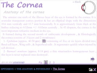
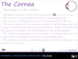
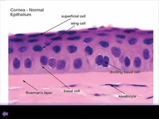
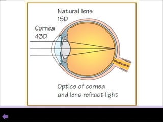
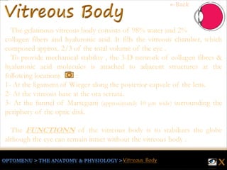
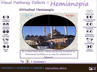
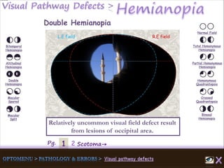
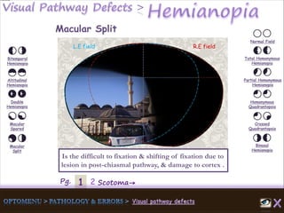
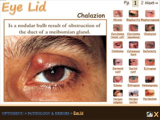
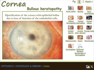
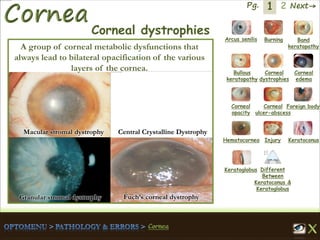
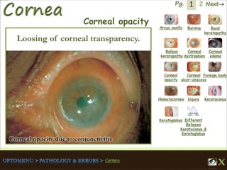
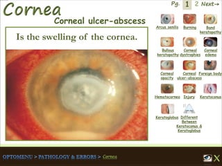
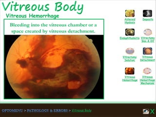
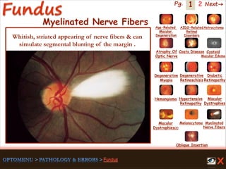
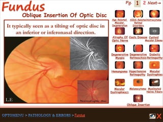
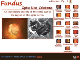
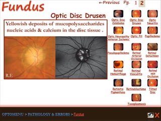
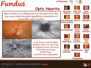
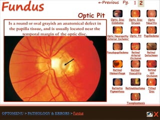
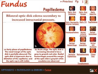
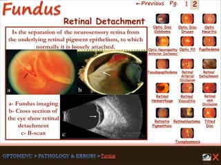
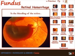
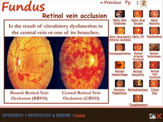
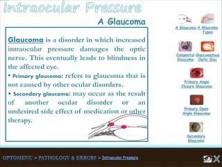
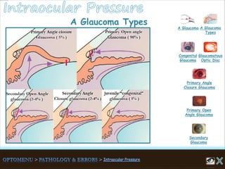
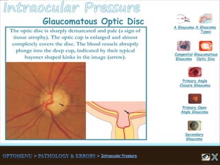
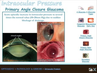
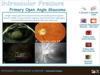
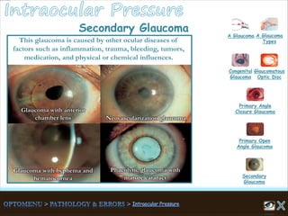
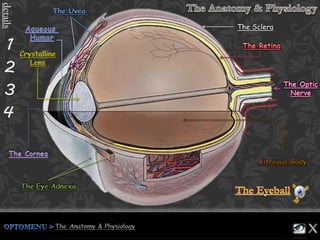
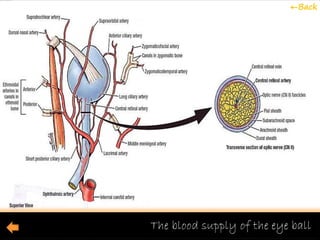
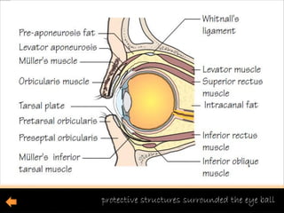
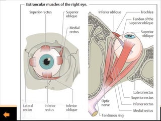
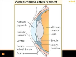
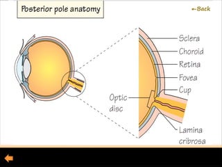
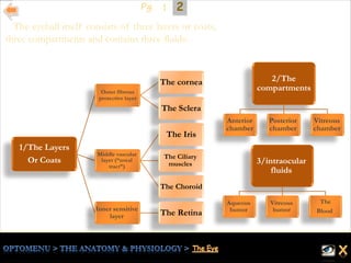
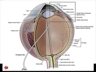
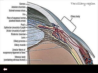
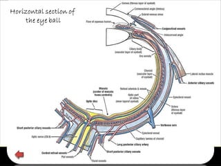
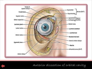
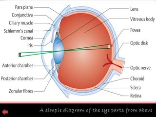
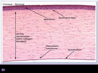
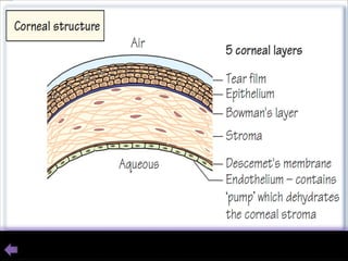
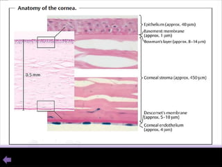
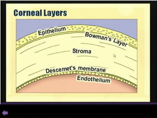
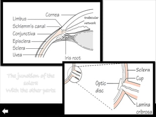
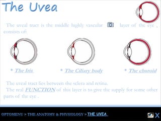
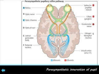
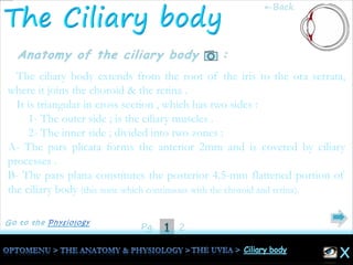
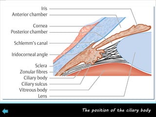
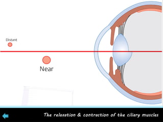
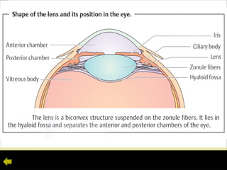
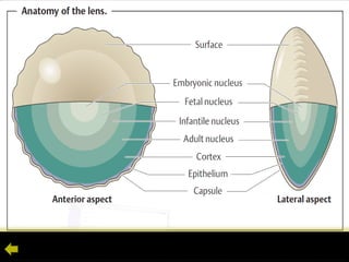
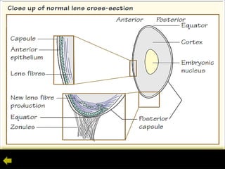
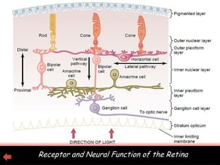
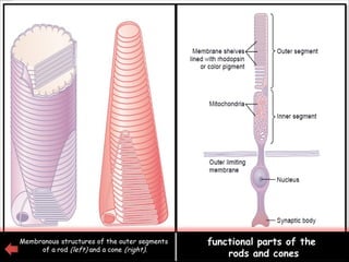
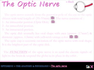
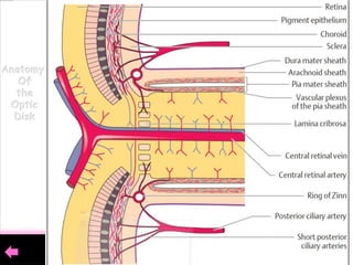
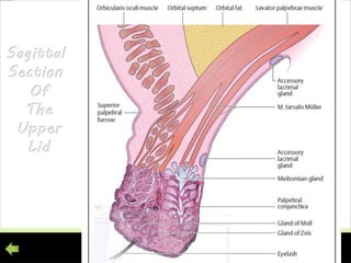
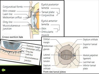
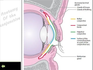
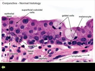
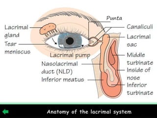
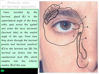
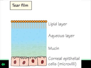
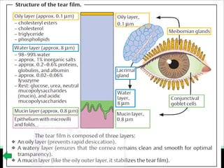
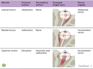
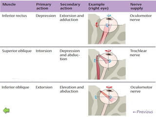
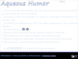
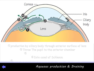
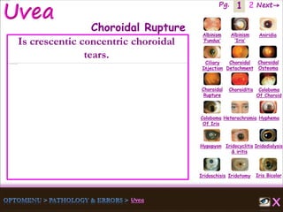
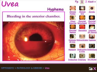
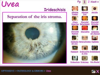
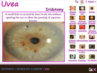
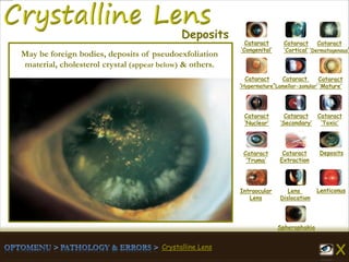
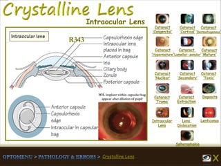
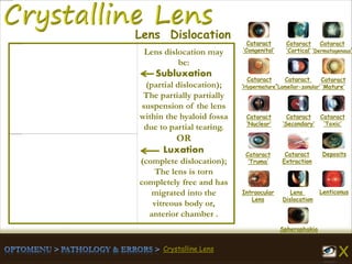
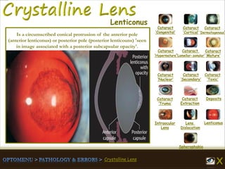
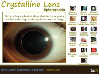
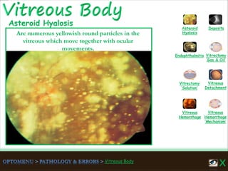
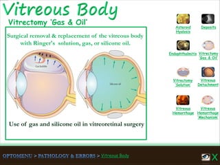
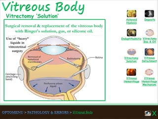
ALNEELAIN UNIVERSITY
THE FACULTY OF OPTICS &
VISUAL SCIENCES
Copyright: Asmaa Jamal, 2011
details
The Sclera
1Pg. 2
The Eye lies in the front half of the orbit surrounded by fat and connective tissue and is
supported by a facial hammock , It has an almost spherical shape with an average
diameter of 23 mm , The optic nerve, which connects the eye with the brain leaves the
orbit at its apex through the optic foramen in which it lies close to the ophthalmic artery.
The Eye ball surrounded by protective structures(orbit, lids, conjunctiva, and lacrimal
apparatus ) , the movement apparatus consisting of the extrinsic ocular muscles
and Tenon’s capsule , and supported by Ophthalmic artery .
Clinically, the eye can be considered to be composed of two segments:
1. Anterior segment – all structures from (and including) the lens forward .
2. Posterior segment – all structures behind the lens .
The FUNCTION of the eye is to form a clear image of objects in our environment.
These images are transmitted to the brain through the optic nerve and the posterior
visual pathways .
←Back
←Back
←Back
The visual
pathway
1/The Layers
Or Coats
Outer fibrous
protective layer
The cornea
The Sclera
Middle vascular
layer (“uveal
tract”)
The Iris
The Ciliary
muscles
The Choroid
Inner sensitive
layer The Retina
2/The
compartments
Anterior
chamber
Posterior
chamber
Vitreous
chamber
3/intraocular
fluids
Aqueous
humor
Vitreous
humor
The
Blood
The eyeball itself consists of three layers or coats,
three compartments and contains three fluids:
21Pg.
presspress
1Pg. 2 3
The anterior one-sixth of the fibrous layer of the eye is formed by the cornea. It is
avascular transparent convex portion & has an elliptical shape with the dimensions
10.6 mm vertically and 11.7 mm horizontally. It is approximately 1mm thick at the
limbus reducing to 0.52mm +/- 0.02mm centrally . At 43 diopters, the cornea is the
most important refractive medium in the eye.
It formed during the second month of embryonic development , & Histologically
the cornea consists of 3 layers and 2 membranes:
1- Epithelium (approx. 40-50 μm): consisting of five or six layers divided into;
Basal cell layer , Wing cells , & Superficial cells . It regenerates quickly when injured by
cell division .
2- Bowman’s membrane (approx. 8-14 μm): a thin structureless homogeneous layer ,
this layer is never replaced once destroyed .
←Back
21Pg. 3
3- Stroma ( approx. 450-500 μm) : consists of approximately 90% of
total corneal thickness. Consists of lamellae of collagen aligned at 90°
to each other and the spacing of the collagen fibrils is highly ordered.,
Between the lamellae are found cells (keratocytes) responsible for the
production of collagen and ground substance Corneal transparency is
due to the regular arrangement and relative dehydration of the collagen
fibres within the stroma..
4- Descemet’s membrane (approx. 5-10 μm) : a thin elastic membrane
resistant to both infection and trauma and relatively strong membrane
but lost tissue is not regenerated .
5- Endothelium (approx. 4 μm) : a single layer of hexagonal not
regenerated cells
31Pg. 2
The primary function of the cornea is refraction (approx. 43 diopters).
The cornea is the eye window that allows to form a clear image .
The cornea is a protective layer to the inner components of the eye .
The water content of the cornea controlled by epithelium(prevent the
passage of fluid into the stroma) and endothelium (pump the fluids into
the aqueous) to provide corneal dehydration to keep its transparency.
The uniform arrangement of the lamellae of collagen fibrils in the
corneal stroma also help to maintain the corneal transparency .
The epithelium and the endothelium attach with the tear that give the
nutrition to the cornea .
1Pg. 2
The Sclera
The whitish opaque outer coat of the eye which form 5/6 of the outer
layer of the eye
formed by the sclera .
All six ocular muscles insert into the sclera .
The sclera is thickest (1mm) anteriorly at the limbus & thinnest
(0.3mm) beneath the insertions of the rectus muscles .
It joins with the :
* Cornea at the limbus ,
* Optic nerve enter the sclera at lamina cribrosa ,
* The angle of the anterior chamber , to form the trabecular network
and the canal of Schlemm .
XThe Sclera
←Back
ScleraSclera
21Pg.
The Sclera
The sclera is fibrous , and consists of :
* layer of loose connective tissue deep to the conjunctiva, over laying
the sclera, called the episclera.
* A cellular connective tissue with a higher water content than the
cornea .
* Stroma consists of irregular lamellae of collagen fibers (this is the
cause of its whitish opaque appearance) .
The most important FUNCTION of the sclera is to protect the eye by
its rigid formation , & cut the light entering the eye off except from the
cornea .
XThe Sclera
The uveal tract is the middle highly vascular layer of the eye ,
consists of:
The uveal tract lies between the sclera and retina.
The real FUNCTION of this layer is to give the supply for some other
parts of the eye .
* The choroid* The Ciliary body* The Iris
1Pg. 2 3
It is a thin circular disc perforated centrally by the pupil.
The iris consists of two layers:
1- The anterior mesodermal stromal layer.
2- The posterior ectodermal pigmented epithelial layer ( an opaque and
protects the eye against excessive incident light).
Its variable pigmentation determines the 'colour' of eyes.
The color of the iris varies in the individual according to the melanin
content of the melanocytes (pigment cells) in the stroma and epithelial
layer.
Eyes with a high melanin content are dark brown, whereas eyes with less
melanin are grayish-blue .
←Back
21Pg. 3
The collarette marks the division of the stroma into pupillary and ciliary
zones.
The ciliary zone in the iris attached peripherally to the anterior surface of
the ciliary body .
The pupil is surrounded by :
1-The circular sphincter muscle, which is supplied by parasympathetic
nerve fibers ( responsible for miosis & contraction ) .
2- The radial dilator pupillae muscle, supplied by sympathetic nerve
fibers ( responsible for mydriasis & dilatation ) .
As similar as fingermarks , the iris has its unique map that never reoccur
with another one .
31Pg. 2
The iris regulate the contraction and dilation of the pupil by its muscles,
so that, it may be regarded as the light regulator of the optical system of
the eye.
It acts as an aperture to improve the quality of the resulting image by
controlling the amount of light that enters the eye.
Sympathetic innervation of pupil
Parasympathetic innervation of pupil
1Pg. 2
The ciliary body extends from the root of the iris to the ora serrata,
where it joins the choroid & the retina .
It is triangular in cross section , which has two sides :
1- The outer side ; is the ciliary muscles .
2- The inner side ; divided into two zones :
A- The pars plicata forms the anterior 2mm and is covered by ciliary
processes .
B- The pars plana constitutes the posterior 4.5-mm flattened portion of
the ciliary body (this zone which continuous with the choroid and retina).
←Back
21Pg.
The suspensory ligament, the zonule, extends from the pars plana and
the intervals between the ciliary processes to the lens capsule.
The ciliary body is covered by a bilaminar epithelium , which is
responsible for the production of aqueous humor.
The contraction of the ciliary muscle is responsible for
accommodation.
The epithelium layer covering the ciliary body produces the aqueous
humor.
The choroid is the middle tunic of the eyeball between the
sclera & the retina . It is highly vascularized, that, the blood flow
through the choroid is the highest in the entire body . In addition to
vessels, it also carries approximately 15–20 ciliary nerves.
The choroid consists of the following :
1- Bruch’s membrane 2 μm ; It consists of basement membrane of
retinal pigmented epithelium cells & elastic and collagenous layers .
2-The choriocapillaris whish supplying the RPE & outer retina .
3- Layer of larger choroidal blood vessels .
4- Pigmented cells .
The FUNCTION of choroid is to regulate temperature and supply
nourishment to the outer layers of the retina, it is the vein of life .
←Back
Sclera
1Pg. 2
Is a transparent, bi-convex structure suspended from the ciliary body by the zonular fibres and
situated between the iris and the vitreous The lens is about 9mm in diameter and about 4mm thick
at the centre. The lens is comprised of 65% water and 35% protein .
Anatomically , the lens consists of :
1- A thick elastic capsule for protection (its elasticity decreases with aged) . The zonule inserts into it
near the equator to suspend the lens by ciliary body.
2- Layer of epithelial cells (just under the anterior capsule). It active metabolically to keep the lens
clean .
3- Lens fibres ; The central lens fibres are called the nucleus and the more peripheral fibres the
cortex .
4- Embryonic nucleus , forms by the growth of primary lens fibers then displaced toward the
center of the lens by the growth secondary one .
5- Fetal nucleus (complete at birth) , & Infantile nucleus ( complete during the 1st and 2nd decades of life)
←Back
21Pg.
The lens is one of the essential refractive media (10–20 diopters, depending
on individual accommodation) of the eye and focuses incident rays of light on
the retina .
Zonule filaments suspend the lens ciliary processes to the ciliary muscle .
When the muscle contracts the filaments relax allowing the lens to
become more convex with a shorter focal length for reading .
The gelatinous vitreous body consists of 98% water and 2%
collagen fibers and hyaluronic acid. It fills the vitreous chamber, which
composed approx. 2/3 of the total volume of the eye .
To provide mechanical stability , the 3-D network of collagen fibres &
hyaluronic acid molecules is attached to adjacent structures at the
following locations :
1- At the ligament of Wieger along the posterior capsule of the lens.
2- At the vitreous base at the ora serrata.
3- At the funnel of Martegiani (approximately 10 μm wide) surrounding the
periphery of the optic disk.
The FUNCTIONN of the vitreous body is to stabilizes the globe
although the eye can remain intact without the vitreous body .
←Back
41Pg. 2 3
The retina is the innermost of three successive layers of the globe. It
comprises two parts:
1- A photoreceptive part (pars optical retinae), comprising the first nine of
the 10 layers of retina .
2- A nonreceptive part (pars caeca retinae), forming the epithelium of the
ciliary body and iris .
Thickness of the retina different from part to another , thinnest at ora
serrata & thickest at optic nerve .
Moving inward along the path of incident light, the individual layers of
the retina are :
←Back
Pg. 421 3
1. Inner limiting membrane (glial cell fibers separating the retina from the vitreous body).
2. Layer of optic nerve fibers (axons of the third neuron).
3. Layer of ganglion cells (cell nuclei of the multipolar ganglion cells of the third neuron).
4. Inner plexiform layer (synapses between the axons of the second neuron and dendrites of
the third neuron).
5. Inner nuclear layer (cell nuclei of the bipolar nerve cells of the second neuron, horizontal
cells, and amacrine cells).
6. Outer plexiform layer (synapses between the axons of the first neuron and dendrites of the
second neuron).
7. Outer nuclear layer (cell nuclei of the rods and cones = first neuron).
8. Outer limiting membrane .
9. Layer of rods and cones (the actual photoreceptors).
10. Retinal pigment epithelium (a single cubic layer of heavily pigmented epithelial cells).
11. Bruch’s membrane (basal membrane of the choroid separating the retina from the choroid).
431Pg. 2
The most sensitive section in the retina is THE
MACULAR :
The macula lutea (yellow spot); is a flattened oval area in the
center of the retina approximately 3–4mm(15 degrees)
temporal to and slightly below the optic disk with (1.7–2 mm)
diameter.
In its center; (the avascular fovea) , the sharpest visual
perception , which contains only cones cell .
41Pg. 2
The retina is the sensitive film of the eye , which receive the visible light
spectrum (wavelengths of 380–760 nm) to send it to the brain as Electric
signals .It has 4 types of Perceptions : Contrast, color, light and shape.
The retina has two types of photoreceptors :
1- The Rods (110–125 million rods) responsible for twilight and night vision,
they are about 500 times more photosensitive than the cones .
2- The Cones (six to seven million cones) responsible for say light vision ,
resolution, and color perception & there are three types of cones:
1- Blue cones, 2-Green cones, 3- Red cones.
* There are nearly 20 rods to every cone.
3
The optic nerve extends from the posterior pole of the eye to the optic
chiasm with total length of (35–55mm) . The nerve consists of :
1- An intraocular portion (Optic Disk) .
2- An intraorbital portion.
3- An intracranial portion.
The optic disk normally has oval shape with area (approx. 2.7mm²) &
diameter (approx. 1.8mm) with yellowish orange color .
The optic cup is eccentric cavitation of the optic nerve with oval shape.
It is the brightest part of the optic disk.
The FUNCTION of the optic nerve is to send the electric signals of
light to the brain & suspend the globe in its place in the orbit .
←Back
Next →
←Previous
The eye adnexa is that parts whish surrounded the eye globe .
It consists of :
The adjacent eye adnexa very important for the safety of the
eye & its movement .
* The Eye Lids * The Lacrimal
System
* The Conjunctiva * Extra-Ocular
Muscles
1Pg. 2
The anatomy of an eyelid can be simplified into four layers :
1- Skin, which is formed by the epidermis and dermis .
2- Striated muscle, formed by the orbicularis oculi to close the eyelids.
3- Tarsal plate which gives the eyelid firmness and shape and
containing the Meibomian glands .
4- Conjunctival mucosa .
On the upper eyelid, approximately 150 eyelashes are arranged in three
or four rows; on the lower eyelid there are about 75 in two rows. As
eyebrows,the eyelashes help prevent dust and sweat from entering the eye.
Different types of muscle groups specialize in eyelid movements, whose
closing takes three forms:
- By blinking, - By reflex, - And consciously.
The superficial
layer
The deep
layer
←Back
21Pg.
Normal width of the palpebral fissure ( the eye opening ) :
- The upper lid covers the superior margin of the cornea by about
(2mm).
- The lower lid close to the inferior margin of the cornea .
- The distance between upper & lower lids is normally (6–10mm) .
- The distance between the lateral and medial angles is (28–30mm) .
The FUNCTION of the eye lids is :
1- protect the eye globe .
2- It contains tear film, which is significant for corneal moisture & healthy
that distributed over the anterior surface by blinking .
1Pg. 2
The conjunctiva is a vascular mucous membrane that covers
the surface of the globe and the lids.
Anatomically, the conjunctiva has continuous regions consist
of :
1-The palpebral conjunctiva : lines the posterior surface of the eyelids.
2-The forniceal conjunctiva : the link between palpebral & bulbar conj.
3-The bulbar conjunctiva : is loosely attached to the anterior of sclera .
4-The limbal conjunctiva : attached strongly to about 3mm from the
cornea .
←Back
21Pg.
Histologically, the conjunctiva consist of :
1- The epithelium layer : contains basal, wing, and superficial cuboidal
layers . & contains goblet cells (mucous glands), langerhans cells, and
melanocytes cells .
2- The stroma : contains nerves, blood vessels, and lymphatic .
The FUNCTION of the conjunctiva is to facilitate the movements of
the globe and lids while protecting the orbital contents from the external
environment.
1Pg. 2
The lacrimal system consists of two sections :
* Structures that secrete tear fluid ( lacrimal gland, Krause and Wolfring glands ).
* Structures that facilitate tear drainage (drainage system ).
The lacrimal gland lies above the outer corner of the eye beneath the
superior temporal margin of the orbital bone in the lacrimal fossa. The
accessory Krause and Wolfring glands located in the conjunctival fornices.
Anatomically, the lacrimal drainage system consists of :
1- The punta (2mm) : lie in papillae at the medial ends of the eyelids.
2- Canaliculi (8mm) : 70% of tears drain through the inferior and 30% through the
superior canaliculus .
3- The lacrimal sac : lies in the lacrimal groove formed by the lacrimal bone and the
frontal process of the maxilla.
4- Nasolacrimal duct (NLD) : is directed downwards, laterally and
backwards to empty into the inferior meatus of the nasal cavity.
21Pg.
The Tear Film :
Is a protein layer consists of three layers :
1- The outer oily layer (approx. 0.1 μm) : produced meibomian glands
and the sebaceous glands and sweat glands of the margin of eye lids . This
layer very important to stabilize the tear film and prevent evapration.
2- The middle watery layer (approx. 8 μm ) : is produced by the lacrimal
gland and the accessory lacrimal glands . It is important for sterilization,
cleaning the surface of the cornea & facilitation of blinking .
3- The inner mucin layer (approx. 0.8 μm) : is secreted by the goblet cells
of the conjunctiva and the lacrimal gland. It helps to stabilize the tear film
and protect the corneal epithelium .
Punta
The external ocular muscles (4 rectus, and 2 oblique) lie in the
fat of the orbit and help to move the eyeball in different
directions.
All the recti muscles attach to the eyeball anterior to the
equator (inserted into the sclera 5-8mm from the limbus) while
the oblique muscles attach behind the equator.
* Table Of
Function
* Anterior View
Of Actions
* Location &
Different view
* Nerve Supply
←Back
Continue →
←Previous
It is an optically clear solution fills the space between the cornea and the
lens with about (0.3 ml.) in volume .
It contains a low concentration of proteins, and high concentration of
ascorbic acid .
It formed by ultrafiltration from the ciliary processes in the posterior
chamber.
Its flow system is :
1-Aqueous flows forward over the anterior surface of the lens .
2-The fluid enters the anterior chamber through the pupil .
3-Drains through the trabecular meshwork into the canal of Schlemm .
4-Then aqueous veins and the conjunctival episceral veins.
Its FUNCTION is to nourish the lens and cornea.
←Back
Ametropia ●
Strabismus ●
● Eye Lids
● Lacrimal System
● Conjunctiva
● Cornea
● Sclera
● Uvea
● Lens
● Intra Ocular Pressure
● Fundus Visual Pathway Defects●
● Vitreous Body
● Red Eye
Emmetropia
Ametropia
Hypermetropia
Myopia
Anisometropia
Astigmatism
Presbyopia
Is an optical condition where light from distant beyond 6 m
focused in front of the retina when accommodation = 0
Is an optical condition where light from distant beyond 6 m
out of focus when accommodation = 0
Is an optical condition where light from distant beyond
6 m focused behind the retina when accommodation = 0
Is an optical condition where light from distant beyond 6 m
not focused the retina when accommodation = 0
Is an optical condition where light from distant beyond 6 m
focused on the retina when accommodation = 0
Is a difference in refractive power between the two eyes
usually > 3 diopters
Is a gradual loss of focusing power due to decreasing of the
elasticity of the lens with increasing age
1 Next→Pg. 2
2←Previous Pg. 1
Emmetropia & Ametropia●● Myopia
Hypermetropia●● Correction Of Myopia
Correction Of Hypermetropia●● Astigmatism
Presbyopia●● Types Of Astigmatism
Emmetropia & Ametropia
2←Previous Pg. 1
Emmetropia & Ametropia●● Myopia
Hypermetropia●● Correction Of Myopia
Correction Of Hypermetropia●● Astigmatism
Presbyopia●● Types Of Astigmatism
Hypermetropia
2←Previous Pg. 1
Correction of hyperopia
(a) Correction with converging
lenses (plus lenses).
(b) Correction of aphakia with
cataract lens, contact lens (c),
anterior chamber intraocular
lens (d, blue) or posterior
chamber intraocular lens(d, red).
Emmetropia & Ametropia●● Myopia
Hypermetropia●● Correction Of Myopia
Correction Of Hypermetropia●● Astigmatism
Presbyopia●● Types Of Astigmatism
Correction Of Hypermetropia
2←Previous Pg. 1
The vision in presbyopic patient
corrected by convex lens (+) for
near vision
Emmetropia & Ametropia●● Myopia
Hypermetropia●● Correction Of Myopia
Correction Of Hypermetropia●● Astigmatism
Presbyopia●● Types Of Astigmatism
Presbyopia
2←Previous Pg. 1
Emmetropia & Ametropia●● Myopia
Hypermetropia●● Correction Of Myopia
Correction Of Hypermetropia●● Astigmatism
Presbyopia●● Types Of Astigmatism
Myopia
2←Previous Pg. 1
Correction of myopia
(a) Correction with
diverging lenses (minus
lenses).
(b) Correction with contact
lens.
(c) Correction by removing
the lens to reduce refractive
power of the eye.
Emmetropia & Ametropia●● Myopia
Hypermetropia●● Correction Of Myopia
Correction Of Hypermetropia●● Astigmatism
Presbyopia●● Types Of Astigmatism
Correction Of Myopia
2←Previous Pg. 1
Emmetropia & Ametropia●● Myopia
Hypermetropia●● Correction Of Myopia
Correction Of Hypermetropia●● Astigmatism
Presbyopia●● Types Of Astigmatism
Astigmatism
2←Previous Pg. 1
Emmetropia & Ametropia●● Myopia
Hypermetropia●● Correction Of Myopia
Correction Of Hypermetropia●● Astigmatism
Presbyopia●● Types Of Astigmatism
Types Of Astigmatism
Esophoria●
Esotropia●
Hypertropia●
Exotropia●
Hypotropia●
Exophoria●
Hyperphoria & Hypophoria●
Cyclophoria●
Nystagmus●
►play
Nystagmus: Bilateral involuntary
rhythmic oscillation of the eyes, may be
latent or manifest .
Nystagmus
►play
Esophoria●
Esotropia●
Hypertropia●
Exotropia●
Hypotropia●
Exophoria●
Hyperphoria & Hypophoria●
Cyclophoria●
Nystagmus●
Esophoria: Latent inward deviation of
the visual axis .
Esophoria
►play
Esophoria●
Esotropia●
Hypertropia●
Exotropia●
Hypotropia●
Exophoria●
Hyperphoria & Hypophoria●
Cyclophoria●
Nystagmus●
Exophoria: Latent outward deviation of
the visual axis .
Exophoria
►play
Esophoria●
Esotropia●
Hypertropia●
Exotropia●
Hypotropia●
Exophoria●
Hyperphoria & Hypophoria●
Cyclophoria●
Nystagmus●
Hyperphoria & Hypophoria
Hyperphoria: latent upward deviation
of the visual axis .
Hypophoria: latent downward deviation
of the visual axis .
►play
Esophoria●
Esotropia●
Hypertropia●
Exotropia●
Hypotropia●
Exophoria●
Hyperphoria & Hypophoria●
Cyclophoria●
Nystagmus●
Cyclophoria: latent rotation of one eye
around its visual axis .
Cyclophoria
Esophoria●
Esotropia●
Hypertropia●
Exotropia●
Hypotropia●
Exophoria●
Hyperphoria & Hypophoria●
Cyclophoria●
Nystagmus●
Esotropia: Manifest convergent
deviation .
Esotropia
Esophoria●
Esotropia●
Hypertropia●
Exotropia●
Hypotropia●
Exophoria●
Hyperphoria & Hypophoria●
Cyclophoria●
Nystagmus●
Exotropia: Manifest convergent
deviation .
Exotropia
Esophoria●
Esotropia●
Hypertropia●
Exotropia●
Hypotropia●
Exophoria●
Hyperphoria & Hypophoria●
Cyclophoria●
Nystagmus●
Hypertropia: Manifest elevated
deviation .
Hypertropia
Esophoria●
Esotropia●
Hypertropia●
Exotropia●
Hypotropia●
Exophoria●
Hyperphoria & Hypophoria●
Cyclophoria●
Nystagmus●
Hypotropia: Manifest depressed
deviation .
Hypotropia
1Pg. 2 Scotoma→
Normal Field
Total Homonymous
Hemianopia
Partial Homonymous
Hemianopia
Homonymous
Quadrantopsia
Crossed
Quadrantopsia
Binasal
Hemianopia
Bitemporal
Hemianopia
Altitudinal
Hemianopia
Double
Hemianopia
Macular
Spared
Macular
Split
Normal Field
Hemianopia is typical binocular sector
defect .
L.E field R.E field
1Pg. 2 Scotoma→
Normal Field
Total Homonymous
Hemianopia
Partial Homonymous
Hemianopia
Homonymous
Quadrantopsia
Crossed
Quadrantopsia
Binasal
Hemianopia
Bitemporal
Hemianopia
Altitudinal
Hemianopia
Double
Hemianopia
Macular
Spared
Macular
Split
Total Homonymous Hemianopia
L.E field R.E field
Is bilateral left or right sided contraction of the
visual field rather than depression.
1Pg. 2 Scotoma→
Normal Field
Total Homonymous
Hemianopia
Partial Homonymous
Hemianopia
Homonymous
Quadrantopsia
Crossed
Quadrantopsia
Binasal
Hemianopia
Bitemporal
Hemianopia
Altitudinal
Hemianopia
Double
Hemianopia
Macular
Spared
Macular
Split
Partial Homonymous Hemianopia
L.E field R.E field
Is bilateral left or right sided defect that one-half of visual field of
each eye is blind or partial blind, congruous or in congruous .
1Pg. 2 Scotoma→
Normal Field
Total Homonymous
Hemianopia
Partial Homonymous
Hemianopia
Homonymous
Quadrantopsia
Crossed
Quadrantopsia
Binasal
Hemianopia
Bitemporal
Hemianopia
Altitudinal
Hemianopia
Double
Hemianopia
Macular
Spared
Macular
Split
Homonymous Quadrantopsia
Is in reality, a form of partial homonymous hemianopia. It is
bilateral left or right sided defect, congruous or in
congruous
L.E field R.E field
1Pg. 2 Scotoma→
Normal Field
Total Homonymous
Hemianopia
Partial Homonymous
Hemianopia
Homonymous
Quadrantopsia
Crossed
Quadrantopsia
Binasal
Hemianopia
Bitemporal
Hemianopia
Altitudinal
Hemianopia
Double
Hemianopia
Macular
Spared
Macular
Split
Crossed Quadrantopsia
Is a defect in visual field in which an upper quadrant of one
field is lost long with the lower quadrant in opposite side of
opposite field.
L.E field R.E field
1Pg. 2 Scotoma→
Normal Field
Total Homonymous
Hemianopia
Partial Homonymous
Hemianopia
Homonymous
Quadrantopsia
Crossed
Quadrantopsia
Binasal
Hemianopia
Bitemporal
Hemianopia
Altitudinal
Hemianopia
Double
Hemianopia
Macular
Spared
Macular
Split
Binasal Hemianopia
Is not a true hemianoptic defect but due to more than one
lesion, irregular, & asymmetrical of the visual field of the
two eyes.
L.E field R.E field
1Pg. 2 Scotoma→
Normal Field
Total Homonymous
Hemianopia
Partial Homonymous
Hemianopia
Homonymous
Quadrantopsia
Crossed
Quadrantopsia
Binasal
Hemianopia
Bitemporal
Hemianopia
Altitudinal
Hemianopia
Double
Hemianopia
Macular
Spared
Macular
Split
Bitemporal Hemianopia
Is a visual field defect in which a part or all temporal
fields is insensitive to the visual stimulus .
L.E field R.E field
1Pg. 2 Scotoma→
Normal Field
Total Homonymous
Hemianopia
Partial Homonymous
Hemianopia
Homonymous
Quadrantopsia
Crossed
Quadrantopsia
Binasal
Hemianopia
Bitemporal
Hemianopia
Altitudinal
Hemianopia
Double
Hemianopia
Macular
Spared
Macular
Split
Altitudinal Hemianopia
Is a visual field defect which divided & bounded by
horizontal meridian, it is may be unilateral or
bilateral
L.E field R.E field
1Pg. 2 Scotoma→
Normal Field
Total Homonymous
Hemianopia
Partial Homonymous
Hemianopia
Homonymous
Quadrantopsia
Crossed
Quadrantopsia
Binasal
Hemianopia
Bitemporal
Hemianopia
Altitudinal
Hemianopia
Double
Hemianopia
Macular
Spared
Macular
Split
Double Hemianopia
Relatively uncommon visual field defect result
from lesions of occipital area.
L.E field R.E field
1Pg. 2 Scotoma→
Normal Field
Total Homonymous
Hemianopia
Partial Homonymous
Hemianopia
Homonymous
Quadrantopsia
Crossed
Quadrantopsia
Binasal
Hemianopia
Bitemporal
Hemianopia
Altitudinal
Hemianopia
Double
Hemianopia
Macular
Spared
Macular
Split
Macular Spared
Is the rule in instances of damage to the
visual cortex .
L.E field R.E field
1Pg. 2 Scotoma→
Normal Field
Total Homonymous
Hemianopia
Partial Homonymous
Hemianopia
Homonymous
Quadrantopsia
Crossed
Quadrantopsia
Binasal
Hemianopia
Bitemporal
Hemianopia
Altitudinal
Hemianopia
Double
Hemianopia
Macular
Spared
Macular
Split
Macular Split
Is the difficult to fixation & shifting of fixation due to
lesion in post-chiasmal pathway, & damage to cortex .
L.E field R.E field
2←Hemianopia Pg. 1
Normal Field
Central Scotoma
Pericentral Scotoma
Paracentral Scotoma
Cecal Scotoma
Nerve Fiber Bundle
Scotoma
Ring ScotomaScotoma is an area of partial or complete blindness within
the confines of a normal or relatively normal visual field .
Normal Field
R.E field
2←Hemianopia Pg. 1
Normal Field
Central Scotoma
Pericentral Scotoma
Paracentral Scotoma
Cecal Scotoma
Nerve Fiber Bundle
Scotoma
Ring ScotomaDefect of the fixation area & the field
immediately surrounding it are involved .
Central Scotoma
R.E field
2←Hemianopia Pg. 1
Normal Field
Central Scotoma
Pericentral Scotoma
Paracentral Scotoma
Cecal Scotoma
Nerve Fiber Bundle
Scotoma
Ring Scotoma
Pericentral Scotoma
R.E field
The fixation area clear & the field immediately
surrounding it shows a depressed visual acuity .
2←Hemianopia Pg. 1
Normal Field
Central Scotoma
Pericentral Scotoma
Paracentral Scotoma
Cecal Scotoma
Nerve Fiber Bundle
Scotoma
Ring Scotoma
Paracentral Scotoma
R.E field
The area of depressed visual acuity is one
side of fixation, which is not involved .
2←Hemianopia Pg. 1
Normal Field
Central Scotoma
Pericentral Scotoma
Paracentral Scotoma
Cecal Scotoma
Nerve Fiber Bundle
Scotoma
Ring Scotoma
Defect of blind spot area .
Cecal Scotoma
R.E field
2←Hemianopia Pg. 1
Normal Field
Central Scotoma
Pericentral Scotoma
Paracentral Scotoma
Cecal Scotoma
Nerve Fiber Bundle
Scotoma
Ring ScotomaThe defect takes form around fixation from the blind
spot to the horizontal raphe in the nasal field .
Nerve Fiber Bundle Scotoma
R.E field
2←Hemianopia Pg. 1
Normal Field
Central Scotoma
Pericentral Scotoma
Paracentral Scotoma
Cecal Scotoma
Nerve Fiber Bundle
Scotoma
Ring ScotomaIrregular circle defect that rings the fixation point at
varying distance & with more less completeness .
Ring Scotoma
R.E field
1 Next→Pg. 2
Abcess Blepharitis Blepharospasm
Carcinoma
'basal cell'
Carcinoma
'squamous'
Chalazion
Coloboma Cutaneous
horn
Deformity
Dermoid
cyst
Ductal
cyst
Ectropion
EntropionEdema Hemangioma
Herpes
simplex
Herpes
zoster
Hordeolum
Collection of pus with severe inflammation,
swelling, and subsequent fluctuation.
Abcess
1 Next→Pg. 2
Blepharitis Abcess Blepharitis Blepharospasm
Carcinoma
'basal cell'
Carcinoma
'squamous'
Chalazion
Coloboma Cutaneous
horn
Deformity
Dermoid
cyst
Ductal
cyst
Ectropion
EntropionEdema Hemangioma
Herpes
simplex
Herpes
zoster
Hordeolum
Is scaly inflammation deposits of eyelids
margin in the roots of the eyelashes.
1 Next→Pg. 2
Blepharospasm Abcess Blepharitis Blepharospasm
Carcinoma
'basal cell'
Carcinoma
'squamous'
Chalazion
Coloboma Cutaneous
horn
Deformity
Dermoid
cyst
Ductal
cyst
Ectropion
EntropionEdema Hemangioma
Herpes
simplex
Herpes
zoster
Hordeolum
Slight involuntary twitching of the eyelids.
1 Next→Pg. 2
Carcinoma'basal cell' Abcess Blepharitis Blepharospasm
Carcinoma
'basal cell'
Carcinoma
'squamous'
Chalazion
Coloboma Cutaneous
horn
Deformity
Dermoid
cyst
Ductal
cyst
Ectropion
EntropionEdema Hemangioma
Herpes
simplex
Herpes
zoster
Hordeolum
Malignant skin tumor derived from the epidermal
basal cells & cells of the outer hair follicle sheaths.
1 Next→Pg. 2
Carcinoma'squamous' Abcess Blepharitis Blepharospasm
Carcinoma
'basal cell'
Carcinoma
'squamous'
Chalazion
Coloboma Cutaneous
horn
Deformity
Dermoid
cyst
Ductal
cyst
Ectropion
EntropionEdema Hemangioma
Herpes
simplex
Herpes
zoster
Hordeolum
Is a malignant tumor, rapidly growing, &
destroys tissue.
1 Next→Pg. 2
Chalazion Abcess Blepharitis Blepharospasm
Carcinoma
'basal cell'
Carcinoma
'squamous'
Chalazion
Coloboma Cutaneous
horn
Deformity
Dermoid
cyst
Ductal
cyst
Ectropion
EntropionEdema Hemangioma
Herpes
simplex
Herpes
zoster
Hordeolum
Is a nodular bulb result of obstruction of
the duct of a meibomian gland.
1 Next→Pg. 2
Coloboma Abcess Blepharitis Blepharospasm
Carcinoma
'basal cell'
Carcinoma
'squamous'
Chalazion
Coloboma Cutaneous
horn
Deformity
Dermoid
cyst
Ductal
cyst
Ectropion
EntropionEdema Hemangioma
Herpes
simplex
Herpes
zoster
Hordeolum
An incomplete eye lid fusion (cut
appearance).
1 Next→Pg. 2
Cutaneous horn Abcess Blepharitis Blepharospasm
Carcinoma
'basal cell'
Carcinoma
'squamous'
Chalazion
Coloboma Cutaneous
horn
Deformity
Dermoid
cyst
Ductal
cyst
Ectropion
EntropionEdema Hemangioma
Herpes
simplex
Herpes
zoster
Hordeolum
A yellow-brown growths of the Skin
consisting of keratin.
1 Next→Pg. 2
Deformity Abcess Blepharitis Blepharospasm
Carcinoma
'basal cell'
Carcinoma
'squamous'
Chalazion
Coloboma Cutaneous
horn
Deformity
Dermoid
cyst
Ductal
cyst
Ectropion
EntropionEdema Hemangioma
Herpes
simplex
Herpes
zoster
Hordeolum
Abnormal or uncompleted growth of eye
lids.
1 Next→Pg. 2
Dermoid cyst Abcess Blepharitis Blepharospasm
Carcinoma
'basal cell'
Carcinoma
'squamous'
Chalazion
Coloboma Cutaneous
horn
Deformity
Dermoid
cyst
Ductal
cyst
Ectropion
EntropionEdema Hemangioma
Herpes
simplex
Herpes
zoster
Hordeolum
Is a benign mature teratoma.
1 Next→Pg. 2
Ductal cyst Abcess Blepharitis Blepharospasm
Carcinoma
'basal cell'
Carcinoma
'squamous'
Chalazion
Coloboma Cutaneous
horn
Deformity
Dermoid
cyst
Ductal
cyst
Ectropion
EntropionEdema Hemangioma
Herpes
simplex
Herpes
zoster
Hordeolum
Local fat metabolism disorder that
produces lipoprotein deposits.
1 Next→Pg. 2
Ectropion Abcess Blepharitis Blepharospasm
Carcinoma
'basal cell'
Carcinoma
'squamous'
Chalazion
Coloboma Cutaneous
horn
Deformity
Dermoid
cyst
Ductal
cyst
Ectropion
EntropionEdema Hemangioma
Herpes
simplex
Herpes
zoster
Hordeolum
Rotation of the eyelid margin
outwards.
1 Next→Pg. 2
Edema Abcess Blepharitis Blepharospasm
Carcinoma
'basal cell'
Carcinoma
'squamous'
Chalazion
Coloboma Cutaneous
horn
Deformity
Dermoid
cyst
Ductal
cyst
Ectropion
EntropionEdema Hemangioma
Herpes
simplex
Herpes
zoster
Hordeolum
Is a swelling of the eyelid due to abnormal
collection of fluid in the subcutaneous
tissue.
1 Next→Pg. 2
Entropion Abcess Blepharitis Blepharospasm
Carcinoma
'basal cell'
Carcinoma
'squamous'
Chalazion
Coloboma Cutaneous
horn
Deformity
Dermoid
cyst
Ductal
cyst
Ectropion
EntropionEdema Hemangioma
Herpes
simplex
Herpes
zoster
Hordeolum
Rotation of eyelid margin inwards
(There trichiasis 'Inturned lashes' in image below) .
Entropion wih trichiasis 'Inturned lashes'
1 Next→Pg. 2
Hemangioma Abcess Blepharitis Blepharospasm
Carcinoma
'basal cell'
Carcinoma
'squamous'
Chalazion
Coloboma Cutaneous
horn
Deformity
Dermoid
cyst
Ductal
cyst
Ectropion
EntropionEdema Hemangioma
Herpes
simplex
Herpes
zoster
Hordeolum
Is a benign neoplasm of blood
vessels.
1 Next→Pg. 2
Herpes simplex Abcess Blepharitis Blepharospasm
Carcinoma
'basal cell'
Carcinoma
'squamous'
Chalazion
Coloboma Cutaneous
horn
Deformity
Dermoid
cyst
Ductal
cyst
Ectropion
EntropionEdema Hemangioma
Herpes
simplex
Herpes
zoster
Hordeolum
Acute, usually unilateral eyelid disorder
accompanied by skin and mucous membrane
vesicles.
1 Next→Pg. 2
Herpes zoster Abcess Blepharitis Blepharospasm
Carcinoma
'basal cell'
Carcinoma
'squamous'
Chalazion
Coloboma Cutaneous
horn
Deformity
Dermoid
cyst
Ductal
cyst
Ectropion
EntropionEdema Hemangioma
Herpes
simplex
Herpes
zoster
Hordeolum
Is a facial rash caused by the varicella-
zoster virus.
1 Next→Pg. 2
Hordeolum Abcess Blepharitis Blepharospasm
Carcinoma
'basal cell'
Carcinoma
'squamous'
Chalazion
Coloboma Cutaneous
horn
Deformity
Dermoid
cyst
Ductal
cyst
Ectropion
EntropionEdema Hemangioma
Herpes
simplex
Herpes
zoster
Hordeolum
Is the result of an acute bacterial infection
of one or more eyelid glands.
2←Previous Pg. 1
Keratoaconthama Lagophthalmos
Molluscum
contagiosum
Louse
infestation
Neurofibroma Ptosis
Xanthelasma
A rapidly growing tumor with a central
keratin mass that opens on the skin surface.
Keratoaconthama
2←Previous Pg. 1
Lagophthalmos
Incomplete lid closure with
infrequent blinking.
Keratoaconthama Lagophthalmos
Molluscum
contagiosum
Louse
infestation
Neurofibroma Ptosis
Xanthelasma
2←Previous Pg. 1
Louse infestation
Infestation of the margin of the eyelid with crab lice
as a result of poor hygienic conditions.
Keratoaconthama Lagophthalmos
Molluscum
contagiosum
Louse
infestation
Neurofibroma Ptosis
Xanthelasma
2←Previous Pg. 1
Molluscum contagiosum
Is non-inflammatory contagious infection
caused by DNA viruses.
Keratoaconthama Lagophthalmos
Molluscum
contagiosum
Louse
infestation
Neurofibroma Ptosis
Xanthelasma
2←Previous Pg. 1
Neurofibroma
A congenital developmental defect of the
neuroectoderm gives rise to neural tumors and
pigment spots.
Keratoaconthama Lagophthalmos
Molluscum
contagiosum
Louse
infestation
Neurofibroma Ptosis
Xanthelasma
2←Previous Pg. 1
Ptosis
Pathological drooping of the upper
lid.
Keratoaconthama Lagophthalmos
Molluscum
contagiosum
Louse
infestation
Neurofibroma Ptosis
Xanthelasma
2←Previous Pg. 1
Xanthelasma
Flat yellowish tumors in the region of the medial
canthus, which are due to deposits of cholesterol in
macrophages.
Keratoaconthama Lagophthalmos
Molluscum
contagiosum
Louse
infestation
Neurofibroma Ptosis
Xanthelasma
Acute
dacryoadenitis
Acute
dacryocystitis
Chronic
dacryocystitis
Lacrimal duct
anamolies
Lacrimal gland
cyst
Lacrimal duct
tearing
Chronic
dacryoadenitis
A sudden unilateral tender swelling of the
lacrimal gland.
Acute dacryoadenitis
Acute
dacryoadenitis
Acute
dacryocystitis
Chronic
dacryocystitis
Lacrimal duct
anamolies
Lacrimal gland
cyst
Lacrimal duct
tearing
Chronic
dacryoadenitis
Painless, slightly inflammatory swelling of
the lacrimal gland, unilateral or bilateral.
Chronic dacryoadenitis
Acute
dacryoadenitis
Acute
dacryocystitis
Chronic
dacryocystitis
Lacrimal duct
anamolies
Lacrimal gland
cyst
Lacrimal duct
tearing
Chronic
dacryoadenitis
Partial or complete obstruction of the
nasolacrimal duct with inflammation.
Acute dacryocystitis
Acute
dacryoadenitis
Acute
dacryocystitis
Chronic
dacryocystitis
Lacrimal duct
anamolies
Lacrimal gland
cyst
Lacrimal duct
tearing
Chronic
dacryoadenitisCongenital or idiopathic obstructions
of the nasolacrimal duct.
Chronic dacryocystitis
Lacrimal duct anamolies
Acute
dacryoadenitis
Acute
dacryocystitis
Chronic
dacryocystitis
Lacrimal duct
anamolies
Lacrimal gland
cyst
Lacrimal duct
tearing
Chronic
dacryoadenitis
Acute
dacryoadenitis
Acute
dacryocystitis
Chronic
dacryocystitis
Lacrimal duct
anamolies
Lacrimal gland
cyst
Lacrimal duct
tearing
Chronic
dacryoadenitis
Often occur together with lid & injuries, particularly
with injuries of the nasal canthus.
Lacrimal duct tearing
Acute
dacryoadenitis
Acute
dacryocystitis
Chronic
dacryocystitis
Lacrimal duct
anamolies
Lacrimal gland
cyst
Lacrimal duct
tearing
Chronic
dacryoadenitis
This is a lacrimal gland fistulas.
Lacrimal gland cyst
1Pg. 2 Next→
Carcinoma Conjunctival
chemosis
Conjunctival
cyst
Conjunctival
hemangioma
Conjunctival
injection
Conjunctival
xerosis
Conjunctivitis
‘Allergic’
Conjunctivitis
‘Bacterial’
Conjunctivitis
‘Epidemic
Viral’
conjunctivitis
Gonoccocal
'neonatal'
Conjunctivitis
’Papillary’
Conjunctivitis
‘Vernal’
Conjunctivitis
‘Woody’
Dermoid
Carcinoma
Malignant tumor whitish, raised, thickened
areas of epithelial tissue.
Conjunctivitis
‘Follicular’
1Pg. 2 Next→
Conjunctival chemosis
Severe white glassy edema and
swelling of the conjunctiva.
Carcinoma Conjunctival
chemosis
Conjunctival
cyst
Conjunctival
hemangioma
Conjunctival
injection
Conjunctival
xerosis
Dermoid
Conjunctivitis
‘Allergic’
Conjunctivitis
‘Bacterial’
Conjunctivitis
‘Epidemic
Viral’
conjunctivitis
Gonoccocal
'neonatal'
Conjunctivitis
’Papillary’
Conjunctivitis
‘Vernal’
Conjunctivitis
‘Woody’
Conjunctivitis
‘Follicular’
1Pg. 2 Next→
Conjunctival cyst Carcinoma Conjunctival
chemosis
Conjunctival
cyst
Conjunctival
hemangioma
Conjunctival
injection
Conjunctival
xerosis
Dermoid
Conjunctivitis
‘Allergic’
Conjunctivitis
‘Bacterial’
Conjunctivitis
‘Epidemic
Viral’
conjunctivitis
Gonoccocal
'neonatal'
Conjunctivitis
’Papillary’
Conjunctivitis
‘Vernal’
Conjunctivitis
‘Woody’
Conjunctivitis
‘Follicular’
Harmless and benign Small, clear, fluid-
filled inclusions of conjunctival epithelium.
1Pg. 2 Next→
Conjunctival hemangioma
Small cavernous proliferations of
blood vessels on the conjunctiva.
Carcinoma Conjunctival
chemosis
Conjunctival
cyst
Conjunctival
hemangioma
Conjunctival
injection
Conjunctival
xerosis
Dermoid
Conjunctivitis
‘Allergic’
Conjunctivitis
‘Bacterial’
Conjunctivitis
‘Epidemic
Viral’
conjunctivitis
Gonoccocal
'neonatal'
Conjunctivitis
’Papillary’
Conjunctivitis
‘Vernal’
Conjunctivitis
‘Woody’
Conjunctivitis
‘Follicular’
1Pg. 2 Next→
Conjunctival injection
Bright red, clearly visible distended vessels that
move with the conjunctiva, decreasing toward the
limbus.
Carcinoma Conjunctival
chemosis
Conjunctival
cyst
Conjunctival
hemangioma
Conjunctival
injection
Conjunctival
xerosis
Dermoid
Conjunctivitis
‘Allergic’
Conjunctivitis
‘Bacterial’
Conjunctivitis
‘Epidemic
Viral’
conjunctivitis
Gonoccocal
'neonatal'
Conjunctivitis
’Papillary’
Conjunctivitis
‘Vernal’
Conjunctivitis
‘Woody’
Conjunctivitis
‘Follicular’
1Pg. 2 Next→
Conjunctival xerosis Carcinoma Conjunctival
chemosis
Conjunctival
cyst
Conjunctival
hemangioma
Conjunctival
injection
Conjunctival
xerosis
Dermoid
Conjunctivitis
‘Allergic’
Conjunctivitis
‘Bacterial’
Conjunctivitis
‘Epidemic
Viral’
conjunctivitis
Gonoccocal
'neonatal'
Conjunctivitis
’Papillary’
Conjunctivitis
‘Vernal’
Conjunctivitis
‘Woody’
Conjunctivitis
‘Follicular’
Desiccation of the conjunctiva due to a
vitamin (A) deficiency.
1Pg. 2 Next→
Allergic conjunctivitis
Conjunctivitis is an inflammatory process involving the
surface of the eye & characterized by vascular dilation,
cellular infiltration, & exudation.
Carcinoma Conjunctival
chemosis
Conjunctival
cyst
Conjunctival
hemangioma
Conjunctival
injection
Conjunctival
xerosis
Dermoid
Conjunctivitis
‘Allergic’
Conjunctivitis
‘Bacterial’
Conjunctivitis
‘Epidemic
Viral’
conjunctivitis
Gonoccocal
'neonatal'
Conjunctivitis
’Papillary’
Conjunctivitis
‘Vernal’
Conjunctivitis
‘Woody’
Conjunctivitis
‘Follicular’
1Pg. 2 Next→
Bacterial conjunctivitis Carcinoma Conjunctival
chemosis
Conjunctival
cyst
Conjunctival
hemangioma
Conjunctival
injection
Conjunctival
xerosis
Dermoid
Conjunctivitis
‘Allergic’
Conjunctivitis
‘Bacterial’
Conjunctivitis
‘Epidemic
Viral’
conjunctivitis
Gonoccocal
'neonatal'
Conjunctivitis
’Papillary’
Conjunctivitis
‘Vernal’
Conjunctivitis
‘Woody’
Conjunctivitis
‘Follicular’
Conjunctivitis is an inflammatory process involving the
surface of the eye & characterized by vascular dilation,
cellular infiltration, & exudation.
1Pg. 2 Next→
Follicular conjunctivitis Carcinoma Conjunctival
chemosis
Conjunctival
cyst
Conjunctival
hemangioma
Conjunctival
injection
Conjunctival
xerosis
Dermoid
Conjunctivitis
‘Allergic’
Conjunctivitis
‘Bacterial’
Conjunctivitis
‘Epidemic
Viral’
conjunctivitis
Gonoccocal
'neonatal'
Conjunctivitis
’Papillary’
Conjunctivitis
‘Vernal’
Conjunctivitis
‘Woody’
Conjunctivitis
‘Follicular’
Conjunctivitis is an inflammatory process involving the
surface of the eye & characterized by vascular dilation,
cellular infiltration, & exudation.
1Pg. 2 Next→
Epidemic viral conjunctivitis Carcinoma Conjunctival
chemosis
Conjunctival
cyst
Conjunctival
hemangioma
Conjunctival
injection
Conjunctival
xerosis
Dermoid
Conjunctivitis
‘Allergic’
Conjunctivitis
‘Bacterial’
Conjunctivitis
‘Epidemic
Viral’
conjunctivitis
Gonoccocal
'neonatal'
Conjunctivitis
’Papillary’
Conjunctivitis
‘Vernal’
Conjunctivitis
‘Woody’
Conjunctivitis
‘Follicular’
Conjunctivitis is an inflammatory process involving the
surface of the eye & characterized by vascular dilation,
cellular infiltration, & exudation.
1Pg. 2 Next→
Gonoccocal 'neonatal‘ conjunctivitis
Carcinoma Conjunctival
chemosis
Conjunctival
cyst
Conjunctival
hemangioma
Conjunctival
injection
Conjunctival
xerosis
Dermoid
Conjunctivitis
‘Allergic’
Conjunctivitis
‘Bacterial’
Conjunctivitis
‘Epidemic
Viral’
conjunctivitis
Gonoccocal
'neonatal'
Conjunctivitis
’Papillary’
Conjunctivitis
‘Vernal’
Conjunctivitis
‘Woody’
Conjunctivitis
‘Follicular’
Conjunctivitis is an inflammatory process involving the
surface of the eye & characterized by vascular dilation,
cellular infiltration, & exudation.
1Pg. 2 Next→
Papillary conjunctivitis Carcinoma Conjunctival
chemosis
Conjunctival
cyst
Conjunctival
hemangioma
Conjunctival
injection
Conjunctival
xerosis
Dermoid
Conjunctivitis
‘Allergic’
Conjunctivitis
‘Bacterial’
Conjunctivitis
‘Epidemic
Viral’
conjunctivitis
Gonoccocal
'neonatal'
Conjunctivitis
’Papillary’
Conjunctivitis
‘Vernal’
Conjunctivitis
‘Woody’
Conjunctivitis
‘Follicular’
Conjunctivitis is an inflammatory process involving the
surface of the eye & characterized by vascular dilation,
cellular infiltration, & exudation.
1Pg. 2 Next→
Vernal conjunctivitis Carcinoma Conjunctival
chemosis
Conjunctival
cyst
Conjunctival
hemangioma
Conjunctival
injection
Conjunctival
xerosis
Dermoid
Conjunctivitis
‘Allergic’
Conjunctivitis
‘Bacterial’
Conjunctivitis
‘Epidemic
Viral’
conjunctivitis
Gonoccocal
'neonatal'
Conjunctivitis
’Papillary’
Conjunctivitis
‘Vernal’
Conjunctivitis
‘Woody’
Conjunctivitis
‘Follicular’
Conjunctivitis is an inflammatory process involving the
surface of the eye & characterized by vascular dilation,
cellular infiltration, & exudation.
1Pg. 2 Next→
Woody conjunctivitis Carcinoma Conjunctival
chemosis
Conjunctival
cyst
Conjunctival
hemangioma
Conjunctival
injection
Conjunctival
xerosis
Dermoid
Conjunctivitis
‘Allergic’
Conjunctivitis
‘Bacterial’
Conjunctivitis
‘Epidemic
Viral’
conjunctivitis
Gonoccocal
'neonatal'
Conjunctivitis
’Papillary’
Conjunctivitis
‘Vernal’
Conjunctivitis
‘Woody’
Conjunctivitis
‘Follicular’
Conjunctivitis is an inflammatory process involving the
surface of the eye & characterized by vascular dilation,
cellular infiltration, & exudation.
1Pg. 2 Next→
Dermoid
Is a round dome-shaped grayish yellow or whitish
congenital tumor. Generally it located on the limbus.
Carcinoma Conjunctival
chemosis
Conjunctival
cyst
Conjunctival
hemangioma
Conjunctival
injection
Conjunctival
xerosis
Dermoid
Conjunctivitis
‘Allergic’
Conjunctivitis
‘Bacterial’
Conjunctivitis
‘Epidemic
Viral’
conjunctivitis
Gonoccocal
'neonatal'
Conjunctivitis
’Papillary’
Conjunctivitis
‘Vernal’
Conjunctivitis
‘Woody’
Conjunctivitis
‘Follicular’
2←Previous 1Pg.
Granuloma Hyperemia
Kaposi’s
sarcoma
Foreign body
Leukoplakia Lymphoma
Malignent
melanoma
Melanosis Ocular-
Mucocutaneous
syndromes
Oncocytoma Papilloma Pingueculum
Pterygium Subconjunctival
hemorrhage
Symblepharon
Tearing Trachoma
Foreign body
Any different floaters or other bodied
& deposits on the ocular surface.
2←Previous 1Pg.
Granuloma
Inflamed nodes of conjunctival stroma with
circumscribed areas of reddening and vascular
injection.
Granuloma Hyperemia
Kaposi’s
sarcoma
Foreign body
Leukoplakia Lymphoma
Malignent
melanoma
Melanosis Ocular-
Mucocutaneous
syndromes
Oncocytoma Papilloma Pingueculum
Pterygium Subconjunctival
hemorrhage
Symblepharon
Tearing Trachoma
2←Previous 1Pg.
Hyperemia
Reddened eyes, non-inflammatory dilatation &
fillings of the conjunctival vessels.
Granuloma Hyperemia
Kaposi’s
sarcoma
Foreign body
Leukoplakia Lymphoma
Malignent
melanoma
Melanosis Ocular-
Mucocutaneous
syndromes
Oncocytoma Papilloma Pingueculum
Pterygium Subconjunctival
hemorrhage
Symblepharon
Tearing Trachoma
2←Previous 1Pg.
Kaposi’s sarcoma
Is a prominent, light to dark red tumor in the conjunctival
fornix or proceeding from the palpebral conjunctiva.
Granuloma Hyperemia
Kaposi’s
sarcoma
Foreign body
Leukoplakia Lymphoma
Malignent
melanoma
Melanosis Ocular-
Mucocutaneous
syndromes
Oncocytoma Papilloma Pingueculum
Pterygium Subconjunctival
hemorrhage
Symblepharon
Tearing Trachoma
2←Previous 1Pg.
Leukoplakia
Keratinization of the epithelium, ''white
spot'' over the conjunctiva.
Granuloma Hyperemia
Kaposi’s
sarcoma
Foreign body
Leukoplakia Lymphoma
Malignent
melanoma
Melanosis Ocular-
Mucocutaneous
syndromes
Oncocytoma Papilloma Pingueculum
Pterygium Subconjunctival
hemorrhage
Symblepharon
Tearing Trachoma
2←Previous 1Pg.
Lymphoma
Typical salmon colored conjunctival tumor
in the inferior fornix.
Granuloma Hyperemia
Kaposi’s
sarcoma
Foreign body
Leukoplakia Lymphoma
Malignent
melanoma
Melanosis Ocular-
Mucocutaneous
syndromes
Oncocytoma Papilloma Pingueculum
Pterygium Subconjunctival
hemorrhage
Symblepharon
Tearing Trachoma
2←Previous 1Pg.
Malignent melanoma
Is the arising of primary acquired
melanosis.
Granuloma Hyperemia
Kaposi’s
sarcoma
Foreign body
Leukoplakia Lymphoma
Malignent
melanoma
Melanosis Ocular-
Mucocutaneous
syndromes
Oncocytoma Papilloma Pingueculum
Pterygium Subconjunctival
hemorrhage
Symblepharon
Tearing Trachoma
2←Previous 1Pg.
Melanosis
Is a pigmented thickening of the
conjunctival epithelium.
Granuloma Hyperemia
Kaposi’s
sarcoma
Foreign body
Leukoplakia Lymphoma
Malignent
melanoma
Melanosis Ocular-
Mucocutaneous
syndromes
Oncocytoma Papilloma Pingueculum
Pterygium Subconjunctival
hemorrhage
Symblepharon
Tearing Trachoma
2←Previous 1Pg.
Ocular-Mucocutaneous syndromes
Are clinical syndromes that involve multiple toxic &
immunologic causative mechanisms.
Granuloma Hyperemia
Kaposi’s
sarcoma
Foreign body
Leukoplakia Lymphoma
Malignent
melanoma
Melanosis Ocular-
Mucocutaneous
syndromes
Oncocytoma Papilloma Pingueculum
Pterygium Subconjunctival
hemorrhage
Symblepharon
Tearing Trachoma
2←Previous 1Pg.
Oncocytoma
Reddish or brownish degeneration of glandular
cells, occurs almost only in the caruncle.
Granuloma Hyperemia
Kaposi’s
sarcoma
Foreign body
Leukoplakia Lymphoma
Malignent
melanoma
Melanosis Ocular-
Mucocutaneous
syndromes
Oncocytoma Papilloma Pingueculum
Pterygium Subconjunctival
hemorrhage
Symblepharon
Tearing Trachoma
2←Previous 1Pg.
Papilloma Granuloma Hyperemia
Kaposi’s
sarcoma
Foreign body
Leukoplakia Lymphoma
Malignent
melanoma
Melanosis Ocular-
Mucocutaneous
syndromes
Oncocytoma Papilloma Pingueculum
Pterygium Subconjunctival
hemorrhage
Symblepharon
Tearing Trachoma
Is a viral origin (HPVs), and may develop
from the bulbar or palpebral conjunctiva.
2←Previous 1Pg.
Pingueculum
Harmless grayish yellow thickening of the
conjunctival epithelium in the palpebral fissure.
Granuloma Hyperemia
Kaposi’s
sarcoma
Foreign body
Leukoplakia Lymphoma
Malignent
melanoma
Melanosis Ocular-
Mucocutaneous
syndromes
Oncocytoma Papilloma Pingueculum
Pterygium Subconjunctival
hemorrhage
Symblepharon
Tearing Trachoma
2←Previous 1Pg.
Pterygium
Triangular fold of conjunctiva that usually grows
from the medial portion of the palpebral fissure
toward the cornea.
Granuloma Hyperemia
Kaposi’s
sarcoma
Foreign body
Leukoplakia Lymphoma
Malignent
melanoma
Melanosis Ocular-
Mucocutaneous
syndromes
Oncocytoma Papilloma Pingueculum
Pterygium Subconjunctival
hemorrhage
Symblepharon
Tearing Trachoma
2←Previous 1Pg.
Subconjunctival hemorrhage
Extensive bleeding under the
conjunctiva.
Granuloma Hyperemia
Kaposi’s
sarcoma
Foreign body
Leukoplakia Lymphoma
Malignent
melanoma
Melanosis Ocular-
Mucocutaneous
syndromes
Oncocytoma Papilloma Pingueculum
Pterygium Subconjunctival
hemorrhage
Symblepharon
Tearing Trachoma
2←Previous 1Pg.
Symblepharon
An abnormal adhesion of bulbar and tarsal
conjunctiva.
Granuloma Hyperemia
Kaposi’s
sarcoma
Foreign body
Leukoplakia Lymphoma
Malignent
melanoma
Melanosis Ocular-
Mucocutaneous
syndromes
Oncocytoma Papilloma Pingueculum
Pterygium Subconjunctival
hemorrhage
Symblepharon
Tearing Trachoma
2←Previous 1Pg.
Tearing
Usually occur due to injury.
Granuloma Hyperemia
Kaposi’s
sarcoma
Foreign body
Leukoplakia Lymphoma
Malignent
melanoma
Melanosis Ocular-
Mucocutaneous
syndromes
Oncocytoma Papilloma Pingueculum
Pterygium Subconjunctival
hemorrhage
Symblepharon
Tearing Trachoma
2←Previous 1Pg.
Trachoma
The most harmful type of conjunctivitis
''Chlamydial type'' lead to loss of eye in final stage.
Granuloma Hyperemia
Kaposi’s
sarcoma
Foreign body
Leukoplakia Lymphoma
Malignent
melanoma
Melanosis Ocular-
Mucocutaneous
syndromes
Oncocytoma Papilloma Pingueculum
Pterygium Subconjunctival
hemorrhage
Symblepharon
Tearing Trachoma
1 Next→Pg. 2
Arcus senilis Burning Band
keratopathy
Bullous
keratopathy
Corneal
dystrophies
Corneal
edema
Corneal
opacity
Corneal
ulcer-abscess
Foreign body
Hematocornea Injury Keratoconus
Keratoglobus Different
Between
Keratoconus &
Keratoglobus
Arcus senilis
Is a grayish-white ring-shaped fatty deposit near the
limbus that can occur at any age but usually appears
in advanced age.
1 Next→Pg. 2
Arcus senilis Burning Band
keratopathy
Bullous
keratopathy
Corneal
dystrophies
Corneal
edema
Corneal
opacity
Corneal
ulcer-abscess
Foreign body
Hematocornea Injury Keratoconus
Keratoglobus Different
Between
Keratoconus &
Keratoglobus
Burning
Injury due to alkali, acid, or
heat.
Chemical burning from hot aluminum
1 Next→Pg. 2
Arcus senilis Burning Band
keratopathy
Bullous
keratopathy
Corneal
dystrophies
Corneal
edema
Corneal
opacity
Corneal
ulcer-abscess
Foreign body
Hematocornea Injury Keratoconus
Keratoglobus Different
Between
Keratoconus &
Keratoglobus
Band keratopathy
Deposition of calcium in the region of the lid
fissure at the level of Bowman's Membrane.
1 Next→Pg. 2
Arcus senilis Burning Band
keratopathy
Bullous
keratopathy
Corneal
dystrophies
Corneal
edema
Corneal
opacity
Corneal
ulcer-abscess
Foreign body
Hematocornea Injury Keratoconus
Keratoglobus Different
Between
Keratoconus &
Keratoglobus
Bullous keratopathy
Opacification of the cornea with epithelial bullae
due to loss of function of the endothelial cells.
1 Next→Pg. 2
Macular stromal dystrophy
Granular stromal dystrophy
Central Crystalline Dystrophy
Fuch’s corneal dystrophy
Arcus senilis Burning Band
keratopathy
Bullous
keratopathy
Corneal
dystrophies
Corneal
edema
Corneal
opacity
Corneal
ulcer-abscess
Foreign body
Hematocornea Injury Keratoconus
Keratoglobus Different
Between
Keratoconus &
Keratoglobus
Corneal dystrophies
A group of corneal metabolic dysfunctions that
always lead to bilateral opacification of the various
layers of the cornea.
1 Next→Pg. 2
Arcus senilis Burning Band
keratopathy
Bullous
keratopathy
Corneal
dystrophies
Corneal
edema
Corneal
opacity
Corneal
ulcer-abscess
Foreign body
Hematocornea Injury Keratoconus
Keratoglobus Different
Between
Keratoconus &
Keratoglobus
Corneal edema
Hydration of the cornea due to enabling of
pumping the water out of the cornea.
1 Next→Pg. 2
Arcus senilis Burning Band
keratopathy
Bullous
keratopathy
Corneal
dystrophies
Corneal
edema
Corneal
opacity
Corneal
ulcer-abscess
Foreign body
Hematocornea Injury Keratoconus
Keratoglobus Different
Between
Keratoconus &
Keratoglobus
Corneal opacity
Loosing of corneal transparency.
Corneal opacity due to conjunctivitis
1 Next→Pg. 2
Arcus senilis Burning Band
keratopathy
Bullous
keratopathy
Corneal
dystrophies
Corneal
edema
Corneal
opacity
Corneal
ulcer-abscess
Foreign body
Hematocornea Injury Keratoconus
Keratoglobus Different
Between
Keratoconus &
Keratoglobus
Corneal ulcer-abscess
Is the swelling of the cornea.
1 Next→Pg. 2
Arcus senilis Burning Band
keratopathy
Bullous
keratopathy
Corneal
dystrophies
Corneal
edema
Corneal
opacity
Corneal
ulcer-abscess
Foreign body
Hematocornea Injury Keratoconus
Keratoglobus Different
Between
Keratoconus &
Keratoglobus
Foreign body
Any different small particles, deposits, &
dust rest on the corneal surface.
1 Next→Pg. 2
Arcus senilis Burning Band
keratopathy
Bullous
keratopathy
Corneal
dystrophies
Corneal
edema
Corneal
opacity
Corneal
ulcer-abscess
Foreign body
Hematocornea Injury Keratoconus
Keratoglobus Different
Between
Keratoconus &
Keratoglobus
Hematocornea
Is a breakdown of hemoglobin and other
products of the erythrocytes.
1 Next→Pg. 2
Arcus senilis Burning Band
keratopathy
Bullous
keratopathy
Corneal
dystrophies
Corneal
edema
Corneal
opacity
Corneal
ulcer-abscess
Foreign body
Hematocornea Injury Keratoconus
Keratoglobus Different
Between
Keratoconus &
Keratoglobus
Injury
Scaring or scratching of the cornea.
1 Next→Pg. 2
Arcus senilis Burning Band
keratopathy
Bullous
keratopathy
Corneal
dystrophies
Corneal
edema
Corneal
opacity
Corneal
ulcer-abscess
Foreign body
Hematocornea Injury Keratoconus
Keratoglobus Different
Between
Keratoconus &
Keratoglobus
Keratoconus
Conical, usually bilateral central deformation, dystrophy
with development of a defect of the collagen in
Bowman's membrane & central corneal thinning.
1 Next→Pg. 2
Arcus senilis Burning Band
keratopathy
Bullous
keratopathy
Corneal
dystrophies
Corneal
edema
Corneal
opacity
Corneal
ulcer-abscess
Foreign body
Hematocornea Injury Keratoconus
Keratoglobus Different
Between
Keratoconus &
Keratoglobus
Keratoglobus
A congenital deformation resulting in hemispherical
protrusion that tends to produce myopia, & flattening of
the cornea that tends to produce hyperopia.
1 Next→Pg. 2
Arcus senilis Burning Band
keratopathy
Bullous
keratopathy
Corneal
dystrophies
Corneal
edema
Corneal
opacity
Corneal
ulcer-abscess
Foreign body
Hematocornea Injury Keratoconus
Keratoglobus Different
Between
Keratoconus &
Keratoglobus
Different Between Keratoconus & Keratoglobus
2←Previous Pg. 1
Keratitis
‘Acanthamoeba’
Keratitis
‘Bacterial’
Keratitis
‘Fungi’
Keratitis
‘Viral’
Keratoplasty
process
LAISK Keratoplasty
Keratotomy
UV Keratopathy
Keratitis ‘Acanthamoeba’
Keratitis is an inflammation of the cornea caused
by infection, trauma, dry eyes, ultraviolet exposure,
contact lens overwear, or degeneration.
2←Previous Pg. 1
Keratitis
‘Acanthamoeba’
Keratitis
‘Bacterial’
Keratitis
‘Fungi’
Keratitis
‘Viral’
Keratoplasty
process
LAISK Keratoplasty
Keratotomy
UV Keratopathy
Keratitis ‘Bacterial’
Keratitis is an inflammation of the cornea caused
by infection, trauma, dry eyes, ultraviolet exposure,
contact lens overwear, or degeneration.
2←Previous Pg. 1
Keratitis
‘Acanthamoeba’
Keratitis
‘Bacterial’
Keratitis
‘Fungi’
Keratitis
‘Viral’
Keratoplasty
process
LAISK Keratoplasty
Keratotomy
UV Keratopathy
Keratitis‘ Fungi’
Keratitis is an inflammation of the cornea caused
by infection, trauma, dry eyes, ultraviolet exposure,
contact lens overwear, or degeneration.
2←Previous Pg. 1
Keratitis
‘Acanthamoeba’
Keratitis
‘Bacterial’
Keratitis
‘Fungi’
Keratitis
‘Viral’
Keratoplasty
process
LAISK Keratoplasty
Keratotomy
UV Keratopathy
Keratitis ‘Viral’
Keratitis is an inflammation of the cornea caused
by infection, trauma, dry eyes, ultraviolet exposure,
contact lens overwear, or degeneration.
Herpes simplex keratitis
following use of steroid eye drops
2←Previous Pg. 1
Keratitis
‘Acanthamoeba’
Keratitis
‘Bacterial’
Keratitis
‘Fungi’
Keratitis
‘Viral’
Keratoplasty
process
LAISK Keratoplasty
Keratotomy
UV Keratopathy
Laser-assisted in intrastromal in situ keratomileusis
Laser epithelium keratomileuais (LASEK) used to correct
the refractive error with preservation of Bowman's layer.
2←Previous Pg. 1
Keratitis
‘Acanthamoeba’
Keratitis
‘Bacterial’
Keratitis
‘Fungi’
Keratitis
‘Viral’
Keratoplasty
process
LAISK Keratoplasty
Keratotomy
UV Keratopathy
Keratoplasty
This is a corneal grafting involves replacement of
diseased corneal tissue with a full thickness corneal
tissue.
2←Previous Pg. 1
Keratitis
‘Acanthamoeba’
Keratitis
‘Bacterial’
Keratitis
‘Fungi’
Keratitis
‘Viral’
Keratoplasty
process
LAISK Keratoplasty
Keratotomy
UV Keratopathy
Keratoplasty process
2←Previous Pg. 1
Keratitis
‘Acanthamoeba’
Keratitis
‘Bacterial’
Keratitis
‘Fungi’
Keratitis
‘Viral’
Keratoplasty
process
LAISK Keratoplasty
Keratotomy
UV Keratopathy
Keratotomy
Correction of myopia by flattening the central dome of
the cornea with four to sixteen radial incisions extending
through as much as 90% of the thickness of the cornea.
2←Previous Pg. 1
Keratitis
‘Acanthamoeba’
Keratitis
‘Bacterial’
Keratitis
‘Fungi’
Keratitis
‘Viral’
Keratoplasty
process
LAISK Keratoplasty
Keratotomy
UV Keratopathy
UV Keratopathy
This is injury due to UV radiation
Superficial punctate keratitis
(stained with fluorescein)
Atrophy Episcleritis Injury
Scleritis
‘Diffuse’
Scleritis
‘Necrotizing’
Scleritis
‘Nodular’
Scleritis
‘Scleromalacia
perforans’
Staphyloma & Ectasia
‘B-scan’
Staphyloma & Ectasia
‘Ophthalmologic Image’
Atrophy
Not a pathology but a fatigue sclera.
Oculodermal
Melanocytosis
Atrophy Episcleritis Injury
Scleritis
‘Diffuse’
Scleritis
‘Necrotizing’
Scleritis
‘Nodular’
Scleritis
‘Scleromalacia
perforans’
Staphyloma & Ectasia
‘B-scan’
Staphyloma & Ectasia
‘Ophthalmologic Image’
Episcleritis
Oculodermal
Melanocytosis
An inflammation of the episclera (connective tissue
between sclera and conjunctiva).
Atrophy Episcleritis Injury
Scleritis
‘Diffuse’
Scleritis
‘Necrotizing’
Scleritis
‘Nodular’
Scleritis
‘Scleromalacia
perforans’
Staphyloma & Ectasia
‘B-scan’
Staphyloma & Ectasia
‘Ophthalmologic Image’
Injury
Oculodermal
Melanocytosis
Rupture & injury of the sclera
usually due to truma.
Atrophy Episcleritis Injury
Scleritis
‘Diffuse’
Scleritis
‘Necrotizing’
Scleritis
‘Nodular’
Scleritis
‘Scleromalacia
perforans’
Staphyloma & Ectasia
‘B-scan’
Staphyloma & Ectasia
‘Ophthalmologic Image’
Oculodermal melanosis ‘Ota’s nevus’
Oculodermal
Melanocytosis
Is characterized by unilateral (very rarely bilateral)
increased melanocytic pigmentation of the lids,
conjunctiva, sclera, iris, ciliary body, and choroid
Scleritis is a diffuse or localized
inflammation of the sclera.
Atrophy Episcleritis Injury
Scleritis
‘Diffuse’
Scleritis
‘Necrotizing’
Scleritis
‘Nodular’
Scleritis
‘Scleromalacia
perforans’
Staphyloma & Ectasia
‘B-scan’
Staphyloma & Ectasia
‘Ophthalmologic Image’
Scleritis ‘Diffuse’
Oculodermal
Melanocytosis
Scleritis is a diffuse or localized
inflammation of the sclera.
Atrophy Episcleritis Injury
Scleritis
‘Diffuse’
Scleritis
‘Necrotizing’
Scleritis
‘Nodular’
Scleritis
‘Scleromalacia
perforans’
Staphyloma & Ectasia
‘B-scan’
Staphyloma & Ectasia
‘Ophthalmologic Image’
Scleritis ‘Necrotizing’
Oculodermal
Melanocytosis
Scleritis is a diffuse or localized
inflammation of the sclera.
Atrophy Episcleritis Injury
Scleritis
‘Diffuse’
Scleritis
‘Necrotizing’
Scleritis
‘Nodular’
Scleritis
‘Scleromalacia
perforans’
Staphyloma & Ectasia
‘B-scan’
Staphyloma & Ectasia
‘Ophthalmologic Image’
Scleritis ‘Nodular’
Oculodermal
Melanocytosis
Scleritis is a diffuse or localized
inflammation of the sclera.
Atrophy Episcleritis Injury
Scleritis
‘Diffuse’
Scleritis
‘Necrotizing’
Scleritis
‘Nodular’
Scleritis
‘Scleromalacia
perforans’
Staphyloma & Ectasia
‘B-scan’
Staphyloma & Ectasia
‘Ophthalmologic Image’
Scleritis ‘Scleromalacia perforans’
Oculodermal
Melanocytosis
Atrophy Episcleritis Injury
Scleritis
‘Diffuse’
Scleritis
‘Necrotizing’
Scleritis
‘Nodular’
Scleritis
‘Scleromalacia
perforans’
Staphyloma & Ectasia
‘B-scan’
Staphyloma & Ectasia
‘Ophthalmologic Image’
Staphyloma & Ectasia ‘Ophthalmologic Image’
Oculodermal
Melanocytosis
Staphyloma is a bulging of the sclera with uveal thinned or
degenerated.
Ectasia is a thinning and bulging of the sclera without uveal
involvement.
Atrophy Episcleritis Injury
Scleritis
‘Diffuse’
Scleritis
‘Necrotizing’
Scleritis
‘Nodular’
Scleritis
‘Scleromalacia
perforans’
Staphyloma & Ectasia
‘B-scan’
Staphyloma & Ectasia
‘Ophthalmologic Image’
Staphyloma & Ectasia ‘B-scan’
Oculodermal
Melanocytosis
Staphyloma is a bulging of the sclera with uveal thinned or
degenerated.
Ectasia is a thinning and bulging of the sclera without uveal
involvement.
1 Next→Pg. 2
Iris Bicolor
Ciliary
Injection
Coloboma
Of Iris
Albinism
‘Fundus’
Albinism
‘Iris’
Aniridia
Choroidal
Detachment
Choroidal
Osteoma
Choroidal
Rupture
Choroiditis Coloboma
Of Choroid
Heterochromia Hyphema
Hypopyon Iridocyclitis
& iritis
Iridodialysis
Iridoschisis Iridotomy
Albinism ‘Fundus’
Albinism is a congenital metabolic disease that leads to
hypopigmenation of the eye due to melanin deficiency.
•The fovea is aplasia which results in significant reduction
in visual and nystagmus.
Choroidal vessels(arrowhead)
Retinal vessels(arrow)
1 Next→Pg. 2
Iris Bicolor
Ciliary
Injection
Coloboma
Of Iris
Albinism
‘Fundus’
Albinism
‘Iris’
Aniridia
Choroidal
Detachment
Choroidal
Osteoma
Choroidal
Rupture
Choroiditis Coloboma
Of Choroid
Heterochromia Hyphema
Hypopyon Iridocyclitis
& iritis
Iridodialysis
Iridoschisis Iridotomy
Albinismb ‘Iris’
Albinism is a congenital metabolic disease that leads to
hypopigmenation of the eye due to melanin deficiency.
•The iris become light blue &appears reddish under
retroillumination due to fundus reflex.
1 Next→Pg. 2
Iris Bicolor
Ciliary
Injection
Coloboma
Of Iris
Albinism
‘Fundus’
Albinism
‘Iris’
Aniridia
Choroidal
Detachment
Choroidal
Osteoma
Choroidal
Rupture
Choroiditis Coloboma
Of Choroid
Heterochromia Hyphema
Hypopyon Iridocyclitis
& iritis
Iridodialysis
Iridoschisis Iridotomy
Aniridia
Is the absence of the iris.
The ciliary (arrow) and the lens are
visible under slit lamp retroillumination
1 Next→Pg. 2
Iris Bicolor
Ciliary
Injection
Coloboma
Of Iris
Albinism
‘Fundus’
Albinism
‘Iris’
Aniridia
Choroidal
Detachment
Choroidal
Osteoma
Choroidal
Rupture
Choroiditis Coloboma
Of Choroid
Heterochromia Hyphema
Hypopyon Iridocyclitis
& iritis
Iridodialysis
Iridoschisis Iridotomy
Ciliary Injection
Is peripheral hyperemia of the anterior ciliary vessels which
produces a deep red or rose color of the corneal stroma, and must
be distinguished from hyperemia of the conjunctival vessels.
1 Next→Pg. 2
Iris Bicolor
Ciliary
Injection
Coloboma
Of Iris
Albinism
‘Fundus’
Albinism
‘Iris’
Aniridia
Choroidal
Detachment
Choroidal
Osteoma
Choroidal
Rupture
Choroiditis Coloboma
Of Choroid
Heterochromia Hyphema
Hypopyon Iridocyclitis
& iritis
Iridodialysis
Iridoschisis Iridotomy
Choroidal Detachment
Is the separation of the choroid from
the sclera.
1 Next→Pg. 2
Iris Bicolor
Ciliary
Injection
Coloboma
Of Iris
Albinism
‘Fundus’
Albinism
‘Iris’
Aniridia
Choroidal
Detachment
Choroidal
Osteoma
Choroidal
Rupture
Choroiditis Coloboma
Of Choroid
Heterochromia Hyphema
Hypopyon Iridocyclitis
& iritis
Iridodialysis
Iridoschisis Iridotomy
Choroidal Osteoma
Is an ossified lesion occurring in the second
to third decade, mainly in women.
1 Next→Pg. 2
Iris Bicolor
Ciliary
Injection
Coloboma
Of Iris
Albinism
‘Fundus’
Albinism
‘Iris’
Aniridia
Choroidal
Detachment
Choroidal
Osteoma
Choroidal
Rupture
Choroiditis Coloboma
Of Choroid
Heterochromia Hyphema
Hypopyon Iridocyclitis
& iritis
Iridodialysis
Iridoschisis Iridotomy
Choroidal Rupture
Is crescentic concentric choroidal
tears.
1 Next→Pg. 2
Multifocal choroiditis Iris Bicolor
Ciliary
Injection
Coloboma
Of Iris
Albinism
‘Fundus’
Albinism
‘Iris’
Aniridia
Choroidal
Detachment
Choroidal
Osteoma
Choroidal
Rupture
Choroiditis Coloboma
Of Choroid
Heterochromia Hyphema
Hypopyon Iridocyclitis
& iritis
Iridodialysis
Iridoschisis Iridotomy
Choroiditis
The inflammation of the choroid.
1 Next→Pg. 2
Coloboma of the retina, choroid, and optic nerve Iris Bicolor
Ciliary
Injection
Coloboma
Of Iris
Albinism
‘Fundus’
Albinism
‘Iris’
Aniridia
Choroidal
Detachment
Choroidal
Osteoma
Choroidal
Rupture
Choroiditis Coloboma
Of Choroid
Heterochromia Hyphema
Hypopyon Iridocyclitis
& iritis
Iridodialysis
Iridoschisis Iridotomy
Coloboma Of Choroid
Congenital anomaly results from incomplete fusion of the
optic fissure during embryogenesis (5th-8th weeks)
causing limited to part of the uveal Tract.
1 Next→Pg. 2
Iris Bicolor
Ciliary
Injection
Coloboma
Of Iris
Albinism
‘Fundus’
Albinism
‘Iris’
Aniridia
Choroidal
Detachment
Choroidal
Osteoma
Choroidal
Rupture
Choroiditis Coloboma
Of Choroid
Heterochromia Hyphema
Hypopyon Iridocyclitis
& iritis
Iridodialysis
Iridoschisis Iridotomy
Coloboma Of Iris
Congenital anomaly results from incomplete fusion of the
optic fissure during embryogenesis (5th-8th weeks)
causing limited to part of the uveal Tract.
1 Next→Pg. 2
Iris Bicolor
Ciliary
Injection
Coloboma
Of Iris
Albinism
‘Fundus’
Albinism
‘Iris’
Aniridia
Choroidal
Detachment
Choroidal
Osteoma
Choroidal
Rupture
Choroiditis Coloboma
Of Choroid
Heterochromia Hyphema
Hypopyon Iridocyclitis
& iritis
Iridodialysis
Iridoschisis Iridotomy
Heterochromia
A congenital or acquired difference in
coloration between the left and right iris.
1 Next→Pg. 2
Iris Bicolor
Ciliary
Injection
Coloboma
Of Iris
Albinism
‘Fundus’
Albinism
‘Iris’
Aniridia
Choroidal
Detachment
Choroidal
Osteoma
Choroidal
Rupture
Choroiditis Coloboma
Of Choroid
Heterochromia Hyphema
Hypopyon Iridocyclitis
& iritis
Iridodialysis
Iridoschisis Iridotomy
Hyphema
Bleeding in the anterior chamber.
1 Next→Pg. 2
Iris Bicolor
Ciliary
Injection
Coloboma
Of Iris
Albinism
‘Fundus’
Albinism
‘Iris’
Aniridia
Choroidal
Detachment
Choroidal
Osteoma
Choroidal
Rupture
Choroiditis Coloboma
Of Choroid
Heterochromia Hyphema
Hypopyon Iridocyclitis
& iritis
Iridodialysis
Iridoschisis Iridotomy
Hypopyon
Collection of pus in the anterior chamber.
(There is cataract in image below).
1 Next→Pg. 2
Iris Bicolor
Ciliary
Injection
Coloboma
Of Iris
Albinism
‘Fundus’
Albinism
‘Iris’
Aniridia
Choroidal
Detachment
Choroidal
Osteoma
Choroidal
Rupture
Choroiditis Coloboma
Of Choroid
Heterochromia Hyphema
Hypopyon Iridocyclitis
& iritis
Iridodialysis
Iridoschisis Iridotomy
Iridocyclitis & iritis
Inflammation of the iris and ciliary Body
(It appears as red eye & small pupil).
1 Next→Pg. 2
Iris Bicolor
Ciliary
Injection
Coloboma
Of Iris
Albinism
‘Fundus’
Albinism
‘Iris’
Aniridia
Choroidal
Detachment
Choroidal
Osteoma
Choroidal
Rupture
Choroiditis Coloboma
Of Choroid
Heterochromia Hyphema
Hypopyon Iridocyclitis
& iritis
Iridodialysis
Iridoschisis Iridotomy
Iridodialysis
Tearing of the root of the iris.
1 Next→Pg. 2
Iris Bicolor
Ciliary
Injection
Coloboma
Of Iris
Albinism
‘Fundus’
Albinism
‘Iris’
Aniridia
Choroidal
Detachment
Choroidal
Osteoma
Choroidal
Rupture
Choroiditis Coloboma
Of Choroid
Heterochromia Hyphema
Hypopyon Iridocyclitis
& iritis
Iridodialysis
Iridoschisis Iridotomy
Iridoschisis
Separation of the iris stroma.
1 Next→Pg. 2
Iris Bicolor
Ciliary
Injection
Coloboma
Of Iris
Albinism
‘Fundus’
Albinism
‘Iris’
Aniridia
Choroidal
Detachment
Choroidal
Osteoma
Choroidal
Rupture
Choroiditis Coloboma
Of Choroid
Heterochromia Hyphema
Hypopyon Iridocyclitis
& iritis
Iridodialysis
Iridoschisis Iridotomy
Iridotomy
A small hole is created by laser in the iris without
opening the eye to allow the passing of aqueous
humor.
1 Next→Pg. 2
Iris Bicolor
Ciliary
Injection
Coloboma
Of Iris
Albinism
‘Fundus’
Albinism
‘Iris’
Aniridia
Choroidal
Detachment
Choroidal
Osteoma
Choroidal
Rupture
Choroiditis Coloboma
Of Choroid
Heterochromia Hyphema
Hypopyon Iridocyclitis
& iritis
Iridodialysis
Iridoschisis Iridotomy
Iris Bicolor
Unilateral or bilateral, hyperpigmentation
or hypopigmentation of the iris.
2←Previous Pg. 1
Melanoma
‘Choroid’
Melanoma
‘B-Scan’
Melanoma
‘Ciliary body’
Melanoma
‘Iris’
Melanosis Panuveitis
Rubeosis
Iridis
Serpiginous
Chorioretinopathy
Sphincter
Tear
Sympathetic
Ophthalmia
Synechia Toxoplasmosis
Retinochoroiditis
Uveal Metastasis
‘Choroid’
Uveal Metastasis
'Iris & Ciliary Body'
Choroidal Melanoma
Pigmented lesions & tumors of the choroid which
may be benign 'nevus' (a), or malignant (b).
a b
2←Previous Pg. 1
a b
Melanoma
‘Choroid’
Melanoma
‘B-Scan’
Melanoma
‘Ciliary body’
Melanoma
‘Iris’
Melanosis Panuveitis
Rubeosis
Iridis
Serpiginous
Chorioretinopathy
Sphincter
Tear
Sympathetic
Ophthalmia
Synechia Toxoplasmosis
Retinochoroiditis
Uveal Metastasis
‘Choroid’
Uveal Metastasis
'Iris & Ciliary Body'
Choroidal Melanoma
Choroidal melanoma with typical mushroom shape;
ultrasound appearance (a), and cross section (b) . .
2←Previous Pg. 1
Melanoma
‘Choroid’
Melanoma
‘B-Scan’
Melanoma
‘Ciliary body’
Melanoma
‘Iris’
Melanosis Panuveitis
Rubeosis
Iridis
Serpiginous
Chorioretinopathy
Sphincter
Tear
Sympathetic
Ophthalmia
Synechia Toxoplasmosis
Retinochoroiditis
Uveal Metastasis
‘Choroid’
Uveal Metastasis
'Iris & Ciliary Body'
Ciliary Body Melanoma
Pigmented tumors of the ciliary
body.
2←Previous Pg. 1
Melanoma
‘Choroid’
Melanoma
‘B-Scan’
Melanoma
‘Ciliary body’
Melanoma
‘Iris’
Melanosis Panuveitis
Rubeosis
Iridis
Serpiginous
Chorioretinopathy
Sphincter
Tear
Sympathetic
Ophthalmia
Synechia Toxoplasmosis
Retinochoroiditis
Uveal Metastasis
‘Choroid’
Uveal Metastasis
'Iris & Ciliary Body'
Iris Melanoma
Pigmented tumors of the iris .
2←Previous Pg. 1
Melanoma
‘Choroid’
Melanoma
‘B-Scan’
Melanoma
‘Ciliary body’
Melanoma
‘Iris’
Melanosis Panuveitis
Rubeosis
Iridis
Serpiginous
Chorioretinopathy
Sphincter
Tear
Sympathetic
Ophthalmia
Synechia Toxoplasmosis
Retinochoroiditis
Uveal Metastasis
‘Choroid’
Uveal Metastasis
'Iris & Ciliary Body'
Melanosis
A dark pigmentation of one iris ( It is a
type of Heterochromia ).
2←Previous Pg. 1
Melanoma
‘Choroid’
Melanoma
‘B-Scan’
Melanoma
‘Ciliary body’
Melanoma
‘Iris’
Melanosis Panuveitis
Rubeosis
Iridis
Serpiginous
Chorioretinopathy
Sphincter
Tear
Sympathetic
Ophthalmia
Synechia Toxoplasmosis
Retinochoroiditis
Uveal Metastasis
‘Choroid’
Uveal Metastasis
'Iris & Ciliary Body'
Panuveitis
Simultaneous anterior and posterior
uveitis.
2←Previous Pg. 1
fluorescein angiogram
shown Rubeosis iridis
Melanoma
‘Choroid’
Melanoma
‘B-Scan’
Melanoma
‘Ciliary body’
Melanoma
‘Iris’
Melanosis Panuveitis
Rubeosis
Iridis
Serpiginous
Chorioretinopathy
Sphincter
Tear
Sympathetic
Ophthalmia
Synechia Toxoplasmosis
Retinochoroiditis
Uveal Metastasis
‘Choroid’
Uveal Metastasis
'Iris & Ciliary Body'
Rubeosis Iridis
The neovascularization of the iris
2←Previous Pg. 1
Fluorescence angiography in
the early phaseActive lesion (arrow)
Fluorescence angiography: diffuse
leakage (arrow) in the late phase
Melanoma
‘Choroid’
Melanoma
‘B-Scan’
Melanoma
‘Ciliary body’
Melanoma
‘Iris’
Melanosis Panuveitis
Rubeosis
Iridis
Serpiginous
Chorioretinopathy
Sphincter
Tear
Sympathetic
Ophthalmia
Synechia Toxoplasmosis
Retinochoroiditis
Uveal Metastasis
‘Choroid’
Uveal Metastasis
'Iris & Ciliary Body'
Serpiginous Chorioretinopathy
Is a rare bilateral
asymmetric to symmetric,
slowly progressive disease
that leads to gradual loss of
the retinal pigment
epithelium and the
choriocapillary layer.
2←Previous Pg. 1
Melanoma
‘Choroid’
Melanoma
‘B-Scan’
Melanoma
‘Ciliary body’
Melanoma
‘Iris’
Melanosis Panuveitis
Rubeosis
Iridis
Serpiginous
Chorioretinopathy
Sphincter
Tear
Sympathetic
Ophthalmia
Synechia Toxoplasmosis
Retinochoroiditis
Uveal Metastasis
‘Choroid’
Uveal Metastasis
'Iris & Ciliary Body'
Sphincter Tear
Tear in the sphincter pupillae with
elongation of the iris.
2←Previous Pg. 1
Melanoma
‘Choroid’
Melanoma
‘B-Scan’
Melanoma
‘Ciliary body’
Melanoma
‘Iris’
Melanosis Panuveitis
Rubeosis
Iridis
Serpiginous
Chorioretinopathy
Sphincter
Tear
Sympathetic
Ophthalmia
Synechia Toxoplasmosis
Retinochoroiditis
Uveal Metastasis
‘Choroid’
Uveal Metastasis
'Iris & Ciliary Body'
Sympathetic Ophthalmia
Specific bilateral inflammation of the uveal tract due to
chronic irritation of one eye, caused by a perforating
wound to the eye or intraocular surgery.
2←Previous Pg. 1
Melanoma
‘Choroid’
Melanoma
‘B-Scan’
Melanoma
‘Ciliary body’
Melanoma
‘Iris’
Melanosis Panuveitis
Rubeosis
Iridis
Serpiginous
Chorioretinopathy
Sphincter
Tear
Sympathetic
Ophthalmia
Synechia Toxoplasmosis
Retinochoroiditis
Uveal Metastasis
‘Choroid’
Uveal Metastasis
'Iris & Ciliary Body'
Synechia
Adhesions of the iris and cornea (Anterior synechia)
or adhesions of the iris & lens (Posterior synechia).
Cloverleaf pupil appears in image
due to posterior synechia
2←Previous Pg. 1
Melanoma
‘Choroid’
Melanoma
‘B-Scan’
Melanoma
‘Ciliary body’
Melanoma
‘Iris’
Melanosis Panuveitis
Rubeosis
Iridis
Serpiginous
Chorioretinopathy
Sphincter
Tear
Sympathetic
Ophthalmia
Synechia Toxoplasmosis
Retinochoroiditis
Uveal Metastasis
‘Choroid’
Uveal Metastasis
'Iris & Ciliary Body'
Toxoplasmosis Retinochoroiditis
Is the most common form of posterior uveitis which caused by
infection with the protozoon toxoplasma gondii that often lead to
bilateral chorioretinal scarring (Appear in the central of image).
2←Previous Pg. 1
Choroidal metastatic breast
carcinoma in an elderly
female patient.
(a) Colour photo.
(b) Fluorescein angiogram.
(c) Ultrasound B-scan
a b
c
Melanoma
‘Choroid’
Melanoma
‘B-Scan’
Melanoma
‘Ciliary body’
Melanoma
‘Iris’
Melanosis Panuveitis
Rubeosis
Iridis
Serpiginous
Chorioretinopathy
Sphincter
Tear
Sympathetic
Ophthalmia
Synechia Toxoplasmosis
Retinochoroiditis
Uveal Metastasis
‘Choroid’
Uveal Metastasis
'Iris & Ciliary Body'
Choroidal Metastasis
Is the result of a wide variety of cancers
including breast and lung cancer.
2←Previous Pg. 1
Melanoma
‘Choroid’
Melanoma
‘B-Scan’
Melanoma
‘Ciliary body’
Melanoma
‘Iris’
Melanosis Panuveitis
Rubeosis
Iridis
Serpiginous
Chorioretinopathy
Sphincter
Tear
Sympathetic
Ophthalmia
Synechia Toxoplasmosis
Retinochoroiditis
Uveal Metastasis
‘Choroid’
Uveal Metastasis
'Iris & Ciliary Body'
Iris & Ciliary Body Metastasis
Is the result of a wide variety of cancers
including breast and lung cancer.
Crystalline Lens
Cataract
‘Congenital’
Cataract
‘Cortical’
Cataract
‘Dermatogenous’
Cataract
‘Hypermature’
Cataract
‘Lamellar-zonular’
Cataract
‘Mature’
Cataract
‘Nuclear’
Cataract
‘Secondary’
Cataract
‘Toxic’
Cataract
‘Truma’
Cataract
Extraction
Deposits
Intraocular
Lens
Lens
Dislocation
Lenticonus
Spherophakia
Congenital Cataract
An expression of genetic changes, ocular malformations,
intrauterine damage, or metabolic disorders.
Cataract is the reduction of the lens
transparency, or any opacity in the lens .
Crystalline Lens
Cataract
‘Congenital’
Cataract
‘Cortical’
Cataract
‘Dermatogenous’
Cataract
‘Hypermature’
Cataract
‘Lamellar-zonular’
Cataract
‘Mature’
Cataract
‘Nuclear’
Cataract
‘Secondary’
Cataract
‘Toxic’
Cataract
‘Truma’
Cataract
Extraction
Deposits
Intraocular
Lens
Lens
Dislocation
Lenticonus
Spherophakia
Cortical Cataract
Cortical cataracts as nuclear one but
patients tend to have acquired hyperopia.
Cataract is the reduction of the lens
transparency, or any opacity in the lens .
Crystalline Lens
Cataract
‘Congenital’
Cataract
‘Cortical’
Cataract
‘Dermatogenous’
Cataract
‘Hypermature’
Cataract
‘Lamellar-zonular’
Cataract
‘Mature’
Cataract
‘Nuclear’
Cataract
‘Secondary’
Cataract
‘Toxic’
Cataract
‘Truma’
Cataract
Extraction
Deposits
Intraocular
Lens
Lens
Dislocation
Lenticonus
Spherophakia
Dermatogenous Cataract
Cataract occurs with chronic neurodermatitis, less
frequently with other skin disorders.
Cataract is the reduction of the lens
transparency, or any opacity in the lens .
Crystalline Lens
Cataract
‘Congenital’
Cataract
‘Cortical’
Cataract
‘Dermatogenous’
Cataract
‘Hypermature’
Cataract
‘Lamellar-zonular’
Cataract
‘Mature’
Cataract
‘Nuclear’
Cataract
‘Secondary’
Cataract
‘Toxic’
Cataract
‘Truma’
Cataract
Extraction
Deposits
Intraocular
Lens
Lens
Dislocation
Lenticonus
Spherophakia
Hypermature cataract
Is the complete liquification of the cortex where the
dense brown nucleus will subside within the capsule.
Cataract is the reduction of the lens
transparency, or any opacity in the lens .
Crystalline Lens
Cataract
‘Congenital’
Cataract
‘Cortical’
Cataract
‘Dermatogenous’
Cataract
‘Hypermature’
Cataract
‘Lamellar-zonular’
Cataract
‘Mature’
Cataract
‘Nuclear’
Cataract
‘Secondary’
Cataract
‘Toxic’
Cataract
‘Truma’
Cataract
Extraction
Deposits
Intraocular
Lens
Lens
Dislocation
Lenticonus
Spherophakia
Lamellar-zonular Cataract
Opacities are located in one layer of lens
fibers as 'riders'.
Cataract is the reduction of the lens
transparency, or any opacity in the lens .
Crystalline Lens
Cataract
‘Congenital’
Cataract
‘Cortical’
Cataract
‘Dermatogenous’
Cataract
‘Hypermature’
Cataract
‘Lamellar-zonular’
Cataract
‘Mature’
Cataract
‘Nuclear’
Cataract
‘Secondary’
Cataract
‘Toxic’
Cataract
‘Truma’
Cataract
Extraction
Deposits
Intraocular
Lens
Lens
Dislocation
Lenticonus
Spherophakia
Mature Cataract
The lens is diffusely white due to complete
opacification of the cortex.
Cataract is the reduction of the lens
transparency, or any opacity in the lens .
Crystalline Lens
Cataract
‘Congenital’
Cataract
‘Cortical’
Cataract
‘Dermatogenous’
Cataract
‘Hypermature’
Cataract
‘Lamellar-zonular’
Cataract
‘Mature’
Cataract
‘Nuclear’
Cataract
‘Secondary’
Cataract
‘Toxic’
Cataract
‘Truma’
Cataract
Extraction
Deposits
Intraocular
Lens
Lens
Dislocation
Lenticonus
Spherophakia
Nuclear Cataract
Yellowish brown opacities of the lens
nucleus.
Cataract is the reduction of the lens
transparency, or any opacity in the lens .
Crystalline Lens
Cataract
‘Congenital’
Cataract
‘Cortical’
Cataract
‘Dermatogenous’
Cataract
‘Hypermature’
Cataract
‘Lamellar-zonular’
Cataract
‘Mature’
Cataract
‘Nuclear’
Cataract
‘Secondary’
Cataract
‘Toxic’
Cataract
‘Truma’
Cataract
Extraction
Deposits
Intraocular
Lens
Lens
Dislocation
Lenticonus
Spherophakia
Secondary cataract
Cataract occurring due to other eye diseases or after
extracapsular cataract extraction.
Cataract is the reduction of the lens
transparency, or any opacity in the lens .
Crystalline Lens
Cataract
‘Congenital’
Cataract
‘Cortical’
Cataract
‘Dermatogenous’
Cataract
‘Hypermature’
Cataract
‘Lamellar-zonular’
Cataract
‘Mature’
Cataract
‘Nuclear’
Cataract
‘Secondary’
Cataract
‘Toxic’
Cataract
‘Truma’
Cataract
Extraction
Deposits
Intraocular
Lens
Lens
Dislocation
Lenticonus
Spherophakia
Toxic cataract
Cataract occurs due to prolonged topical or
systemic therapy.
Cataract is the reduction of the lens
transparency, or any opacity in the lens .
posterior capsule opacity due to Cortisone therapy
Crystalline Lens
Cataract
‘Congenital’
Cataract
‘Cortical’
Cataract
‘Dermatogenous’
Cataract
‘Hypermature’
Cataract
‘Lamellar-zonular’
Cataract
‘Mature’
Cataract
‘Nuclear’
Cataract
‘Secondary’
Cataract
‘Toxic’
Cataract
‘Truma’
Cataract
Extraction
Deposits
Intraocular
Lens
Lens
Dislocation
Lenticonus
Spherophakia
Traumatic Cataract
Is a traumatic lens opacity.
Cataract is the reduction of the lens
transparency, or any opacity in the lens .
Crystalline Lens
Entering the anterior chamber with
a keratome from temporal side
Circular opening of the anterior
lens capsule (capsulorhexis)
A posterior chamber intraocular
lens is implanted in the capsular
bag.
Ultrasonic destruction of the lens
nucleus in the capsular sac
Cataract
‘Congenital’
Cataract
‘Cortical’
Cataract
‘Dermatogenous’
Cataract
‘Hypermature’
Cataract
‘Lamellar-zonular’
Cataract
‘Mature’
Cataract
‘Nuclear’
Cataract
‘Secondary’
Cataract
‘Toxic’
Cataract
‘Truma’
Cataract
Extraction
Deposits
Intraocular
Lens
Lens
Dislocation
Lenticonus
Spherophakia
Cataract Extraction
Crystalline Lens
Cataract
‘Congenital’
Cataract
‘Cortical’
Cataract
‘Dermatogenous’
Cataract
‘Hypermature’
Cataract
‘Lamellar-zonular’
Cataract
‘Mature’
Cataract
‘Nuclear’
Cataract
‘Secondary’
Cataract
‘Toxic’
Cataract
‘Truma’
Cataract
Extraction
Deposits
Intraocular
Lens
Lens
Dislocation
Lenticonus
Spherophakia
Deposits
May be foreign bodies, deposits of pseudoexfoliation
material, cholesterol crystal (appear below) & others.
Crystalline Lens
IOL implant within capsular bag
appear after dilation of pupil
Cataract
‘Congenital’
Cataract
‘Cortical’
Cataract
‘Dermatogenous’
Cataract
‘Hypermature’
Cataract
‘Lamellar-zonular’
Cataract
‘Mature’
Cataract
‘Nuclear’
Cataract
‘Secondary’
Cataract
‘Toxic’
Cataract
‘Truma’
Cataract
Extraction
Deposits
Intraocular
Lens
Lens
Dislocation
Lenticonus
Spherophakia
Intraocular Lens
R343
Crystalline Lens
Lens dislocation may
be:
Subluxation
(partial dislocation);
The partially partially
suspension of the lens
within the hyaloid fossa
due to partial tearing.
OR
Luxation
(complete dislocation);
The lens is torn
completely free and has
migrated into the
vitreous body or,
anterior chamber .
Cataract
‘Congenital’
Cataract
‘Cortical’
Cataract
‘Dermatogenous’
Cataract
‘Hypermature’
Cataract
‘Lamellar-zonular’
Cataract
‘Mature’
Cataract
‘Nuclear’
Cataract
‘Secondary’
Cataract
‘Toxic’
Cataract
‘Truma’
Cataract
Extraction
Deposits
Intraocular
Lens
Lens
Dislocation
Lenticonus
Spherophakia
Lens Dislocation
Crystalline Lens
Cataract
‘Congenital’
Cataract
‘Cortical’
Cataract
‘Dermatogenous’
Cataract
‘Hypermature’
Cataract
‘Lamellar-zonular’
Cataract
‘Mature’
Cataract
‘Nuclear’
Cataract
‘Secondary’
Cataract
‘Toxic’
Cataract
‘Truma’
Cataract
Extraction
Deposits
Intraocular
Lens
Lens
Dislocation
Lenticonus
Spherophakia
Lenticonus
Is a circumscribed conical protrusion of the anterior pole
(anterior lenticonus) or posterior pole (posterior lenticouns) 'seen
in image associated with a posterior subcapsular opacity'.
Crystalline Lens
Cataract
‘Congenital’
Cataract
‘Cortical’
Cataract
‘Dermatogenous’
Cataract
‘Hypermature’
Cataract
‘Lamellar-zonular’
Cataract
‘Mature’
Cataract
‘Nuclear’
Cataract
‘Secondary’
Cataract
‘Toxic’
Cataract
‘Truma’
Cataract
Extraction
Deposits
Intraocular
Lens
Lens
Dislocation
Lenticonus
Spherophakia
Spherophakia
The lens has a spherical shape that the lens equator
is visible at the edge of the pupil as shown in image.
Vitreous Body
Vitreous
Hemorrhage
‘Mechanism’
Vitreous
Hemorrhage
Vitreous
Detachment
Vitrectomy
‘Solution’
Vitrectomy
‘Gas & Oil’
Endophthalmitis
Asteroid
Hyalosis
Deposits
Are numerous yellowish round particles in the
vitreous which move together with ocular
movements.
Asteroid Hyalosis
Vitreous Body
Vitreous
Hemorrhage
‘Mechanism’
Vitreous
Hemorrhage
Vitreous
Detachment
Vitrectomy
‘Solution’
Vitrectomy
‘Gas & Oil’
Endophthalmitis
Asteroid
Hyalosis
Deposits
Calcium deposits & dead cells (common), or may be
foreign body (rare). 'Retinal detachment appear in image'.
Deposits
Vitreous Body
Vitreous
Hemorrhage
‘Mechanism’
Vitreous
Hemorrhage
Vitreous
Detachment
Vitrectomy
‘Solution’
Vitrectomy
‘Gas & Oil’
Endophthalmitis
Asteroid
Hyalosis
Deposits
Endophthalmitis
Inflammatory changes in the posterior uvea extend
into the vitreous (Any intraocular inflammation).
Vitreous Body
Use of gas and silicone oil in vitreoretinal surgery
Vitreous
Hemorrhage
‘Mechanism’
Vitreous
Hemorrhage
Vitreous
Detachment
Vitrectomy
‘Solution’
Vitrectomy
‘Gas & Oil’
Endophthalmitis
Asteroid
Hyalosis
Deposits
Surgical removal & replacement of the vitreous body
with Ringer's solution, gas, or silicone oil.
Vitrectomy ‘Gas & Oil’
Vitreous Body
Vitreous
Hemorrhage
‘Mechanism’
Vitreous
Hemorrhage
Vitreous
Detachment
Vitrectomy
‘Solution’
Vitrectomy
‘Gas & Oil’
Endophthalmitis
Asteroid
Hyalosis
Deposits
Surgical removal & replacement of the vitreous body
with Ringer's solution, gas, or silicone oil.
Vitrectomy ‘Solution’
Vitreous Body
Complete posterior vitreous detachment (arrows)
Vitreous
Hemorrhage
‘Mechanism’
Vitreous
Hemorrhage
Vitreous
Detachment
Vitrectomy
‘Solution’
Vitrectomy
‘Gas & Oil’
Endophthalmitis
Asteroid
Hyalosis
Deposits
- Complete or partial detachment of the
vitreous body from its underlying tissue.
Vitreous Detachment
Vitreous Body
Vitreous
Hemorrhage
‘Mechanism’
Vitreous
Hemorrhage
Vitreous
Detachment
Vitrectomy
‘Solution’
Vitrectomy
‘Gas & Oil’
Endophthalmitis
Asteroid
Hyalosis
Deposits
Bleeding into the vitreous chamber or a
space created by vitreous detachment.
Vitreous Hemorrhage
Vitreous Body
Vitreous
Hemorrhage
‘Mechanism’
Vitreous
Hemorrhage
Vitreous
Detachment
Vitrectomy
‘Solution’
Vitrectomy
‘Gas & Oil’
Endophthalmitis
Asteroid
Hyalosis
Deposits
Vitreous Hemorrhage ‘Mechanism’
1 Next→Pg. 2
Age-Related
Macular
Degeneration
AIDS-Related
Retinal
Disorders
Astrocytoma
Atrophy Of
Optic Nerve
Coats Disease Cystoid
Macular Edema
Degenerative
Myopia
Degenerative
Retinoschisis
Diabetic
Retinopathy
Hemangioma Hypertensive
Retinopathy
Macular
Dystrophies(2)
Macular
Dystrophies
Melanocytoma Myelinated
Nerve Fibers
Oblique Insertion
Age-Related Macular Degeneration
Dry ARMDWet ARMD
Hard DrusenSoft Drusen
Progressive degeneration of the macula in elderly
patients (Drusen-deposits of protein & lipid containing
material beneath the retinal pigment epithelium).
1 Next→Pg. 2
AIDS-Related Retinal Disorders
Cytomegalovirus retinitis
Retinal disorders in AIDS involve either AIDS-
associated microangiopathy or infection.
Age-Related
Macular
Degeneration
AIDS-Related
Retinal
Disorders
Astrocytoma
Atrophy Of
Optic Nerve
Coats Disease Cystoid
Macular Edema
Degenerative
Myopia
Degenerative
Retinoschisis
Diabetic
Retinopathy
Hemangioma Hypertensive
Retinopathy
Macular
Dystrophies(2)
Macular
Dystrophies
Melanocytoma Myelinated
Nerve Fibers
Oblique Insertion
1 Next→Pg. 2
Astrocytoma
A benign tumor of optic disc appears as
white reflecting ‘mulberry tumor’ .
Age-Related
Macular
Degeneration
AIDS-Related
Retinal
Disorders
Astrocytoma
Atrophy Of
Optic Nerve
Coats Disease Cystoid
Macular Edema
Degenerative
Myopia
Degenerative
Retinoschisis
Diabetic
Retinopathy
Hemangioma Hypertensive
Retinopathy
Macular
Dystrophies(2)
Macular
Dystrophies
Melanocytoma Myelinated
Nerve Fibers
Oblique Insertion
1 Next→Pg. 2
Atrophy Of Optic Nerve
Primary atrophy
The optic disk is well
defined and pale.
Secondary atrophy
The optic disk is
elevated and pale due to
proliferation of
astrocytes.
Irreversible loss of axons in the region of the third neuron
(from the retinal layer of ganglion cells to the lateral
geniculate body).
Age-Related
Macular
Degeneration
AIDS-Related
Retinal
Disorders
Astrocytoma
Atrophy Of
Optic Nerve
Coats Disease Cystoid
Macular Edema
Degenerative
Myopia
Degenerative
Retinoschisis
Diabetic
Retinopathy
Hemangioma Hypertensive
Retinopathy
Macular
Dystrophies(2)
Macular
Dystrophies
Melanocytoma Myelinated
Nerve Fibers
Oblique Insertion
1 Next→Pg. 2
Coats Disease
Typical vascular changes of telangiectasia (arrow)
accompanied by exudative retinal detachment with
numerous lipid deposits (arrowheads).
A congenital retinal vascular anamoly that
affects mostly boys .
Age-Related
Macular
Degeneration
AIDS-Related
Retinal
Disorders
Astrocytoma
Atrophy Of
Optic Nerve
Coats Disease Cystoid
Macular Edema
Degenerative
Myopia
Degenerative
Retinoschisis
Diabetic
Retinopathy
Hemangioma Hypertensive
Retinopathy
Macular
Dystrophies(2)
Macular
Dystrophies
Melanocytoma Myelinated
Nerve Fibers
Oblique Insertion
1 Next→Pg. 2
Cystoid Macular Edema
Same old Pt. with
cystoid macular
edema in the left
eye.
(a)Fundusoscopy
imaging
(b) infrared imaging
(c) Fluorescence
Angiography.
OCT shown cystoid macular edema
a b c
Originates in a disturbance of vascular permeability in the perifoveal
capillaries and/or in the retinal pigment epithelium that appears as an
accumulation of fluid in the outer plexiform layer of the retina
Age-Related
Macular
Degeneration
AIDS-Related
Retinal
Disorders
Astrocytoma
Atrophy Of
Optic Nerve
Coats Disease Cystoid
Macular Edema
Degenerative
Myopia
Degenerative
Retinoschisis
Diabetic
Retinopathy
Hemangioma Hypertensive
Retinopathy
Macular
Dystrophies(2)
Macular
Dystrophies
Melanocytoma Myelinated
Nerve Fibers
Oblique Insertion
1 Next→Pg. 2
Degenerative Myopia
The fundus in degenerative myopia is characterized
by abnormal chorioretinal atrophy (arrows in image)
Age-Related
Macular
Degeneration
AIDS-Related
Retinal
Disorders
Astrocytoma
Atrophy Of
Optic Nerve
Coats Disease Cystoid
Macular Edema
Degenerative
Myopia
Degenerative
Retinoschisis
Diabetic
Retinopathy
Hemangioma Hypertensive
Retinopathy
Macular
Dystrophies(2)
Macular
Dystrophies
Melanocytoma Myelinated
Nerve Fibers
Oblique Insertion
1 Next→Pg. 2
Degenerative Retinoschisis
A frequently bilateral split in an inner and
outer layer of the retina.
Age-Related
Macular
Degeneration
AIDS-Related
Retinal
Disorders
Astrocytoma
Atrophy Of
Optic Nerve
Coats Disease Cystoid
Macular Edema
Degenerative
Myopia
Degenerative
Retinoschisis
Diabetic
Retinopathy
Hemangioma Hypertensive
Retinopathy
Macular
Dystrophies(2)
Macular
Dystrophies
Melanocytoma Myelinated
Nerve Fibers
Oblique Insertion
1 Next→Pg. 2
Diabetic Retinopathy
Proliferative DR with neovascularization
(arrows)
Nonproliferative DR with hard exudates
(arrows)
Is an ocular microangiopathy.
Age-Related
Macular
Degeneration
AIDS-Related
Retinal
Disorders
Astrocytoma
Atrophy Of
Optic Nerve
Coats Disease Cystoid
Macular Edema
Degenerative
Myopia
Degenerative
Retinoschisis
Diabetic
Retinopathy
Hemangioma Hypertensive
Retinopathy
Macular
Dystrophies(2)
Macular
Dystrophies
Melanocytoma Myelinated
Nerve Fibers
Oblique Insertion
1 Next→Pg. 2
Hemangioma
A hemangioblastoma (arrow)
With hard exudate (arrowhead).
Capillary hemangiomas or hemangioblastomas
occur in angiomatosis retinae (von Hippel-Lindau
disease).
Age-Related
Macular
Degeneration
AIDS-Related
Retinal
Disorders
Astrocytoma
Atrophy Of
Optic Nerve
Coats Disease Cystoid
Macular Edema
Degenerative
Myopia
Degenerative
Retinoschisis
Diabetic
Retinopathy
Hemangioma Hypertensive
Retinopathy
Macular
Dystrophies(2)
Macular
Dystrophies
Melanocytoma Myelinated
Nerve Fibers
Oblique Insertion
1 Next→Pg. 2
Hypertensive Retinopathy
Cotton wool (arrow)
Arterial changes in hypertension are primarily
caused by vasospasm; in arteriosclerosis they are the
result of thickening of the wall of the arteriole.
Age-Related
Macular
Degeneration
AIDS-Related
Retinal
Disorders
Astrocytoma
Atrophy Of
Optic Nerve
Coats Disease Cystoid
Macular Edema
Degenerative
Myopia
Degenerative
Retinoschisis
Diabetic
Retinopathy
Hemangioma Hypertensive
Retinopathy
Macular
Dystrophies(2)
Macular
Dystrophies
Melanocytoma Myelinated
Nerve Fibers
Oblique Insertion
1 Next→Pg. 2
Macular Dystrophies
Stargardt Disease
Best’s vitelliform dystrophy Cone Dystrophy
Macular dystrophies are disorders of the macula
that usually occur bilaterally and manifest
themselves between the ages of 10 and 30.
Age-Related
Macular
Degeneration
AIDS-Related
Retinal
Disorders
Astrocytoma
Atrophy Of
Optic Nerve
Coats Disease Cystoid
Macular Edema
Degenerative
Myopia
Degenerative
Retinoschisis
Diabetic
Retinopathy
Hemangioma Hypertensive
Retinopathy
Macular
Dystrophies(2)
Macular
Dystrophies
Melanocytoma Myelinated
Nerve Fibers
Oblique Insertion
1 Next→Pg. 2
Macular Dystrophies
The Macular Degeneration of Myopia
Pattern Dystrophy
Macular dystrophies are disorders of the macula
that usually occur bilaterally and manifest
themselves between the ages of 10 and 30.
Age-Related
Macular
Degeneration
AIDS-Related
Retinal
Disorders
Astrocytoma
Atrophy Of
Optic Nerve
Coats Disease Cystoid
Macular Edema
Degenerative
Myopia
Degenerative
Retinoschisis
Diabetic
Retinopathy
Hemangioma Hypertensive
Retinopathy
Macular
Dystrophies(2)
Macular
Dystrophies
Melanocytoma Myelinated
Nerve Fibers
Oblique Insertion
1 Next→Pg. 2
Melanocytoma
A benign pigmented, deep black, prominent
abnormalities in the region of the disc & extending
beyond it .
Age-Related
Macular
Degeneration
AIDS-Related
Retinal
Disorders
Astrocytoma
Atrophy Of
Optic Nerve
Coats Disease Cystoid
Macular Edema
Degenerative
Myopia
Degenerative
Retinoschisis
Diabetic
Retinopathy
Hemangioma Hypertensive
Retinopathy
Macular
Dystrophies(2)
Macular
Dystrophies
Melanocytoma Myelinated
Nerve Fibers
Oblique Insertion
1 Next→Pg. 2
Myelinated Nerve Fibers
Whitish, striated appearing of nerve fibers & can
simulate segmental blurring of the margin .
Age-Related
Macular
Degeneration
AIDS-Related
Retinal
Disorders
Astrocytoma
Atrophy Of
Optic Nerve
Coats Disease Cystoid
Macular Edema
Degenerative
Myopia
Degenerative
Retinoschisis
Diabetic
Retinopathy
Hemangioma Hypertensive
Retinopathy
Macular
Dystrophies(2)
Macular
Dystrophies
Melanocytoma Myelinated
Nerve Fibers
Oblique Insertion
1 Next→Pg. 2
Oblique Insertion Of Optic Disc
L.E
It typically seen as a tilting of optic disc in
an inferior or inferonasal direction.
Age-Related
Macular
Degeneration
AIDS-Related
Retinal
Disorders
Astrocytoma
Atrophy Of
Optic Nerve
Coats Disease Cystoid
Macular Edema
Degenerative
Myopia
Degenerative
Retinoschisis
Diabetic
Retinopathy
Hemangioma Hypertensive
Retinopathy
Macular
Dystrophies(2)
Macular
Dystrophies
Melanocytoma Myelinated
Nerve Fibers
Oblique Insertion
2←Previous Pg. 1
Toxoplasmosis
Retinal
Vasculitis
Tilted
Disc
Pseudopapilledema
Optic Disc
Drusen
Papilledema
Optic
Neuritis
Optic Pit
Optic Disc
Coloboma
Optic Neuropathy
‘Anterior Ischemic’
Retinal
Detachmen
Retinal
Hemorrhage
Retinal
vein
Occlusion
Retinal
Arterial
Occlusion
Retinitis
Pigmentosa
Retinoblastoma
L.E
Optic Disc Coloboma
An incomplete closure of the optic cup in
the region of the optic nerve.
2←Previous Pg. 1
R.E
Toxoplasmosis
Retinal
Vasculitis
Tilted
Disc
Pseudopapilledema
Optic Disc
Drusen
Papilledema
Optic
Neuritis
Optic Pit
Optic Disc
Coloboma
Optic Neuropathy
‘Anterior Ischemic’
Retinal
Detachmen
Retinal
Hemorrhage
Retinal
vein
Occlusion
Retinal
Arterial
Occlusion
Retinitis
Pigmentosa
Retinoblastoma
Optic Disc Drusen
Yellowish deposits of mucopolysaccharides
nucleic acids & calcium in the disc tissue .
2←Previous Pg. 1
a
c
b
Same patient with left sided
Papillitis (where the optic disc
& cupping become obscured)
(a) Funduscopy image
(b) IR image
(c) Fluorescein angiography
Toxoplasmosis
Retinal
Vasculitis
Tilted
Disc
Pseudopapilledema
Optic Disc
Drusen
Papilledema
Optic
Neuritis
Optic Pit
Optic Disc
Coloboma
Optic Neuropathy
‘Anterior Ischemic’
Retinal
Detachmen
Retinal
Hemorrhage
Retinal
vein
Occlusion
Retinal
Arterial
Occlusion
Retinitis
Pigmentosa
Retinoblastoma
Optic Neuritis
Optic neuritis is an inflammation of the optic nerve that
may occur within the globe (papillitis) or posterior to it
(retrobulbar optic neuritis).
2←Previous Pg. 1
a b
c
Old patient with R.E
Anterior Ischemic Optic
Neuropathy
(a) Funduscopy image
(b) IR image
(c) Fluorescein angiography
Toxoplasmosis
Retinal
Vasculitis
Tilted
Disc
Pseudopapilledema
Optic Disc
Drusen
Papilledema
Optic
Neuritis
Optic Pit
Optic Disc
Coloboma
Optic Neuropathy
‘Anterior Ischemic’
Retinal
Detachmen
Retinal
Hemorrhage
Retinal
vein
Occlusion
Retinal
Arterial
Occlusion
Retinitis
Pigmentosa
Retinoblastoma
Optic Neuropathy ‘Anterior Ischemic’
An acute disruption of the blood supply to
the optic disc .
2←Previous Pg. 1
Toxoplasmosis
Retinal
Vasculitis
Tilted
Disc
Pseudopapilledema
Optic Disc
Drusen
Papilledema
Optic
Neuritis
Optic Pit
Optic Disc
Coloboma
Optic Neuropathy
‘Anterior Ischemic’
Retinal
Detachmen
Retinal
Hemorrhage
Retinal
vein
Occlusion
Retinal
Arterial
Occlusion
Retinitis
Pigmentosa
Retinoblastoma
Optic Pit
Is a round or oval grayish an anatomical defect in
the papilla tissue, and is usually located near the
temporal margin of the optic disc.
2←Previous Pg. 1
a b
(a) Early phase of papilledema:
The nasal margin of the optic
disk is partially obscured. The
optic disk is hyperemic due to
dilatation of the capillaries, and
the optic cup is still visible.
(b) Acute stage: The optic disk is
increasing elevated & has a
gray to grayish red color. Radial
hemorrhages around the margin
of the optic disk & grayish white
exudates are observed.
Toxoplasmosis
Retinal
Vasculitis
Tilted
Disc
Pseudopapilledema
Optic Disc
Drusen
Papilledema
Optic
Neuritis
Optic Pit
Optic Disc
Coloboma
Optic Neuropathy
‘Anterior Ischemic’
Retinal
Detachmen
Retinal
Hemorrhage
Retinal
vein
Occlusion
Retinal
Arterial
Occlusion
Retinitis
Pigmentosa
Retinoblastoma
Papilledema
Bilateral optic disk edema secondary to
increased intracranial pressure.
2←Previous Pg. 1
Toxoplasmosis
Retinal
Vasculitis
Tilted
Disc
Pseudopapilledema
Optic Disc
Drusen
Papilledema
Optic
Neuritis
Optic Pit
Optic Disc
Coloboma
Optic Neuropathy
‘Anterior Ischemic’
Retinal
Detachmen
Retinal
Hemorrhage
Retinal
vein
Occlusion
Retinal
Arterial
Occlusion
Retinitis
Pigmentosa
Retinoblastoma
Pseudopapilledema
Is due to a narrow scleral canal. Because of the constriction, the
nerve fibers are tightly compressed. The optic disk is elevated &
the full circle of the margin obscured. The optic cup is absent.
2←Previous Pg. 1
Branch Retinal Artery Occlusion (BRAO)
Toxoplasmosis
Retinal
Vasculitis
Tilted
Disc
Pseudopapilledema
Optic Disc
Drusen
Papilledema
Optic
Neuritis
Optic Pit
Optic Disc
Coloboma
Optic Neuropathy
‘Anterior Ischemic’
Retinal
Detachmen
Retinal
Hemorrhage
Retinal
vein
Occlusion
Retinal
Arterial
Occlusion
Retinitis
Pigmentosa
Retinoblastoma
Retinal arterial occlusion
Retinal infarction due to occlusion of an artery in
lamina cribrosa or a branch retinal artery occlusion.
Central Retinal Artery Occlusion (CRAO)
2←Previous Pg. 1
Toxoplasmosis
Retinal
Vasculitis
Tilted
Disc
Pseudopapilledema
Optic Disc
Drusen
Papilledema
Optic
Neuritis
Optic Pit
Optic Disc
Coloboma
Optic Neuropathy
‘Anterior Ischemic’
Retinal
Detachment
Retinal
Hemorrhage
Retinal
vein
Occlusion
Retinal
Arterial
Occlusion
Retinitis
Pigmentosa
Retinoblastoma
Retinal Detachment
Is the separation of the neurosensory retina from
the underlying retinal pigment epithelium, to which
normally it is loosely attached.
a- Fundus imaging
b- Cross section of
the eye show retinal
detachment
c- B-scan c
ba
2←Previous Pg. 1
Toxoplasmosis
Retinal
Vasculitis
Tilted
Disc
Pseudopapilledema
Optic Disc
Drusen
Papilledema
Optic
Neuritis
Optic Pit
Optic Disc
Coloboma
Optic Neuropathy
‘Anterior Ischemic’
Retinal
Detachmen
Retinal
Hemorrhage
Retinal
vein
Occlusion
Retinal
Arterial
Occlusion
Retinitis
Pigmentosa
Retinoblastoma
Retinal Hemorrhage
Is the bleeding of the retina .
2←Previous Pg. 1
Toxoplasmosis
Retinal
Vasculitis
Tilted
Disc
Pseudopapilledema
Optic Disc
Drusen
Papilledema
Optic
Neuritis
Optic Pit
Optic Disc
Coloboma
Optic Neuropathy
‘Anterior Ischemic’
Retinal
Detachmen
Retinal
Hemorrhage
Retinal
vein
Occlusion
Retinal
Arterial
Occlusion
Retinitis
Pigmentosa
Retinoblastoma
Retinal Vasculitis
Is an inflammation of the retinal
vasculature.
vitreous infiltrates (arrow).
2←Previous Pg. 1
Branch Retinal Vein
Occlusion (BRVO)
Central Retinal Vein
Occlusion (CRVO)
Toxoplasmosis
Retinal
Vasculitis
Tilted
Disc
Pseudopapilledema
Optic Disc
Drusen
Papilledema
Optic
Neuritis
Optic Pit
Optic Disc
Coloboma
Optic Neuropathy
‘Anterior Ischemic’
Retinal
Detachmen
Retinal
Hemorrhage
Retinal
Vein
Occlusion
Retinal
Arterial
Occlusion
Retinitis
Pigmentosa
Retinoblastoma
Retinal vein occlusion
Is the result of circulatory dysfunction in
the central vein or one of its branches.
2←Previous Pg. 1
Toxoplasmosis
Retinal
Vasculitis
Tilted
Disc
Pseudopapilledema
Optic Disc
Drusen
Papilledema
Optic
Neuritis
Optic Pit
Optic Disc
Coloboma
Optic Neuropathy
‘Anterior Ischemic’
Retinal
Detachmen
Retinal
Hemorrhage
Retinal
vein
Occlusion
Retinal
Arterial
Occlusion
Retinitis
Pigmentosa
Retinoblastoma
Retinitis Pigmentosa
Is a pigment deposits that lead to progressive loss of
visual acuity, visual field defects, & night blindness.
2←Previous Pg. 1
Toxoplasmosis
Retinal
Vasculitis
Tilted
Disc
Pseudopapilledema
Optic Disc
Drusen
Papilledema
Optic
Neuritis
Optic Pit
Optic Disc
Coloboma
Optic Neuropathy
‘Anterior Ischemic’
Retinal
Detachmen
Retinal
Hemorrhage
Retinal
vein
Occlusion
Retinal
Arterial
Occlusion
Retinitis
Pigmentosa
Retinoblastoma
Retinoblastoma
A malignant tumor of early childhood that
develops from immature retinal cells .
2←Previous Pg. 1
Toxoplasmosis
Retinal
Vasculitis
Tilted
Disc
Pseudopapilledema
Optic Disc
Drusen
Papilledema
Optic
Neuritis
Optic Pit
Optic Disc
Coloboma
Optic Neuropathy
‘Anterior Ischemic’
Retinal
Detachmen
Retinal
Hemorrhage
Retinal
vein
Occlusion
Retinal
Arterial
Occlusion
Retinitis
Pigmentosa
Retinoblastoma
Tilted Disc
Is a congenital anomaly in which the optic
nerve enters the globe obliquely .
2←Previous Pg. 1
Acute grayish
white chorioretinal
focal lesion
(arrow) and
brownish white
chorioretinal
scars (arrowhead).
Toxoplasmosis
Retinal
Vasculitis
Tilted
Disc
Pseudopapilledema
Optic Disc
Drusen
Papilledema
Optic
Neuritis
Optic Pit
Optic Disc
Coloboma
Optic Neuropathy
‘Anterior Ischemic’
Retinal
Detachmen
Retinal
Hemorrhage
Retinal
vein
Occlusion
Retinal
Arterial
Occlusion
Retinitis
Pigmentosa
Retinoblastoma
Toxoplasmosis
Is a focal choroioretinal inflammation
caused by infection .
A Glaucoma A Glaucoma
Types
Congenital
Glaucoma
Glaucomatous
Optic Disc
Secondary
Glaucoma
Primary Open
Angle Glaucoma
Primary Angle
Closure Glaucoma
Glaucoma is a disorder in which increased
intraocular pressure damages the optic
nerve. This eventually leads to blindness in
the affected eye.
• Primary glaucoma: refers to glaucoma that is
not caused by other ocular disorders.
• Secondary glaucoma: may occur as the result
of another ocular disorder or an
undesired side effect of medication or other
therapy.
A Glaucoma
A Glaucoma Types
A Glaucoma A Glaucoma
Types
Congenital
Glaucoma
Glaucomatous
Optic Disc
Secondary
Glaucoma
Primary Open
Angle Glaucoma
Primary Angle
Closure Glaucoma
Congenital Glaucoma
A Glaucoma A Glaucoma
Types
Congenital
Glaucoma
Glaucomatous
Optic Disc
Secondary
Glaucoma
Primary Open
Angle Glaucoma
Primary Angle
Closure Glaucoma
Any abnormal
increase in
intraocular
pressure during
the first years of
life will cause
dilatation of the
wall of the globe,
and especially of
the cornea
(buphthalmos) .
The optic disc is sharply demarcated and pale (a sign of
tissue atrophy). The optic cup is enlarged and almost
completely covers the disc. The blood vessels abruptly
plunge into the deep cup, indicated by their typical
bayonet shaped kinks in the image (arrow).
Glaucomatous Optic Disc
A Glaucoma A Glaucoma
Types
Congenital
Glaucoma
Glaucomatous
Optic Disc
Secondary
Glaucoma
Primary Open
Angle Glaucoma
Primary Angle
Closure Glaucoma
closed angles
Acute angle closure
Primary Angle Closure Glaucoma
A Glaucoma A Glaucoma
Types
Congenital
Glaucoma
Glaucomatous
Optic Disc
Secondary
Glaucoma
Primary Open
Angle Glaucoma
Primary Angle
Closure Glaucoma
pupillary block
Acute episodic increase in intraocular pressure to several
times the normal value (10–20mm Hg) due to sudden
blockage of drainage.
open angles
a
d
b- cup/disc ratio 1.0
Simple chronic glaucoma characterized by intraocular pressure greater
than 21 mmHg, an open chamber angle(a), characteristic disc cupping(b),
with visual field defects(c), & nerve fiber layer defect (d).
Primary Open Angle Glaucoma
A Glaucoma A Glaucoma
Types
Congenital
Glaucoma
Glaucomatous
Optic Disc
Secondary
Glaucoma
Primary Open
Angle Glaucoma
Primary Angle
Closure Glaucoma
Phacolytic glaucoma with
mature cataract
Glaucoma with hyphema and
hematocornea
Glaucoma with anterior
chamber lens Neovascularization glaucoma
This glaucoma is caused by other ocular diseases of
factors such as inflammation, trauma, bleeding, tumors,
medication, and physical or chemical influences.
Secondary Glaucoma
A Glaucoma A Glaucoma
Types
Congenital
Glaucoma
Glaucomatous
Optic Disc
Secondary
Glaucoma
Primary Open
Angle Glaucoma
Primary Angle
Closure Glaucoma
Endoph-
thalmitis
Keratitis
Scleritis
Marginal
Keratitis
Uveitis
Foreign
Body
Abrasion
Pterygium
Conjunctivitis
Subconjunctival
Hemorrhage
Episcleritis Blepharitis
Acute Glaucoma
Acute angle-closure glaucoma is a sudden
elevation in intraocular pressure that occurs
when the iris blocks the eye's drainage channel
(the trabecular meshwork).
Endoph-
thalmitis
Keratitis
Scleritis
Marginal
Keratitis
Uveitis
Foreign
Body
Abrasion
Pterygium
Conjunctivitis
Subconjunctival
Hemorrhage
Episcleritis Blepharitis
Allergy
Allergic red eye occur when the body’s
immune system over reacts to a substance in the
environment that is normally harmless.
Endoph-
thalmitis
Keratitis
Scleritis
Marginal
Keratitis
Uveitis
Foreign
Body
Abrasion
Pterygium
Conjunctivitis
Subconjunctival
Hemorrhage
Episcleritis Blepharitis
Conjunctivitis
Conjunctivitis is an inflammatory process
involving the surface of the eye & characterized
by vascular dilation, cellular infiltration,
exudation.
Endoph-
thalmitis
Keratitis
Scleritis
Marginal
Keratitis
Uveitis
Foreign
Body
Abrasion
Pterygium
Conjunctivitis
Subconjunctival
Hemorrhage
Episcleritis Blepharitis
Keratitis
Keratitis is an inflammation of the cornea
caused by infection, trauma, dry eyes, ultraviolet
exposure, contact lens over wear, or
degeneration.
Endoph-
thalmitis
Keratitis
Scleritis
Marginal
Keratitis
Uveitis
Foreign
Body
Abrasion
Pterygium
Conjunctivitis
Subconjunctival
Hemorrhage
Episcleritis Blepharitis
Endophthalmitis
Endophthalmitis is an infection of
the inside of the eye.
Endoph-
thalmitis
Keratitis
Scleritis
Marginal
Keratitis
Uveitis
Foreign
Body
Abrasion
Pterygium
Conjunctivitis
Subconjunctival
Hemorrhage
Episcleritis Blepharitis
Anterior Uveitis
Anterior uveitis (iritis, iridocyclitis)
is an inflammation of the iris and
ciliary muscle.
Endoph-
thalmitis
Keratitis
Scleritis
Marginal
Keratitis
Uveitis
Foreign
Body
Abrasion
Pterygium
Conjunctivitis
Subconjunctival
Hemorrhage
Episcleritis Blepharitis
Episcleritis
Episcleritis is a focal inflammation of
the deep subconjunctival (episcleral)
tissue.
Endoph-
thalmitis
Keratitis
Scleritis
Marginal
Keratitis
Uveitis
Foreign
Body
Abrasion
Pterygium
Conjunctivitis
Subconjunctival
Hemorrhage
Episcleritis Blepharitis
Marginal Keratitis
Marginal keratitis is a
Staphylococcal hypersensitivity
Endoph-
thalmitis
Keratitis
Scleritis
Marginal
Keratitis
Uveitis
Foreign
Body
Abrasion
Pterygium
Conjunctivitis
Subconjunctival
Hemorrhage
Episcleritis Blepharitis
Scleritis
Scleritis is a focal or diffuse
inflammation of the sclera.
Endoph-
thalmitis
Keratitis
Scleritis
Marginal
Keratitis
Uveitis
Foreign
Body
Abrasion
Pterygium
Conjunctivitis
Subconjunctival
Hemorrhage
Episcleritis Blepharitis
Corneal Abrasion
Corneal abrasion is a medical
condition involving the loss of the
surface epithelial layer of the cornea
Endoph-
thalmitis
Keratitis
Scleritis
Marginal
Keratitis
Uveitis
Foreign
Body
Abrasion
Pterygium
Conjunctivitis
Subconjunctival
Hemorrhage
Episcleritis Blepharitis
Foreign Body
Airborne foreign bodies & metal
splinters from grinding or cutting disk,
or other particular cause eye redness.
Endoph-
thalmitis
Keratitis
Scleritis
Marginal
Keratitis
Uveitis
Foreign
Body
Abrasion
Pterygium
Conjunctivitis
Subconjunctival
Hemorrhage
Episcleritis Blepharitis
Subconjunctival Hemorrhage
Subconjunctival hemorrhage occur due
to burst of the weak-walled conjunctival
vessels, especially in the elderly.
Endoph-
thalmitis
Keratitis
Scleritis
Marginal
Keratitis
Uveitis
Foreign
Body
Abrasion
Pterygium
Conjunctivitis
Subconjunctival
Hemorrhage
Episcleritis Blepharitis
Blepharitis
Blepharitis is a diffuse inflammation
of the sebaceous glands or lash
follicles of the eyelids.
Endoph-
thalmitis
Keratitis
Scleritis
Marginal
Keratitis
Uveitis
Foreign
Body
Abrasion
Pterygium
Conjunctivitis
Subconjunctival
Hemorrhage
Episcleritis Blepharitis
Pterygium
Pterygium is a fibrovascular proliferation of the
nasal (rarely, temporal) bulbar conjunctiva that
grows toward the cornea and eventually over its
surface.
Fundus Photography●
Scanning Laser Ophthalmoscopes●
● External Photography
● Slit Lamp Biomicrography
● Corneal Topography
● Confocal Microscopy
● Anterior Segment (OCT)
Optical Coherence Tomography
- Specular Micrography
Optical Coherence Tomography●- Gonioscopy
● Ultrasonography
● Computerized Axial Tomography (CAT)
● Magnetic Resonance Imaging (MRI)
Ultrasonography●
Computerized Axial Tomography (CAT)●
Magnetic Resonance Imaging (MRI)●
Angiography -
Is the use of a conventional camera to photograph
external ocular & surrounding features .
SLR (Single lens Reflex) Camera
press
Types of External
Photography :
①Close-up
photography of the eye
& ②Its adnexa
③Binocular
photography
④Portraiture.
❶
❹❸
❷
←Back
Parts of the Slit Lamp
Biomicrography
1.Cable guide.
2.Flash housing.
3.Flash intensity changer for
background illumination.
4.Objective tube.
5.Camera body.
6.Eye-piece with double cross hair
reticle.
7.Mirror housing.
8.Background illumination.
9.Mirror and Diffusion filter.
10.Cold light source.
11.Shutter release bar.
12.Photo control unit.
Parts of the Slit Lamp
Biomicrography
Is the photography of the
structures of the eye with a
specially designed horizontally
mounted microscope.
press
1
23
4
5
6
7
8
9
10
12
11
Diffuse illumination
Blue filter
Indirect illumination Retro-illumination
Sclerotic scatter
Direct illumination
From
Iris
From
Retina
Illumination
techniques
in slit lamp
←Back
Tangential
illumination
←Back
Is a special instrument used to observe the corneal
endothelial cells .
Corneal endothelial cells image
obtained by specular microscopy:
a- Wide angle view
b- Magnified view
a b
Is the using of goniolens in conjugate with slit lamp or
operating microscope to observe the iridcorneal angle .
←Back
Image of normal angle structures
obtained by using of goniolens
Observing by
goniolens
(a) Indirect gonioscopy
(b) Direct gonioscopy
a
b
Placido Topographer
Is a noninvasive medical imaging technique for mapping the
surface curvature of the cornea & the outer structure of the eye.
It gives; the axial curvature, tangential curvature, and elevation.
press
Placido
rings
Corneal
surface
Regular corneal astigmatism in
a normal cornea
Irregular corneal astigmatism
in keratoconus ←Back
Confocal microscope
An optical imaging
technique used to
increase optical
resolution and contrast of
a micrograph by using
point illumination and a
spatial pinhole to
eliminate out of focus
light in specimens that
are thicker than the focal
plane.
press
Epithelium Bowman’s lamina
Stroma Endothelium
Corneal layers images by confocal microscpe←Back
Optical coherence tomographer
Anterior segment
optical coherence
tomography (OCT) is
a non-contact optical
method allowing
cross-sectional and
3D imaging at a high
resolution of the
cornea and anterior
segment of the eye
press
←Back
a
b
Optical coherence tomographer
images showing :
(a) Anterior chamber
(b) Crystalline lens
Ultrasound Scanner
Is the using of high frequency mechanical pulses (sound waves more than
20,000 Hz) to produce pictures for anterior eye segment by calculate the time
for the reflected sound (Echo)
A-scan ultrasonography measures the optical axis as corneal thickness,
anterior chamber depth, lens thickness and axial length.
B-scan ultrasonography scans ocular structures . press
←Back
Anterior chamber imaging by
Ultrasound Scanner
Computerized tomographer
Is the Emitting of
several simultaneous
X-rays from different
angles.
X-rays which have
high energy, short
wavelength and able to
pass through tissue;
passed through the
body creating a cross-
sectional image.
press
←Back
(a) CT of a retrobulbar cavernous
hemangioma on the left.
(b) CT image of a patient with
Graves’ disease.
(c) CT of a fracture of the right
orbital floor and medial orbital wall;
opacity of the maxillary sinus and
ethmoid cells due to hemorrhage .
ba
c
Magnetic resonance imager
Is the using of
electromagnetic waves
combined with the
reception of weak radio
signals to record the
density or concentration
of hydrogen (or other)
nuclei in the body to get
high resolution image .
press
(a) MRI of head section across
the ocular globe .
(b) High resolution scan of the
eyes .
(c) MRI of a lacrimal gland
tumor on the left . ←Back
a
b
c
Fundus Camera
Is the
photographing
of the ocular
fundus .
press
Fundus camera imaging
shown:
(A) Colour photo of
Choroidal metastatic
breast carcinoma .
(B) Same image with
Red-free photo
(C) Fundus mosaic
C←Back
C←Back
The stages of fluorescein (A) and indocyanine green (B)
angiography in a patient with idiopathic choroidal vasculopathy
A B
Is the study of blood vessels by injecting a dye (e.g. fluorescein,
indocyanine green “useful in observing choroidal circulation” ) .
Optos Panoramic200 Ophthalmoscope
Optos Panoramic200
Is a scanning laser ophthalmoscope that uses a wide ellipsoidal mirror to
image the retina through an undilated pupil . It is very useful in peripheral
retinal lesions imaging which is difficult to image with conventional retinal
cameras .
press
Heidelberg Retinal Tomograph (HRT)
Is a confocal scanning laser ophthalmoscope that provides objective
quantitative measurements of the optic nerve head and surrounding retinal
nerve fiber layer. it gives high quality stereo photographs of the optic disc .
Heidelberg Retinal Tomograph
Panoramic200 simulated colour (a), green (retinal layers – (b)), and
red (choroidal layers – (c)) of a healthy fundus.
←Back
a cb
Heidelberg Retinal
Tomographer image
of the optic disc
Is a non-invasive optical method allowing cross-sectional imaging
through the retinal layers, particularly in the macular region .
Optical coherence tomographer
press
OCT imaging showing :
(a) Cystoid macular edema
(b) A retinal slice
(c) 3D composite of 50 slices
←Back
a
c
b
Is the using of high frequency mechanical pulses (sound waves more
than 20,000 Hz) to produce pictures of the eye by calculate the time
for the reflected sound (Echo)
A-scan ultrasonography measures the optical axis as axial length.
B-scan ultrasonography scans ocular structures .
Ultrasound Scanner
press
Ultrasound
Images(top)
Shape of Echos
(bottom)
←Back
Cornea Lens
Orbital
tissues
Sclera
Retina
Copyright: Asmaa Jamal, 2011
1- A Textbook of Clinical Ophthalmology: Ronald P. Crick, Peng T. Khaw, 3rd Ed,
World Scientific Publishing Co. Ltd. , London, New Jersey, Singapore, Hong Kong,
2003 .
2- Color Atlas of Ophthalmology: Arthur L. Ming, Ian J. Constable, 3rd Ed, World
Science .
3- Common Eye Diseases and their Management: N. R. Galloway, W. M. K.
Amoaku, P. H. Galloway, and A. C. Browning, 3rd Ed, Springer Verlag London Ltd. ,
UK, 2006 .
4- Fluorescence Angiography in Ophthalmology: S. Dithmer, F. G. Holz, Springer
Verlog Heidelberg, 2008 .
5-Grant’s Atlas of Anatomy: Anne M. R. Agur, Arthur F. Dalley, 12th Ed, Wolters
Klumer, Lippincott Williams & Wilikins Co. , 2009 .
Pg. 1 2 3
Copyright: Asmaa Jamal, 2011
6- Medical Retina: Frank G. Holz, Richard F. Spadia, Springer Verlag Heidelberg,
2005 .
7- Miracle in the Eye: Harun Yahya, Michael Daventry translation, 3rd Ed, Istanbul,
2006 .
8- Ophthalmic Imaging: James Wolffsohn, Elsevier Ltd. , 2008 .
9- Ophthalmic Pathology an illustrated guide for Clinicians: K. Weng Sehu, William
R. Lee, Blackwell publishing Ltd. , 2005 .
10- Ophthalmology a short textbook: Gerhard K. Lang, George Thieme Verlag,
New York , 2000 .
11- Ophthalmology at a Glance: J. Oliver, L. Cassidy, Blackwell Science Ltd. , 2005 .
Pg. 1 2 3
Copyright: Asmaa Jamal, 2011
12- Pocket Atlas of Ophthalmology: T. Schlote, J. Rohrbach, M. Grueb, J. Mielke,
George Thieme Verlag, New York, 2006 .
13- Slit lamp imaging guide: Haag – Streit International , slit lamp BX 900® .
14- Textbook of Medical Physiology: Arthur C. Guyton, John E. Hall, 11th Ed, Elsevier
Saunders, Philadelphia, 2006 .
15- The Visual Fields a Textbook and Atlas of Clinical Perimetery: David O. Harrington, 5th
Ed, Mosby Co. , London, 1981 .
16- www.aao.org
17- www.eyeatlas.com
No
Image
Pg. 1 2 3

The eye atlas

  • 2.
    ALNEELAIN UNIVERSITY THE FACULTYOF OPTICS & VISUAL SCIENCES Copyright: Asmaa Jamal, 2011
  • 3.
  • 4.
  • 5.
  • 6.
    1Pg. 2 The Eyelies in the front half of the orbit surrounded by fat and connective tissue and is supported by a facial hammock , It has an almost spherical shape with an average diameter of 23 mm , The optic nerve, which connects the eye with the brain leaves the orbit at its apex through the optic foramen in which it lies close to the ophthalmic artery. The Eye ball surrounded by protective structures(orbit, lids, conjunctiva, and lacrimal apparatus ) , the movement apparatus consisting of the extrinsic ocular muscles and Tenon’s capsule , and supported by Ophthalmic artery . Clinically, the eye can be considered to be composed of two segments: 1. Anterior segment – all structures from (and including) the lens forward . 2. Posterior segment – all structures behind the lens . The FUNCTION of the eye is to form a clear image of objects in our environment. These images are transmitted to the brain through the optic nerve and the posterior visual pathways .
  • 7.
  • 10.
  • 11.
  • 12.
  • 13.
    1/The Layers Or Coats Outerfibrous protective layer The cornea The Sclera Middle vascular layer (“uveal tract”) The Iris The Ciliary muscles The Choroid Inner sensitive layer The Retina 2/The compartments Anterior chamber Posterior chamber Vitreous chamber 3/intraocular fluids Aqueous humor Vitreous humor The Blood The eyeball itself consists of three layers or coats, three compartments and contains three fluids: 21Pg.
  • 14.
  • 19.
    1Pg. 2 3 Theanterior one-sixth of the fibrous layer of the eye is formed by the cornea. It is avascular transparent convex portion & has an elliptical shape with the dimensions 10.6 mm vertically and 11.7 mm horizontally. It is approximately 1mm thick at the limbus reducing to 0.52mm +/- 0.02mm centrally . At 43 diopters, the cornea is the most important refractive medium in the eye. It formed during the second month of embryonic development , & Histologically the cornea consists of 3 layers and 2 membranes: 1- Epithelium (approx. 40-50 μm): consisting of five or six layers divided into; Basal cell layer , Wing cells , & Superficial cells . It regenerates quickly when injured by cell division . 2- Bowman’s membrane (approx. 8-14 μm): a thin structureless homogeneous layer , this layer is never replaced once destroyed . ←Back
  • 20.
    21Pg. 3 3- Stroma( approx. 450-500 μm) : consists of approximately 90% of total corneal thickness. Consists of lamellae of collagen aligned at 90° to each other and the spacing of the collagen fibrils is highly ordered., Between the lamellae are found cells (keratocytes) responsible for the production of collagen and ground substance Corneal transparency is due to the regular arrangement and relative dehydration of the collagen fibres within the stroma.. 4- Descemet’s membrane (approx. 5-10 μm) : a thin elastic membrane resistant to both infection and trauma and relatively strong membrane but lost tissue is not regenerated . 5- Endothelium (approx. 4 μm) : a single layer of hexagonal not regenerated cells
  • 21.
    31Pg. 2 The primaryfunction of the cornea is refraction (approx. 43 diopters). The cornea is the eye window that allows to form a clear image . The cornea is a protective layer to the inner components of the eye . The water content of the cornea controlled by epithelium(prevent the passage of fluid into the stroma) and endothelium (pump the fluids into the aqueous) to provide corneal dehydration to keep its transparency. The uniform arrangement of the lamellae of collagen fibrils in the corneal stroma also help to maintain the corneal transparency . The epithelium and the endothelium attach with the tear that give the nutrition to the cornea .
  • 28.
    1Pg. 2 The Sclera Thewhitish opaque outer coat of the eye which form 5/6 of the outer layer of the eye formed by the sclera . All six ocular muscles insert into the sclera . The sclera is thickest (1mm) anteriorly at the limbus & thinnest (0.3mm) beneath the insertions of the rectus muscles . It joins with the : * Cornea at the limbus , * Optic nerve enter the sclera at lamina cribrosa , * The angle of the anterior chamber , to form the trabecular network and the canal of Schlemm . XThe Sclera ←Back
  • 29.
  • 30.
    21Pg. The Sclera The sclerais fibrous , and consists of : * layer of loose connective tissue deep to the conjunctiva, over laying the sclera, called the episclera. * A cellular connective tissue with a higher water content than the cornea . * Stroma consists of irregular lamellae of collagen fibers (this is the cause of its whitish opaque appearance) . The most important FUNCTION of the sclera is to protect the eye by its rigid formation , & cut the light entering the eye off except from the cornea . XThe Sclera
  • 31.
    The uveal tractis the middle highly vascular layer of the eye , consists of: The uveal tract lies between the sclera and retina. The real FUNCTION of this layer is to give the supply for some other parts of the eye . * The choroid* The Ciliary body* The Iris
  • 33.
    1Pg. 2 3 Itis a thin circular disc perforated centrally by the pupil. The iris consists of two layers: 1- The anterior mesodermal stromal layer. 2- The posterior ectodermal pigmented epithelial layer ( an opaque and protects the eye against excessive incident light). Its variable pigmentation determines the 'colour' of eyes. The color of the iris varies in the individual according to the melanin content of the melanocytes (pigment cells) in the stroma and epithelial layer. Eyes with a high melanin content are dark brown, whereas eyes with less melanin are grayish-blue . ←Back
  • 34.
    21Pg. 3 The collarettemarks the division of the stroma into pupillary and ciliary zones. The ciliary zone in the iris attached peripherally to the anterior surface of the ciliary body . The pupil is surrounded by : 1-The circular sphincter muscle, which is supplied by parasympathetic nerve fibers ( responsible for miosis & contraction ) . 2- The radial dilator pupillae muscle, supplied by sympathetic nerve fibers ( responsible for mydriasis & dilatation ) . As similar as fingermarks , the iris has its unique map that never reoccur with another one .
  • 35.
    31Pg. 2 The irisregulate the contraction and dilation of the pupil by its muscles, so that, it may be regarded as the light regulator of the optical system of the eye. It acts as an aperture to improve the quality of the resulting image by controlling the amount of light that enters the eye.
  • 38.
  • 39.
  • 40.
    1Pg. 2 The ciliarybody extends from the root of the iris to the ora serrata, where it joins the choroid & the retina . It is triangular in cross section , which has two sides : 1- The outer side ; is the ciliary muscles . 2- The inner side ; divided into two zones : A- The pars plicata forms the anterior 2mm and is covered by ciliary processes . B- The pars plana constitutes the posterior 4.5-mm flattened portion of the ciliary body (this zone which continuous with the choroid and retina). ←Back
  • 41.
    21Pg. The suspensory ligament,the zonule, extends from the pars plana and the intervals between the ciliary processes to the lens capsule. The ciliary body is covered by a bilaminar epithelium , which is responsible for the production of aqueous humor. The contraction of the ciliary muscle is responsible for accommodation. The epithelium layer covering the ciliary body produces the aqueous humor.
  • 44.
    The choroid isthe middle tunic of the eyeball between the sclera & the retina . It is highly vascularized, that, the blood flow through the choroid is the highest in the entire body . In addition to vessels, it also carries approximately 15–20 ciliary nerves. The choroid consists of the following : 1- Bruch’s membrane 2 μm ; It consists of basement membrane of retinal pigmented epithelium cells & elastic and collagenous layers . 2-The choriocapillaris whish supplying the RPE & outer retina . 3- Layer of larger choroidal blood vessels . 4- Pigmented cells . The FUNCTION of choroid is to regulate temperature and supply nourishment to the outer layers of the retina, it is the vein of life . ←Back
  • 46.
  • 47.
    1Pg. 2 Is atransparent, bi-convex structure suspended from the ciliary body by the zonular fibres and situated between the iris and the vitreous The lens is about 9mm in diameter and about 4mm thick at the centre. The lens is comprised of 65% water and 35% protein . Anatomically , the lens consists of : 1- A thick elastic capsule for protection (its elasticity decreases with aged) . The zonule inserts into it near the equator to suspend the lens by ciliary body. 2- Layer of epithelial cells (just under the anterior capsule). It active metabolically to keep the lens clean . 3- Lens fibres ; The central lens fibres are called the nucleus and the more peripheral fibres the cortex . 4- Embryonic nucleus , forms by the growth of primary lens fibers then displaced toward the center of the lens by the growth secondary one . 5- Fetal nucleus (complete at birth) , & Infantile nucleus ( complete during the 1st and 2nd decades of life) ←Back
  • 48.
    21Pg. The lens isone of the essential refractive media (10–20 diopters, depending on individual accommodation) of the eye and focuses incident rays of light on the retina . Zonule filaments suspend the lens ciliary processes to the ciliary muscle . When the muscle contracts the filaments relax allowing the lens to become more convex with a shorter focal length for reading .
  • 53.
    The gelatinous vitreousbody consists of 98% water and 2% collagen fibers and hyaluronic acid. It fills the vitreous chamber, which composed approx. 2/3 of the total volume of the eye . To provide mechanical stability , the 3-D network of collagen fibres & hyaluronic acid molecules is attached to adjacent structures at the following locations : 1- At the ligament of Wieger along the posterior capsule of the lens. 2- At the vitreous base at the ora serrata. 3- At the funnel of Martegiani (approximately 10 μm wide) surrounding the periphery of the optic disk. The FUNCTIONN of the vitreous body is to stabilizes the globe although the eye can remain intact without the vitreous body . ←Back
  • 55.
    41Pg. 2 3 Theretina is the innermost of three successive layers of the globe. It comprises two parts: 1- A photoreceptive part (pars optical retinae), comprising the first nine of the 10 layers of retina . 2- A nonreceptive part (pars caeca retinae), forming the epithelium of the ciliary body and iris . Thickness of the retina different from part to another , thinnest at ora serrata & thickest at optic nerve . Moving inward along the path of incident light, the individual layers of the retina are : ←Back
  • 56.
    Pg. 421 3 1.Inner limiting membrane (glial cell fibers separating the retina from the vitreous body). 2. Layer of optic nerve fibers (axons of the third neuron). 3. Layer of ganglion cells (cell nuclei of the multipolar ganglion cells of the third neuron). 4. Inner plexiform layer (synapses between the axons of the second neuron and dendrites of the third neuron). 5. Inner nuclear layer (cell nuclei of the bipolar nerve cells of the second neuron, horizontal cells, and amacrine cells). 6. Outer plexiform layer (synapses between the axons of the first neuron and dendrites of the second neuron). 7. Outer nuclear layer (cell nuclei of the rods and cones = first neuron). 8. Outer limiting membrane . 9. Layer of rods and cones (the actual photoreceptors). 10. Retinal pigment epithelium (a single cubic layer of heavily pigmented epithelial cells). 11. Bruch’s membrane (basal membrane of the choroid separating the retina from the choroid).
  • 57.
    431Pg. 2 The mostsensitive section in the retina is THE MACULAR : The macula lutea (yellow spot); is a flattened oval area in the center of the retina approximately 3–4mm(15 degrees) temporal to and slightly below the optic disk with (1.7–2 mm) diameter. In its center; (the avascular fovea) , the sharpest visual perception , which contains only cones cell .
  • 58.
    41Pg. 2 The retinais the sensitive film of the eye , which receive the visible light spectrum (wavelengths of 380–760 nm) to send it to the brain as Electric signals .It has 4 types of Perceptions : Contrast, color, light and shape. The retina has two types of photoreceptors : 1- The Rods (110–125 million rods) responsible for twilight and night vision, they are about 500 times more photosensitive than the cones . 2- The Cones (six to seven million cones) responsible for say light vision , resolution, and color perception & there are three types of cones: 1- Blue cones, 2-Green cones, 3- Red cones. * There are nearly 20 rods to every cone. 3
  • 66.
    The optic nerveextends from the posterior pole of the eye to the optic chiasm with total length of (35–55mm) . The nerve consists of : 1- An intraocular portion (Optic Disk) . 2- An intraorbital portion. 3- An intracranial portion. The optic disk normally has oval shape with area (approx. 2.7mm²) & diameter (approx. 1.8mm) with yellowish orange color . The optic cup is eccentric cavitation of the optic nerve with oval shape. It is the brightest part of the optic disk. The FUNCTION of the optic nerve is to send the electric signals of light to the brain & suspend the globe in its place in the orbit . ←Back
  • 69.
  • 70.
  • 72.
    The eye adnexais that parts whish surrounded the eye globe . It consists of : The adjacent eye adnexa very important for the safety of the eye & its movement . * The Eye Lids * The Lacrimal System * The Conjunctiva * Extra-Ocular Muscles
  • 73.
    1Pg. 2 The anatomyof an eyelid can be simplified into four layers : 1- Skin, which is formed by the epidermis and dermis . 2- Striated muscle, formed by the orbicularis oculi to close the eyelids. 3- Tarsal plate which gives the eyelid firmness and shape and containing the Meibomian glands . 4- Conjunctival mucosa . On the upper eyelid, approximately 150 eyelashes are arranged in three or four rows; on the lower eyelid there are about 75 in two rows. As eyebrows,the eyelashes help prevent dust and sweat from entering the eye. Different types of muscle groups specialize in eyelid movements, whose closing takes three forms: - By blinking, - By reflex, - And consciously. The superficial layer The deep layer ←Back
  • 74.
    21Pg. Normal width ofthe palpebral fissure ( the eye opening ) : - The upper lid covers the superior margin of the cornea by about (2mm). - The lower lid close to the inferior margin of the cornea . - The distance between upper & lower lids is normally (6–10mm) . - The distance between the lateral and medial angles is (28–30mm) . The FUNCTION of the eye lids is : 1- protect the eye globe . 2- It contains tear film, which is significant for corneal moisture & healthy that distributed over the anterior surface by blinking .
  • 79.
    1Pg. 2 The conjunctivais a vascular mucous membrane that covers the surface of the globe and the lids. Anatomically, the conjunctiva has continuous regions consist of : 1-The palpebral conjunctiva : lines the posterior surface of the eyelids. 2-The forniceal conjunctiva : the link between palpebral & bulbar conj. 3-The bulbar conjunctiva : is loosely attached to the anterior of sclera . 4-The limbal conjunctiva : attached strongly to about 3mm from the cornea . ←Back
  • 80.
    21Pg. Histologically, the conjunctivaconsist of : 1- The epithelium layer : contains basal, wing, and superficial cuboidal layers . & contains goblet cells (mucous glands), langerhans cells, and melanocytes cells . 2- The stroma : contains nerves, blood vessels, and lymphatic . The FUNCTION of the conjunctiva is to facilitate the movements of the globe and lids while protecting the orbital contents from the external environment.
  • 83.
    1Pg. 2 The lacrimalsystem consists of two sections : * Structures that secrete tear fluid ( lacrimal gland, Krause and Wolfring glands ). * Structures that facilitate tear drainage (drainage system ). The lacrimal gland lies above the outer corner of the eye beneath the superior temporal margin of the orbital bone in the lacrimal fossa. The accessory Krause and Wolfring glands located in the conjunctival fornices. Anatomically, the lacrimal drainage system consists of : 1- The punta (2mm) : lie in papillae at the medial ends of the eyelids. 2- Canaliculi (8mm) : 70% of tears drain through the inferior and 30% through the superior canaliculus . 3- The lacrimal sac : lies in the lacrimal groove formed by the lacrimal bone and the frontal process of the maxilla. 4- Nasolacrimal duct (NLD) : is directed downwards, laterally and backwards to empty into the inferior meatus of the nasal cavity.
  • 84.
    21Pg. The Tear Film: Is a protein layer consists of three layers : 1- The outer oily layer (approx. 0.1 μm) : produced meibomian glands and the sebaceous glands and sweat glands of the margin of eye lids . This layer very important to stabilize the tear film and prevent evapration. 2- The middle watery layer (approx. 8 μm ) : is produced by the lacrimal gland and the accessory lacrimal glands . It is important for sterilization, cleaning the surface of the cornea & facilitation of blinking . 3- The inner mucin layer (approx. 0.8 μm) : is secreted by the goblet cells of the conjunctiva and the lacrimal gland. It helps to stabilize the tear film and protect the corneal epithelium .
  • 85.
  • 89.
    The external ocularmuscles (4 rectus, and 2 oblique) lie in the fat of the orbit and help to move the eyeball in different directions. All the recti muscles attach to the eyeball anterior to the equator (inserted into the sclera 5-8mm from the limbus) while the oblique muscles attach behind the equator. * Table Of Function * Anterior View Of Actions * Location & Different view * Nerve Supply ←Back
  • 93.
  • 94.
  • 95.
    It is anoptically clear solution fills the space between the cornea and the lens with about (0.3 ml.) in volume . It contains a low concentration of proteins, and high concentration of ascorbic acid . It formed by ultrafiltration from the ciliary processes in the posterior chamber. Its flow system is : 1-Aqueous flows forward over the anterior surface of the lens . 2-The fluid enters the anterior chamber through the pupil . 3-Drains through the trabecular meshwork into the canal of Schlemm . 4-Then aqueous veins and the conjunctival episceral veins. Its FUNCTION is to nourish the lens and cornea. ←Back
  • 98.
    Ametropia ● Strabismus ● ●Eye Lids ● Lacrimal System ● Conjunctiva ● Cornea ● Sclera ● Uvea ● Lens ● Intra Ocular Pressure ● Fundus Visual Pathway Defects● ● Vitreous Body ● Red Eye
  • 100.
    Emmetropia Ametropia Hypermetropia Myopia Anisometropia Astigmatism Presbyopia Is an opticalcondition where light from distant beyond 6 m focused in front of the retina when accommodation = 0 Is an optical condition where light from distant beyond 6 m out of focus when accommodation = 0 Is an optical condition where light from distant beyond 6 m focused behind the retina when accommodation = 0 Is an optical condition where light from distant beyond 6 m not focused the retina when accommodation = 0 Is an optical condition where light from distant beyond 6 m focused on the retina when accommodation = 0 Is a difference in refractive power between the two eyes usually > 3 diopters Is a gradual loss of focusing power due to decreasing of the elasticity of the lens with increasing age 1 Next→Pg. 2
  • 101.
    2←Previous Pg. 1 Emmetropia& Ametropia●● Myopia Hypermetropia●● Correction Of Myopia Correction Of Hypermetropia●● Astigmatism Presbyopia●● Types Of Astigmatism Emmetropia & Ametropia
  • 102.
    2←Previous Pg. 1 Emmetropia& Ametropia●● Myopia Hypermetropia●● Correction Of Myopia Correction Of Hypermetropia●● Astigmatism Presbyopia●● Types Of Astigmatism Hypermetropia
  • 103.
    2←Previous Pg. 1 Correctionof hyperopia (a) Correction with converging lenses (plus lenses). (b) Correction of aphakia with cataract lens, contact lens (c), anterior chamber intraocular lens (d, blue) or posterior chamber intraocular lens(d, red). Emmetropia & Ametropia●● Myopia Hypermetropia●● Correction Of Myopia Correction Of Hypermetropia●● Astigmatism Presbyopia●● Types Of Astigmatism Correction Of Hypermetropia
  • 104.
    2←Previous Pg. 1 Thevision in presbyopic patient corrected by convex lens (+) for near vision Emmetropia & Ametropia●● Myopia Hypermetropia●● Correction Of Myopia Correction Of Hypermetropia●● Astigmatism Presbyopia●● Types Of Astigmatism Presbyopia
  • 105.
    2←Previous Pg. 1 Emmetropia& Ametropia●● Myopia Hypermetropia●● Correction Of Myopia Correction Of Hypermetropia●● Astigmatism Presbyopia●● Types Of Astigmatism Myopia
  • 106.
    2←Previous Pg. 1 Correctionof myopia (a) Correction with diverging lenses (minus lenses). (b) Correction with contact lens. (c) Correction by removing the lens to reduce refractive power of the eye. Emmetropia & Ametropia●● Myopia Hypermetropia●● Correction Of Myopia Correction Of Hypermetropia●● Astigmatism Presbyopia●● Types Of Astigmatism Correction Of Myopia
  • 107.
    2←Previous Pg. 1 Emmetropia& Ametropia●● Myopia Hypermetropia●● Correction Of Myopia Correction Of Hypermetropia●● Astigmatism Presbyopia●● Types Of Astigmatism Astigmatism
  • 108.
    2←Previous Pg. 1 Emmetropia& Ametropia●● Myopia Hypermetropia●● Correction Of Myopia Correction Of Hypermetropia●● Astigmatism Presbyopia●● Types Of Astigmatism Types Of Astigmatism
  • 109.
  • 110.
  • 111.
  • 112.
    ►play Esophoria● Esotropia● Hypertropia● Exotropia● Hypotropia● Exophoria● Hyperphoria & Hypophoria● Cyclophoria● Nystagmus● Hyperphoria& Hypophoria Hyperphoria: latent upward deviation of the visual axis . Hypophoria: latent downward deviation of the visual axis .
  • 113.
  • 114.
  • 115.
  • 116.
  • 117.
  • 118.
    1Pg. 2 Scotoma→ NormalField Total Homonymous Hemianopia Partial Homonymous Hemianopia Homonymous Quadrantopsia Crossed Quadrantopsia Binasal Hemianopia Bitemporal Hemianopia Altitudinal Hemianopia Double Hemianopia Macular Spared Macular Split Normal Field Hemianopia is typical binocular sector defect . L.E field R.E field
  • 119.
    1Pg. 2 Scotoma→ NormalField Total Homonymous Hemianopia Partial Homonymous Hemianopia Homonymous Quadrantopsia Crossed Quadrantopsia Binasal Hemianopia Bitemporal Hemianopia Altitudinal Hemianopia Double Hemianopia Macular Spared Macular Split Total Homonymous Hemianopia L.E field R.E field Is bilateral left or right sided contraction of the visual field rather than depression.
  • 120.
    1Pg. 2 Scotoma→ NormalField Total Homonymous Hemianopia Partial Homonymous Hemianopia Homonymous Quadrantopsia Crossed Quadrantopsia Binasal Hemianopia Bitemporal Hemianopia Altitudinal Hemianopia Double Hemianopia Macular Spared Macular Split Partial Homonymous Hemianopia L.E field R.E field Is bilateral left or right sided defect that one-half of visual field of each eye is blind or partial blind, congruous or in congruous .
  • 121.
    1Pg. 2 Scotoma→ NormalField Total Homonymous Hemianopia Partial Homonymous Hemianopia Homonymous Quadrantopsia Crossed Quadrantopsia Binasal Hemianopia Bitemporal Hemianopia Altitudinal Hemianopia Double Hemianopia Macular Spared Macular Split Homonymous Quadrantopsia Is in reality, a form of partial homonymous hemianopia. It is bilateral left or right sided defect, congruous or in congruous L.E field R.E field
  • 122.
    1Pg. 2 Scotoma→ NormalField Total Homonymous Hemianopia Partial Homonymous Hemianopia Homonymous Quadrantopsia Crossed Quadrantopsia Binasal Hemianopia Bitemporal Hemianopia Altitudinal Hemianopia Double Hemianopia Macular Spared Macular Split Crossed Quadrantopsia Is a defect in visual field in which an upper quadrant of one field is lost long with the lower quadrant in opposite side of opposite field. L.E field R.E field
  • 123.
    1Pg. 2 Scotoma→ NormalField Total Homonymous Hemianopia Partial Homonymous Hemianopia Homonymous Quadrantopsia Crossed Quadrantopsia Binasal Hemianopia Bitemporal Hemianopia Altitudinal Hemianopia Double Hemianopia Macular Spared Macular Split Binasal Hemianopia Is not a true hemianoptic defect but due to more than one lesion, irregular, & asymmetrical of the visual field of the two eyes. L.E field R.E field
  • 124.
    1Pg. 2 Scotoma→ NormalField Total Homonymous Hemianopia Partial Homonymous Hemianopia Homonymous Quadrantopsia Crossed Quadrantopsia Binasal Hemianopia Bitemporal Hemianopia Altitudinal Hemianopia Double Hemianopia Macular Spared Macular Split Bitemporal Hemianopia Is a visual field defect in which a part or all temporal fields is insensitive to the visual stimulus . L.E field R.E field
  • 125.
    1Pg. 2 Scotoma→ NormalField Total Homonymous Hemianopia Partial Homonymous Hemianopia Homonymous Quadrantopsia Crossed Quadrantopsia Binasal Hemianopia Bitemporal Hemianopia Altitudinal Hemianopia Double Hemianopia Macular Spared Macular Split Altitudinal Hemianopia Is a visual field defect which divided & bounded by horizontal meridian, it is may be unilateral or bilateral L.E field R.E field
  • 126.
    1Pg. 2 Scotoma→ NormalField Total Homonymous Hemianopia Partial Homonymous Hemianopia Homonymous Quadrantopsia Crossed Quadrantopsia Binasal Hemianopia Bitemporal Hemianopia Altitudinal Hemianopia Double Hemianopia Macular Spared Macular Split Double Hemianopia Relatively uncommon visual field defect result from lesions of occipital area. L.E field R.E field
  • 127.
    1Pg. 2 Scotoma→ NormalField Total Homonymous Hemianopia Partial Homonymous Hemianopia Homonymous Quadrantopsia Crossed Quadrantopsia Binasal Hemianopia Bitemporal Hemianopia Altitudinal Hemianopia Double Hemianopia Macular Spared Macular Split Macular Spared Is the rule in instances of damage to the visual cortex . L.E field R.E field
  • 128.
    1Pg. 2 Scotoma→ NormalField Total Homonymous Hemianopia Partial Homonymous Hemianopia Homonymous Quadrantopsia Crossed Quadrantopsia Binasal Hemianopia Bitemporal Hemianopia Altitudinal Hemianopia Double Hemianopia Macular Spared Macular Split Macular Split Is the difficult to fixation & shifting of fixation due to lesion in post-chiasmal pathway, & damage to cortex . L.E field R.E field
  • 129.
    2←Hemianopia Pg. 1 NormalField Central Scotoma Pericentral Scotoma Paracentral Scotoma Cecal Scotoma Nerve Fiber Bundle Scotoma Ring ScotomaScotoma is an area of partial or complete blindness within the confines of a normal or relatively normal visual field . Normal Field R.E field
  • 130.
    2←Hemianopia Pg. 1 NormalField Central Scotoma Pericentral Scotoma Paracentral Scotoma Cecal Scotoma Nerve Fiber Bundle Scotoma Ring ScotomaDefect of the fixation area & the field immediately surrounding it are involved . Central Scotoma R.E field
  • 131.
    2←Hemianopia Pg. 1 NormalField Central Scotoma Pericentral Scotoma Paracentral Scotoma Cecal Scotoma Nerve Fiber Bundle Scotoma Ring Scotoma Pericentral Scotoma R.E field The fixation area clear & the field immediately surrounding it shows a depressed visual acuity .
  • 132.
    2←Hemianopia Pg. 1 NormalField Central Scotoma Pericentral Scotoma Paracentral Scotoma Cecal Scotoma Nerve Fiber Bundle Scotoma Ring Scotoma Paracentral Scotoma R.E field The area of depressed visual acuity is one side of fixation, which is not involved .
  • 133.
    2←Hemianopia Pg. 1 NormalField Central Scotoma Pericentral Scotoma Paracentral Scotoma Cecal Scotoma Nerve Fiber Bundle Scotoma Ring Scotoma Defect of blind spot area . Cecal Scotoma R.E field
  • 134.
    2←Hemianopia Pg. 1 NormalField Central Scotoma Pericentral Scotoma Paracentral Scotoma Cecal Scotoma Nerve Fiber Bundle Scotoma Ring ScotomaThe defect takes form around fixation from the blind spot to the horizontal raphe in the nasal field . Nerve Fiber Bundle Scotoma R.E field
  • 135.
    2←Hemianopia Pg. 1 NormalField Central Scotoma Pericentral Scotoma Paracentral Scotoma Cecal Scotoma Nerve Fiber Bundle Scotoma Ring ScotomaIrregular circle defect that rings the fixation point at varying distance & with more less completeness . Ring Scotoma R.E field
  • 136.
    1 Next→Pg. 2 AbcessBlepharitis Blepharospasm Carcinoma 'basal cell' Carcinoma 'squamous' Chalazion Coloboma Cutaneous horn Deformity Dermoid cyst Ductal cyst Ectropion EntropionEdema Hemangioma Herpes simplex Herpes zoster Hordeolum Collection of pus with severe inflammation, swelling, and subsequent fluctuation. Abcess
  • 137.
    1 Next→Pg. 2 BlepharitisAbcess Blepharitis Blepharospasm Carcinoma 'basal cell' Carcinoma 'squamous' Chalazion Coloboma Cutaneous horn Deformity Dermoid cyst Ductal cyst Ectropion EntropionEdema Hemangioma Herpes simplex Herpes zoster Hordeolum Is scaly inflammation deposits of eyelids margin in the roots of the eyelashes.
  • 138.
    1 Next→Pg. 2 BlepharospasmAbcess Blepharitis Blepharospasm Carcinoma 'basal cell' Carcinoma 'squamous' Chalazion Coloboma Cutaneous horn Deformity Dermoid cyst Ductal cyst Ectropion EntropionEdema Hemangioma Herpes simplex Herpes zoster Hordeolum Slight involuntary twitching of the eyelids.
  • 139.
    1 Next→Pg. 2 Carcinoma'basalcell' Abcess Blepharitis Blepharospasm Carcinoma 'basal cell' Carcinoma 'squamous' Chalazion Coloboma Cutaneous horn Deformity Dermoid cyst Ductal cyst Ectropion EntropionEdema Hemangioma Herpes simplex Herpes zoster Hordeolum Malignant skin tumor derived from the epidermal basal cells & cells of the outer hair follicle sheaths.
  • 140.
    1 Next→Pg. 2 Carcinoma'squamous'Abcess Blepharitis Blepharospasm Carcinoma 'basal cell' Carcinoma 'squamous' Chalazion Coloboma Cutaneous horn Deformity Dermoid cyst Ductal cyst Ectropion EntropionEdema Hemangioma Herpes simplex Herpes zoster Hordeolum Is a malignant tumor, rapidly growing, & destroys tissue.
  • 141.
    1 Next→Pg. 2 ChalazionAbcess Blepharitis Blepharospasm Carcinoma 'basal cell' Carcinoma 'squamous' Chalazion Coloboma Cutaneous horn Deformity Dermoid cyst Ductal cyst Ectropion EntropionEdema Hemangioma Herpes simplex Herpes zoster Hordeolum Is a nodular bulb result of obstruction of the duct of a meibomian gland.
  • 142.
    1 Next→Pg. 2 ColobomaAbcess Blepharitis Blepharospasm Carcinoma 'basal cell' Carcinoma 'squamous' Chalazion Coloboma Cutaneous horn Deformity Dermoid cyst Ductal cyst Ectropion EntropionEdema Hemangioma Herpes simplex Herpes zoster Hordeolum An incomplete eye lid fusion (cut appearance).
  • 143.
    1 Next→Pg. 2 Cutaneoushorn Abcess Blepharitis Blepharospasm Carcinoma 'basal cell' Carcinoma 'squamous' Chalazion Coloboma Cutaneous horn Deformity Dermoid cyst Ductal cyst Ectropion EntropionEdema Hemangioma Herpes simplex Herpes zoster Hordeolum A yellow-brown growths of the Skin consisting of keratin.
  • 144.
    1 Next→Pg. 2 DeformityAbcess Blepharitis Blepharospasm Carcinoma 'basal cell' Carcinoma 'squamous' Chalazion Coloboma Cutaneous horn Deformity Dermoid cyst Ductal cyst Ectropion EntropionEdema Hemangioma Herpes simplex Herpes zoster Hordeolum Abnormal or uncompleted growth of eye lids.
  • 145.
    1 Next→Pg. 2 Dermoidcyst Abcess Blepharitis Blepharospasm Carcinoma 'basal cell' Carcinoma 'squamous' Chalazion Coloboma Cutaneous horn Deformity Dermoid cyst Ductal cyst Ectropion EntropionEdema Hemangioma Herpes simplex Herpes zoster Hordeolum Is a benign mature teratoma.
  • 146.
    1 Next→Pg. 2 Ductalcyst Abcess Blepharitis Blepharospasm Carcinoma 'basal cell' Carcinoma 'squamous' Chalazion Coloboma Cutaneous horn Deformity Dermoid cyst Ductal cyst Ectropion EntropionEdema Hemangioma Herpes simplex Herpes zoster Hordeolum Local fat metabolism disorder that produces lipoprotein deposits.
  • 147.
    1 Next→Pg. 2 EctropionAbcess Blepharitis Blepharospasm Carcinoma 'basal cell' Carcinoma 'squamous' Chalazion Coloboma Cutaneous horn Deformity Dermoid cyst Ductal cyst Ectropion EntropionEdema Hemangioma Herpes simplex Herpes zoster Hordeolum Rotation of the eyelid margin outwards.
  • 148.
    1 Next→Pg. 2 EdemaAbcess Blepharitis Blepharospasm Carcinoma 'basal cell' Carcinoma 'squamous' Chalazion Coloboma Cutaneous horn Deformity Dermoid cyst Ductal cyst Ectropion EntropionEdema Hemangioma Herpes simplex Herpes zoster Hordeolum Is a swelling of the eyelid due to abnormal collection of fluid in the subcutaneous tissue.
  • 149.
    1 Next→Pg. 2 EntropionAbcess Blepharitis Blepharospasm Carcinoma 'basal cell' Carcinoma 'squamous' Chalazion Coloboma Cutaneous horn Deformity Dermoid cyst Ductal cyst Ectropion EntropionEdema Hemangioma Herpes simplex Herpes zoster Hordeolum Rotation of eyelid margin inwards (There trichiasis 'Inturned lashes' in image below) . Entropion wih trichiasis 'Inturned lashes'
  • 150.
    1 Next→Pg. 2 HemangiomaAbcess Blepharitis Blepharospasm Carcinoma 'basal cell' Carcinoma 'squamous' Chalazion Coloboma Cutaneous horn Deformity Dermoid cyst Ductal cyst Ectropion EntropionEdema Hemangioma Herpes simplex Herpes zoster Hordeolum Is a benign neoplasm of blood vessels.
  • 151.
    1 Next→Pg. 2 Herpessimplex Abcess Blepharitis Blepharospasm Carcinoma 'basal cell' Carcinoma 'squamous' Chalazion Coloboma Cutaneous horn Deformity Dermoid cyst Ductal cyst Ectropion EntropionEdema Hemangioma Herpes simplex Herpes zoster Hordeolum Acute, usually unilateral eyelid disorder accompanied by skin and mucous membrane vesicles.
  • 152.
    1 Next→Pg. 2 Herpeszoster Abcess Blepharitis Blepharospasm Carcinoma 'basal cell' Carcinoma 'squamous' Chalazion Coloboma Cutaneous horn Deformity Dermoid cyst Ductal cyst Ectropion EntropionEdema Hemangioma Herpes simplex Herpes zoster Hordeolum Is a facial rash caused by the varicella- zoster virus.
  • 153.
    1 Next→Pg. 2 HordeolumAbcess Blepharitis Blepharospasm Carcinoma 'basal cell' Carcinoma 'squamous' Chalazion Coloboma Cutaneous horn Deformity Dermoid cyst Ductal cyst Ectropion EntropionEdema Hemangioma Herpes simplex Herpes zoster Hordeolum Is the result of an acute bacterial infection of one or more eyelid glands.
  • 154.
    2←Previous Pg. 1 KeratoaconthamaLagophthalmos Molluscum contagiosum Louse infestation Neurofibroma Ptosis Xanthelasma A rapidly growing tumor with a central keratin mass that opens on the skin surface. Keratoaconthama
  • 155.
    2←Previous Pg. 1 Lagophthalmos Incompletelid closure with infrequent blinking. Keratoaconthama Lagophthalmos Molluscum contagiosum Louse infestation Neurofibroma Ptosis Xanthelasma
  • 156.
    2←Previous Pg. 1 Louseinfestation Infestation of the margin of the eyelid with crab lice as a result of poor hygienic conditions. Keratoaconthama Lagophthalmos Molluscum contagiosum Louse infestation Neurofibroma Ptosis Xanthelasma
  • 157.
    2←Previous Pg. 1 Molluscumcontagiosum Is non-inflammatory contagious infection caused by DNA viruses. Keratoaconthama Lagophthalmos Molluscum contagiosum Louse infestation Neurofibroma Ptosis Xanthelasma
  • 158.
    2←Previous Pg. 1 Neurofibroma Acongenital developmental defect of the neuroectoderm gives rise to neural tumors and pigment spots. Keratoaconthama Lagophthalmos Molluscum contagiosum Louse infestation Neurofibroma Ptosis Xanthelasma
  • 159.
    2←Previous Pg. 1 Ptosis Pathologicaldrooping of the upper lid. Keratoaconthama Lagophthalmos Molluscum contagiosum Louse infestation Neurofibroma Ptosis Xanthelasma
  • 160.
    2←Previous Pg. 1 Xanthelasma Flatyellowish tumors in the region of the medial canthus, which are due to deposits of cholesterol in macrophages. Keratoaconthama Lagophthalmos Molluscum contagiosum Louse infestation Neurofibroma Ptosis Xanthelasma
  • 161.
    Acute dacryoadenitis Acute dacryocystitis Chronic dacryocystitis Lacrimal duct anamolies Lacrimal gland cyst Lacrimalduct tearing Chronic dacryoadenitis A sudden unilateral tender swelling of the lacrimal gland. Acute dacryoadenitis
  • 162.
    Acute dacryoadenitis Acute dacryocystitis Chronic dacryocystitis Lacrimal duct anamolies Lacrimal gland cyst Lacrimalduct tearing Chronic dacryoadenitis Painless, slightly inflammatory swelling of the lacrimal gland, unilateral or bilateral. Chronic dacryoadenitis
  • 163.
    Acute dacryoadenitis Acute dacryocystitis Chronic dacryocystitis Lacrimal duct anamolies Lacrimal gland cyst Lacrimalduct tearing Chronic dacryoadenitis Partial or complete obstruction of the nasolacrimal duct with inflammation. Acute dacryocystitis
  • 164.
    Acute dacryoadenitis Acute dacryocystitis Chronic dacryocystitis Lacrimal duct anamolies Lacrimal gland cyst Lacrimalduct tearing Chronic dacryoadenitisCongenital or idiopathic obstructions of the nasolacrimal duct. Chronic dacryocystitis
  • 165.
    Lacrimal duct anamolies Acute dacryoadenitis Acute dacryocystitis Chronic dacryocystitis Lacrimalduct anamolies Lacrimal gland cyst Lacrimal duct tearing Chronic dacryoadenitis
  • 166.
    Acute dacryoadenitis Acute dacryocystitis Chronic dacryocystitis Lacrimal duct anamolies Lacrimal gland cyst Lacrimalduct tearing Chronic dacryoadenitis Often occur together with lid & injuries, particularly with injuries of the nasal canthus. Lacrimal duct tearing
  • 167.
    Acute dacryoadenitis Acute dacryocystitis Chronic dacryocystitis Lacrimal duct anamolies Lacrimal gland cyst Lacrimalduct tearing Chronic dacryoadenitis This is a lacrimal gland fistulas. Lacrimal gland cyst
  • 168.
    1Pg. 2 Next→ CarcinomaConjunctival chemosis Conjunctival cyst Conjunctival hemangioma Conjunctival injection Conjunctival xerosis Conjunctivitis ‘Allergic’ Conjunctivitis ‘Bacterial’ Conjunctivitis ‘Epidemic Viral’ conjunctivitis Gonoccocal 'neonatal' Conjunctivitis ’Papillary’ Conjunctivitis ‘Vernal’ Conjunctivitis ‘Woody’ Dermoid Carcinoma Malignant tumor whitish, raised, thickened areas of epithelial tissue. Conjunctivitis ‘Follicular’
  • 169.
    1Pg. 2 Next→ Conjunctivalchemosis Severe white glassy edema and swelling of the conjunctiva. Carcinoma Conjunctival chemosis Conjunctival cyst Conjunctival hemangioma Conjunctival injection Conjunctival xerosis Dermoid Conjunctivitis ‘Allergic’ Conjunctivitis ‘Bacterial’ Conjunctivitis ‘Epidemic Viral’ conjunctivitis Gonoccocal 'neonatal' Conjunctivitis ’Papillary’ Conjunctivitis ‘Vernal’ Conjunctivitis ‘Woody’ Conjunctivitis ‘Follicular’
  • 170.
    1Pg. 2 Next→ Conjunctivalcyst Carcinoma Conjunctival chemosis Conjunctival cyst Conjunctival hemangioma Conjunctival injection Conjunctival xerosis Dermoid Conjunctivitis ‘Allergic’ Conjunctivitis ‘Bacterial’ Conjunctivitis ‘Epidemic Viral’ conjunctivitis Gonoccocal 'neonatal' Conjunctivitis ’Papillary’ Conjunctivitis ‘Vernal’ Conjunctivitis ‘Woody’ Conjunctivitis ‘Follicular’ Harmless and benign Small, clear, fluid- filled inclusions of conjunctival epithelium.
  • 171.
    1Pg. 2 Next→ Conjunctivalhemangioma Small cavernous proliferations of blood vessels on the conjunctiva. Carcinoma Conjunctival chemosis Conjunctival cyst Conjunctival hemangioma Conjunctival injection Conjunctival xerosis Dermoid Conjunctivitis ‘Allergic’ Conjunctivitis ‘Bacterial’ Conjunctivitis ‘Epidemic Viral’ conjunctivitis Gonoccocal 'neonatal' Conjunctivitis ’Papillary’ Conjunctivitis ‘Vernal’ Conjunctivitis ‘Woody’ Conjunctivitis ‘Follicular’
  • 172.
    1Pg. 2 Next→ Conjunctivalinjection Bright red, clearly visible distended vessels that move with the conjunctiva, decreasing toward the limbus. Carcinoma Conjunctival chemosis Conjunctival cyst Conjunctival hemangioma Conjunctival injection Conjunctival xerosis Dermoid Conjunctivitis ‘Allergic’ Conjunctivitis ‘Bacterial’ Conjunctivitis ‘Epidemic Viral’ conjunctivitis Gonoccocal 'neonatal' Conjunctivitis ’Papillary’ Conjunctivitis ‘Vernal’ Conjunctivitis ‘Woody’ Conjunctivitis ‘Follicular’
  • 173.
    1Pg. 2 Next→ Conjunctivalxerosis Carcinoma Conjunctival chemosis Conjunctival cyst Conjunctival hemangioma Conjunctival injection Conjunctival xerosis Dermoid Conjunctivitis ‘Allergic’ Conjunctivitis ‘Bacterial’ Conjunctivitis ‘Epidemic Viral’ conjunctivitis Gonoccocal 'neonatal' Conjunctivitis ’Papillary’ Conjunctivitis ‘Vernal’ Conjunctivitis ‘Woody’ Conjunctivitis ‘Follicular’ Desiccation of the conjunctiva due to a vitamin (A) deficiency.
  • 174.
    1Pg. 2 Next→ Allergicconjunctivitis Conjunctivitis is an inflammatory process involving the surface of the eye & characterized by vascular dilation, cellular infiltration, & exudation. Carcinoma Conjunctival chemosis Conjunctival cyst Conjunctival hemangioma Conjunctival injection Conjunctival xerosis Dermoid Conjunctivitis ‘Allergic’ Conjunctivitis ‘Bacterial’ Conjunctivitis ‘Epidemic Viral’ conjunctivitis Gonoccocal 'neonatal' Conjunctivitis ’Papillary’ Conjunctivitis ‘Vernal’ Conjunctivitis ‘Woody’ Conjunctivitis ‘Follicular’
  • 175.
    1Pg. 2 Next→ Bacterialconjunctivitis Carcinoma Conjunctival chemosis Conjunctival cyst Conjunctival hemangioma Conjunctival injection Conjunctival xerosis Dermoid Conjunctivitis ‘Allergic’ Conjunctivitis ‘Bacterial’ Conjunctivitis ‘Epidemic Viral’ conjunctivitis Gonoccocal 'neonatal' Conjunctivitis ’Papillary’ Conjunctivitis ‘Vernal’ Conjunctivitis ‘Woody’ Conjunctivitis ‘Follicular’ Conjunctivitis is an inflammatory process involving the surface of the eye & characterized by vascular dilation, cellular infiltration, & exudation.
  • 176.
    1Pg. 2 Next→ Follicularconjunctivitis Carcinoma Conjunctival chemosis Conjunctival cyst Conjunctival hemangioma Conjunctival injection Conjunctival xerosis Dermoid Conjunctivitis ‘Allergic’ Conjunctivitis ‘Bacterial’ Conjunctivitis ‘Epidemic Viral’ conjunctivitis Gonoccocal 'neonatal' Conjunctivitis ’Papillary’ Conjunctivitis ‘Vernal’ Conjunctivitis ‘Woody’ Conjunctivitis ‘Follicular’ Conjunctivitis is an inflammatory process involving the surface of the eye & characterized by vascular dilation, cellular infiltration, & exudation.
  • 177.
    1Pg. 2 Next→ Epidemicviral conjunctivitis Carcinoma Conjunctival chemosis Conjunctival cyst Conjunctival hemangioma Conjunctival injection Conjunctival xerosis Dermoid Conjunctivitis ‘Allergic’ Conjunctivitis ‘Bacterial’ Conjunctivitis ‘Epidemic Viral’ conjunctivitis Gonoccocal 'neonatal' Conjunctivitis ’Papillary’ Conjunctivitis ‘Vernal’ Conjunctivitis ‘Woody’ Conjunctivitis ‘Follicular’ Conjunctivitis is an inflammatory process involving the surface of the eye & characterized by vascular dilation, cellular infiltration, & exudation.
  • 178.
    1Pg. 2 Next→ Gonoccocal'neonatal‘ conjunctivitis Carcinoma Conjunctival chemosis Conjunctival cyst Conjunctival hemangioma Conjunctival injection Conjunctival xerosis Dermoid Conjunctivitis ‘Allergic’ Conjunctivitis ‘Bacterial’ Conjunctivitis ‘Epidemic Viral’ conjunctivitis Gonoccocal 'neonatal' Conjunctivitis ’Papillary’ Conjunctivitis ‘Vernal’ Conjunctivitis ‘Woody’ Conjunctivitis ‘Follicular’ Conjunctivitis is an inflammatory process involving the surface of the eye & characterized by vascular dilation, cellular infiltration, & exudation.
  • 179.
    1Pg. 2 Next→ Papillaryconjunctivitis Carcinoma Conjunctival chemosis Conjunctival cyst Conjunctival hemangioma Conjunctival injection Conjunctival xerosis Dermoid Conjunctivitis ‘Allergic’ Conjunctivitis ‘Bacterial’ Conjunctivitis ‘Epidemic Viral’ conjunctivitis Gonoccocal 'neonatal' Conjunctivitis ’Papillary’ Conjunctivitis ‘Vernal’ Conjunctivitis ‘Woody’ Conjunctivitis ‘Follicular’ Conjunctivitis is an inflammatory process involving the surface of the eye & characterized by vascular dilation, cellular infiltration, & exudation.
  • 180.
    1Pg. 2 Next→ Vernalconjunctivitis Carcinoma Conjunctival chemosis Conjunctival cyst Conjunctival hemangioma Conjunctival injection Conjunctival xerosis Dermoid Conjunctivitis ‘Allergic’ Conjunctivitis ‘Bacterial’ Conjunctivitis ‘Epidemic Viral’ conjunctivitis Gonoccocal 'neonatal' Conjunctivitis ’Papillary’ Conjunctivitis ‘Vernal’ Conjunctivitis ‘Woody’ Conjunctivitis ‘Follicular’ Conjunctivitis is an inflammatory process involving the surface of the eye & characterized by vascular dilation, cellular infiltration, & exudation.
  • 181.
    1Pg. 2 Next→ Woodyconjunctivitis Carcinoma Conjunctival chemosis Conjunctival cyst Conjunctival hemangioma Conjunctival injection Conjunctival xerosis Dermoid Conjunctivitis ‘Allergic’ Conjunctivitis ‘Bacterial’ Conjunctivitis ‘Epidemic Viral’ conjunctivitis Gonoccocal 'neonatal' Conjunctivitis ’Papillary’ Conjunctivitis ‘Vernal’ Conjunctivitis ‘Woody’ Conjunctivitis ‘Follicular’ Conjunctivitis is an inflammatory process involving the surface of the eye & characterized by vascular dilation, cellular infiltration, & exudation.
  • 182.
    1Pg. 2 Next→ Dermoid Isa round dome-shaped grayish yellow or whitish congenital tumor. Generally it located on the limbus. Carcinoma Conjunctival chemosis Conjunctival cyst Conjunctival hemangioma Conjunctival injection Conjunctival xerosis Dermoid Conjunctivitis ‘Allergic’ Conjunctivitis ‘Bacterial’ Conjunctivitis ‘Epidemic Viral’ conjunctivitis Gonoccocal 'neonatal' Conjunctivitis ’Papillary’ Conjunctivitis ‘Vernal’ Conjunctivitis ‘Woody’ Conjunctivitis ‘Follicular’
  • 183.
    2←Previous 1Pg. Granuloma Hyperemia Kaposi’s sarcoma Foreignbody Leukoplakia Lymphoma Malignent melanoma Melanosis Ocular- Mucocutaneous syndromes Oncocytoma Papilloma Pingueculum Pterygium Subconjunctival hemorrhage Symblepharon Tearing Trachoma Foreign body Any different floaters or other bodied & deposits on the ocular surface.
  • 184.
    2←Previous 1Pg. Granuloma Inflamed nodesof conjunctival stroma with circumscribed areas of reddening and vascular injection. Granuloma Hyperemia Kaposi’s sarcoma Foreign body Leukoplakia Lymphoma Malignent melanoma Melanosis Ocular- Mucocutaneous syndromes Oncocytoma Papilloma Pingueculum Pterygium Subconjunctival hemorrhage Symblepharon Tearing Trachoma
  • 185.
    2←Previous 1Pg. Hyperemia Reddened eyes,non-inflammatory dilatation & fillings of the conjunctival vessels. Granuloma Hyperemia Kaposi’s sarcoma Foreign body Leukoplakia Lymphoma Malignent melanoma Melanosis Ocular- Mucocutaneous syndromes Oncocytoma Papilloma Pingueculum Pterygium Subconjunctival hemorrhage Symblepharon Tearing Trachoma
  • 186.
    2←Previous 1Pg. Kaposi’s sarcoma Isa prominent, light to dark red tumor in the conjunctival fornix or proceeding from the palpebral conjunctiva. Granuloma Hyperemia Kaposi’s sarcoma Foreign body Leukoplakia Lymphoma Malignent melanoma Melanosis Ocular- Mucocutaneous syndromes Oncocytoma Papilloma Pingueculum Pterygium Subconjunctival hemorrhage Symblepharon Tearing Trachoma
  • 187.
    2←Previous 1Pg. Leukoplakia Keratinization ofthe epithelium, ''white spot'' over the conjunctiva. Granuloma Hyperemia Kaposi’s sarcoma Foreign body Leukoplakia Lymphoma Malignent melanoma Melanosis Ocular- Mucocutaneous syndromes Oncocytoma Papilloma Pingueculum Pterygium Subconjunctival hemorrhage Symblepharon Tearing Trachoma
  • 188.
    2←Previous 1Pg. Lymphoma Typical salmoncolored conjunctival tumor in the inferior fornix. Granuloma Hyperemia Kaposi’s sarcoma Foreign body Leukoplakia Lymphoma Malignent melanoma Melanosis Ocular- Mucocutaneous syndromes Oncocytoma Papilloma Pingueculum Pterygium Subconjunctival hemorrhage Symblepharon Tearing Trachoma
  • 189.
    2←Previous 1Pg. Malignent melanoma Isthe arising of primary acquired melanosis. Granuloma Hyperemia Kaposi’s sarcoma Foreign body Leukoplakia Lymphoma Malignent melanoma Melanosis Ocular- Mucocutaneous syndromes Oncocytoma Papilloma Pingueculum Pterygium Subconjunctival hemorrhage Symblepharon Tearing Trachoma
  • 190.
    2←Previous 1Pg. Melanosis Is apigmented thickening of the conjunctival epithelium. Granuloma Hyperemia Kaposi’s sarcoma Foreign body Leukoplakia Lymphoma Malignent melanoma Melanosis Ocular- Mucocutaneous syndromes Oncocytoma Papilloma Pingueculum Pterygium Subconjunctival hemorrhage Symblepharon Tearing Trachoma
  • 191.
    2←Previous 1Pg. Ocular-Mucocutaneous syndromes Areclinical syndromes that involve multiple toxic & immunologic causative mechanisms. Granuloma Hyperemia Kaposi’s sarcoma Foreign body Leukoplakia Lymphoma Malignent melanoma Melanosis Ocular- Mucocutaneous syndromes Oncocytoma Papilloma Pingueculum Pterygium Subconjunctival hemorrhage Symblepharon Tearing Trachoma
  • 192.
    2←Previous 1Pg. Oncocytoma Reddish orbrownish degeneration of glandular cells, occurs almost only in the caruncle. Granuloma Hyperemia Kaposi’s sarcoma Foreign body Leukoplakia Lymphoma Malignent melanoma Melanosis Ocular- Mucocutaneous syndromes Oncocytoma Papilloma Pingueculum Pterygium Subconjunctival hemorrhage Symblepharon Tearing Trachoma
  • 193.
    2←Previous 1Pg. Papilloma GranulomaHyperemia Kaposi’s sarcoma Foreign body Leukoplakia Lymphoma Malignent melanoma Melanosis Ocular- Mucocutaneous syndromes Oncocytoma Papilloma Pingueculum Pterygium Subconjunctival hemorrhage Symblepharon Tearing Trachoma Is a viral origin (HPVs), and may develop from the bulbar or palpebral conjunctiva.
  • 194.
    2←Previous 1Pg. Pingueculum Harmless grayishyellow thickening of the conjunctival epithelium in the palpebral fissure. Granuloma Hyperemia Kaposi’s sarcoma Foreign body Leukoplakia Lymphoma Malignent melanoma Melanosis Ocular- Mucocutaneous syndromes Oncocytoma Papilloma Pingueculum Pterygium Subconjunctival hemorrhage Symblepharon Tearing Trachoma
  • 195.
    2←Previous 1Pg. Pterygium Triangular foldof conjunctiva that usually grows from the medial portion of the palpebral fissure toward the cornea. Granuloma Hyperemia Kaposi’s sarcoma Foreign body Leukoplakia Lymphoma Malignent melanoma Melanosis Ocular- Mucocutaneous syndromes Oncocytoma Papilloma Pingueculum Pterygium Subconjunctival hemorrhage Symblepharon Tearing Trachoma
  • 196.
    2←Previous 1Pg. Subconjunctival hemorrhage Extensivebleeding under the conjunctiva. Granuloma Hyperemia Kaposi’s sarcoma Foreign body Leukoplakia Lymphoma Malignent melanoma Melanosis Ocular- Mucocutaneous syndromes Oncocytoma Papilloma Pingueculum Pterygium Subconjunctival hemorrhage Symblepharon Tearing Trachoma
  • 197.
    2←Previous 1Pg. Symblepharon An abnormaladhesion of bulbar and tarsal conjunctiva. Granuloma Hyperemia Kaposi’s sarcoma Foreign body Leukoplakia Lymphoma Malignent melanoma Melanosis Ocular- Mucocutaneous syndromes Oncocytoma Papilloma Pingueculum Pterygium Subconjunctival hemorrhage Symblepharon Tearing Trachoma
  • 198.
    2←Previous 1Pg. Tearing Usually occurdue to injury. Granuloma Hyperemia Kaposi’s sarcoma Foreign body Leukoplakia Lymphoma Malignent melanoma Melanosis Ocular- Mucocutaneous syndromes Oncocytoma Papilloma Pingueculum Pterygium Subconjunctival hemorrhage Symblepharon Tearing Trachoma
  • 199.
    2←Previous 1Pg. Trachoma The mostharmful type of conjunctivitis ''Chlamydial type'' lead to loss of eye in final stage. Granuloma Hyperemia Kaposi’s sarcoma Foreign body Leukoplakia Lymphoma Malignent melanoma Melanosis Ocular- Mucocutaneous syndromes Oncocytoma Papilloma Pingueculum Pterygium Subconjunctival hemorrhage Symblepharon Tearing Trachoma
  • 200.
    1 Next→Pg. 2 Arcussenilis Burning Band keratopathy Bullous keratopathy Corneal dystrophies Corneal edema Corneal opacity Corneal ulcer-abscess Foreign body Hematocornea Injury Keratoconus Keratoglobus Different Between Keratoconus & Keratoglobus Arcus senilis Is a grayish-white ring-shaped fatty deposit near the limbus that can occur at any age but usually appears in advanced age.
  • 201.
    1 Next→Pg. 2 Arcussenilis Burning Band keratopathy Bullous keratopathy Corneal dystrophies Corneal edema Corneal opacity Corneal ulcer-abscess Foreign body Hematocornea Injury Keratoconus Keratoglobus Different Between Keratoconus & Keratoglobus Burning Injury due to alkali, acid, or heat. Chemical burning from hot aluminum
  • 202.
    1 Next→Pg. 2 Arcussenilis Burning Band keratopathy Bullous keratopathy Corneal dystrophies Corneal edema Corneal opacity Corneal ulcer-abscess Foreign body Hematocornea Injury Keratoconus Keratoglobus Different Between Keratoconus & Keratoglobus Band keratopathy Deposition of calcium in the region of the lid fissure at the level of Bowman's Membrane.
  • 203.
    1 Next→Pg. 2 Arcussenilis Burning Band keratopathy Bullous keratopathy Corneal dystrophies Corneal edema Corneal opacity Corneal ulcer-abscess Foreign body Hematocornea Injury Keratoconus Keratoglobus Different Between Keratoconus & Keratoglobus Bullous keratopathy Opacification of the cornea with epithelial bullae due to loss of function of the endothelial cells.
  • 204.
    1 Next→Pg. 2 Macularstromal dystrophy Granular stromal dystrophy Central Crystalline Dystrophy Fuch’s corneal dystrophy Arcus senilis Burning Band keratopathy Bullous keratopathy Corneal dystrophies Corneal edema Corneal opacity Corneal ulcer-abscess Foreign body Hematocornea Injury Keratoconus Keratoglobus Different Between Keratoconus & Keratoglobus Corneal dystrophies A group of corneal metabolic dysfunctions that always lead to bilateral opacification of the various layers of the cornea.
  • 205.
    1 Next→Pg. 2 Arcussenilis Burning Band keratopathy Bullous keratopathy Corneal dystrophies Corneal edema Corneal opacity Corneal ulcer-abscess Foreign body Hematocornea Injury Keratoconus Keratoglobus Different Between Keratoconus & Keratoglobus Corneal edema Hydration of the cornea due to enabling of pumping the water out of the cornea.
  • 206.
    1 Next→Pg. 2 Arcussenilis Burning Band keratopathy Bullous keratopathy Corneal dystrophies Corneal edema Corneal opacity Corneal ulcer-abscess Foreign body Hematocornea Injury Keratoconus Keratoglobus Different Between Keratoconus & Keratoglobus Corneal opacity Loosing of corneal transparency. Corneal opacity due to conjunctivitis
  • 207.
    1 Next→Pg. 2 Arcussenilis Burning Band keratopathy Bullous keratopathy Corneal dystrophies Corneal edema Corneal opacity Corneal ulcer-abscess Foreign body Hematocornea Injury Keratoconus Keratoglobus Different Between Keratoconus & Keratoglobus Corneal ulcer-abscess Is the swelling of the cornea.
  • 208.
    1 Next→Pg. 2 Arcussenilis Burning Band keratopathy Bullous keratopathy Corneal dystrophies Corneal edema Corneal opacity Corneal ulcer-abscess Foreign body Hematocornea Injury Keratoconus Keratoglobus Different Between Keratoconus & Keratoglobus Foreign body Any different small particles, deposits, & dust rest on the corneal surface.
  • 209.
    1 Next→Pg. 2 Arcussenilis Burning Band keratopathy Bullous keratopathy Corneal dystrophies Corneal edema Corneal opacity Corneal ulcer-abscess Foreign body Hematocornea Injury Keratoconus Keratoglobus Different Between Keratoconus & Keratoglobus Hematocornea Is a breakdown of hemoglobin and other products of the erythrocytes.
  • 210.
    1 Next→Pg. 2 Arcussenilis Burning Band keratopathy Bullous keratopathy Corneal dystrophies Corneal edema Corneal opacity Corneal ulcer-abscess Foreign body Hematocornea Injury Keratoconus Keratoglobus Different Between Keratoconus & Keratoglobus Injury Scaring or scratching of the cornea.
  • 211.
    1 Next→Pg. 2 Arcussenilis Burning Band keratopathy Bullous keratopathy Corneal dystrophies Corneal edema Corneal opacity Corneal ulcer-abscess Foreign body Hematocornea Injury Keratoconus Keratoglobus Different Between Keratoconus & Keratoglobus Keratoconus Conical, usually bilateral central deformation, dystrophy with development of a defect of the collagen in Bowman's membrane & central corneal thinning.
  • 212.
    1 Next→Pg. 2 Arcussenilis Burning Band keratopathy Bullous keratopathy Corneal dystrophies Corneal edema Corneal opacity Corneal ulcer-abscess Foreign body Hematocornea Injury Keratoconus Keratoglobus Different Between Keratoconus & Keratoglobus Keratoglobus A congenital deformation resulting in hemispherical protrusion that tends to produce myopia, & flattening of the cornea that tends to produce hyperopia.
  • 213.
    1 Next→Pg. 2 Arcussenilis Burning Band keratopathy Bullous keratopathy Corneal dystrophies Corneal edema Corneal opacity Corneal ulcer-abscess Foreign body Hematocornea Injury Keratoconus Keratoglobus Different Between Keratoconus & Keratoglobus Different Between Keratoconus & Keratoglobus
  • 214.
    2←Previous Pg. 1 Keratitis ‘Acanthamoeba’ Keratitis ‘Bacterial’ Keratitis ‘Fungi’ Keratitis ‘Viral’ Keratoplasty process LAISKKeratoplasty Keratotomy UV Keratopathy Keratitis ‘Acanthamoeba’ Keratitis is an inflammation of the cornea caused by infection, trauma, dry eyes, ultraviolet exposure, contact lens overwear, or degeneration.
  • 215.
    2←Previous Pg. 1 Keratitis ‘Acanthamoeba’ Keratitis ‘Bacterial’ Keratitis ‘Fungi’ Keratitis ‘Viral’ Keratoplasty process LAISKKeratoplasty Keratotomy UV Keratopathy Keratitis ‘Bacterial’ Keratitis is an inflammation of the cornea caused by infection, trauma, dry eyes, ultraviolet exposure, contact lens overwear, or degeneration.
  • 216.
    2←Previous Pg. 1 Keratitis ‘Acanthamoeba’ Keratitis ‘Bacterial’ Keratitis ‘Fungi’ Keratitis ‘Viral’ Keratoplasty process LAISKKeratoplasty Keratotomy UV Keratopathy Keratitis‘ Fungi’ Keratitis is an inflammation of the cornea caused by infection, trauma, dry eyes, ultraviolet exposure, contact lens overwear, or degeneration.
  • 217.
    2←Previous Pg. 1 Keratitis ‘Acanthamoeba’ Keratitis ‘Bacterial’ Keratitis ‘Fungi’ Keratitis ‘Viral’ Keratoplasty process LAISKKeratoplasty Keratotomy UV Keratopathy Keratitis ‘Viral’ Keratitis is an inflammation of the cornea caused by infection, trauma, dry eyes, ultraviolet exposure, contact lens overwear, or degeneration. Herpes simplex keratitis following use of steroid eye drops
  • 218.
    2←Previous Pg. 1 Keratitis ‘Acanthamoeba’ Keratitis ‘Bacterial’ Keratitis ‘Fungi’ Keratitis ‘Viral’ Keratoplasty process LAISKKeratoplasty Keratotomy UV Keratopathy Laser-assisted in intrastromal in situ keratomileusis Laser epithelium keratomileuais (LASEK) used to correct the refractive error with preservation of Bowman's layer.
  • 219.
    2←Previous Pg. 1 Keratitis ‘Acanthamoeba’ Keratitis ‘Bacterial’ Keratitis ‘Fungi’ Keratitis ‘Viral’ Keratoplasty process LAISKKeratoplasty Keratotomy UV Keratopathy Keratoplasty This is a corneal grafting involves replacement of diseased corneal tissue with a full thickness corneal tissue.
  • 220.
  • 221.
    2←Previous Pg. 1 Keratitis ‘Acanthamoeba’ Keratitis ‘Bacterial’ Keratitis ‘Fungi’ Keratitis ‘Viral’ Keratoplasty process LAISKKeratoplasty Keratotomy UV Keratopathy Keratotomy Correction of myopia by flattening the central dome of the cornea with four to sixteen radial incisions extending through as much as 90% of the thickness of the cornea.
  • 222.
    2←Previous Pg. 1 Keratitis ‘Acanthamoeba’ Keratitis ‘Bacterial’ Keratitis ‘Fungi’ Keratitis ‘Viral’ Keratoplasty process LAISKKeratoplasty Keratotomy UV Keratopathy UV Keratopathy This is injury due to UV radiation Superficial punctate keratitis (stained with fluorescein)
  • 223.
    Atrophy Episcleritis Injury Scleritis ‘Diffuse’ Scleritis ‘Necrotizing’ Scleritis ‘Nodular’ Scleritis ‘Scleromalacia perforans’ Staphyloma& Ectasia ‘B-scan’ Staphyloma & Ectasia ‘Ophthalmologic Image’ Atrophy Not a pathology but a fatigue sclera. Oculodermal Melanocytosis
  • 224.
    Atrophy Episcleritis Injury Scleritis ‘Diffuse’ Scleritis ‘Necrotizing’ Scleritis ‘Nodular’ Scleritis ‘Scleromalacia perforans’ Staphyloma& Ectasia ‘B-scan’ Staphyloma & Ectasia ‘Ophthalmologic Image’ Episcleritis Oculodermal Melanocytosis An inflammation of the episclera (connective tissue between sclera and conjunctiva).
  • 225.
    Atrophy Episcleritis Injury Scleritis ‘Diffuse’ Scleritis ‘Necrotizing’ Scleritis ‘Nodular’ Scleritis ‘Scleromalacia perforans’ Staphyloma& Ectasia ‘B-scan’ Staphyloma & Ectasia ‘Ophthalmologic Image’ Injury Oculodermal Melanocytosis Rupture & injury of the sclera usually due to truma.
  • 226.
    Atrophy Episcleritis Injury Scleritis ‘Diffuse’ Scleritis ‘Necrotizing’ Scleritis ‘Nodular’ Scleritis ‘Scleromalacia perforans’ Staphyloma& Ectasia ‘B-scan’ Staphyloma & Ectasia ‘Ophthalmologic Image’ Oculodermal melanosis ‘Ota’s nevus’ Oculodermal Melanocytosis Is characterized by unilateral (very rarely bilateral) increased melanocytic pigmentation of the lids, conjunctiva, sclera, iris, ciliary body, and choroid
  • 227.
    Scleritis is adiffuse or localized inflammation of the sclera. Atrophy Episcleritis Injury Scleritis ‘Diffuse’ Scleritis ‘Necrotizing’ Scleritis ‘Nodular’ Scleritis ‘Scleromalacia perforans’ Staphyloma & Ectasia ‘B-scan’ Staphyloma & Ectasia ‘Ophthalmologic Image’ Scleritis ‘Diffuse’ Oculodermal Melanocytosis
  • 228.
    Scleritis is adiffuse or localized inflammation of the sclera. Atrophy Episcleritis Injury Scleritis ‘Diffuse’ Scleritis ‘Necrotizing’ Scleritis ‘Nodular’ Scleritis ‘Scleromalacia perforans’ Staphyloma & Ectasia ‘B-scan’ Staphyloma & Ectasia ‘Ophthalmologic Image’ Scleritis ‘Necrotizing’ Oculodermal Melanocytosis
  • 229.
    Scleritis is adiffuse or localized inflammation of the sclera. Atrophy Episcleritis Injury Scleritis ‘Diffuse’ Scleritis ‘Necrotizing’ Scleritis ‘Nodular’ Scleritis ‘Scleromalacia perforans’ Staphyloma & Ectasia ‘B-scan’ Staphyloma & Ectasia ‘Ophthalmologic Image’ Scleritis ‘Nodular’ Oculodermal Melanocytosis
  • 230.
    Scleritis is adiffuse or localized inflammation of the sclera. Atrophy Episcleritis Injury Scleritis ‘Diffuse’ Scleritis ‘Necrotizing’ Scleritis ‘Nodular’ Scleritis ‘Scleromalacia perforans’ Staphyloma & Ectasia ‘B-scan’ Staphyloma & Ectasia ‘Ophthalmologic Image’ Scleritis ‘Scleromalacia perforans’ Oculodermal Melanocytosis
  • 231.
    Atrophy Episcleritis Injury Scleritis ‘Diffuse’ Scleritis ‘Necrotizing’ Scleritis ‘Nodular’ Scleritis ‘Scleromalacia perforans’ Staphyloma& Ectasia ‘B-scan’ Staphyloma & Ectasia ‘Ophthalmologic Image’ Staphyloma & Ectasia ‘Ophthalmologic Image’ Oculodermal Melanocytosis Staphyloma is a bulging of the sclera with uveal thinned or degenerated. Ectasia is a thinning and bulging of the sclera without uveal involvement.
  • 232.
    Atrophy Episcleritis Injury Scleritis ‘Diffuse’ Scleritis ‘Necrotizing’ Scleritis ‘Nodular’ Scleritis ‘Scleromalacia perforans’ Staphyloma& Ectasia ‘B-scan’ Staphyloma & Ectasia ‘Ophthalmologic Image’ Staphyloma & Ectasia ‘B-scan’ Oculodermal Melanocytosis Staphyloma is a bulging of the sclera with uveal thinned or degenerated. Ectasia is a thinning and bulging of the sclera without uveal involvement.
  • 233.
    1 Next→Pg. 2 IrisBicolor Ciliary Injection Coloboma Of Iris Albinism ‘Fundus’ Albinism ‘Iris’ Aniridia Choroidal Detachment Choroidal Osteoma Choroidal Rupture Choroiditis Coloboma Of Choroid Heterochromia Hyphema Hypopyon Iridocyclitis & iritis Iridodialysis Iridoschisis Iridotomy Albinism ‘Fundus’ Albinism is a congenital metabolic disease that leads to hypopigmenation of the eye due to melanin deficiency. •The fovea is aplasia which results in significant reduction in visual and nystagmus. Choroidal vessels(arrowhead) Retinal vessels(arrow)
  • 234.
    1 Next→Pg. 2 IrisBicolor Ciliary Injection Coloboma Of Iris Albinism ‘Fundus’ Albinism ‘Iris’ Aniridia Choroidal Detachment Choroidal Osteoma Choroidal Rupture Choroiditis Coloboma Of Choroid Heterochromia Hyphema Hypopyon Iridocyclitis & iritis Iridodialysis Iridoschisis Iridotomy Albinismb ‘Iris’ Albinism is a congenital metabolic disease that leads to hypopigmenation of the eye due to melanin deficiency. •The iris become light blue &appears reddish under retroillumination due to fundus reflex.
  • 235.
    1 Next→Pg. 2 IrisBicolor Ciliary Injection Coloboma Of Iris Albinism ‘Fundus’ Albinism ‘Iris’ Aniridia Choroidal Detachment Choroidal Osteoma Choroidal Rupture Choroiditis Coloboma Of Choroid Heterochromia Hyphema Hypopyon Iridocyclitis & iritis Iridodialysis Iridoschisis Iridotomy Aniridia Is the absence of the iris. The ciliary (arrow) and the lens are visible under slit lamp retroillumination
  • 236.
    1 Next→Pg. 2 IrisBicolor Ciliary Injection Coloboma Of Iris Albinism ‘Fundus’ Albinism ‘Iris’ Aniridia Choroidal Detachment Choroidal Osteoma Choroidal Rupture Choroiditis Coloboma Of Choroid Heterochromia Hyphema Hypopyon Iridocyclitis & iritis Iridodialysis Iridoschisis Iridotomy Ciliary Injection Is peripheral hyperemia of the anterior ciliary vessels which produces a deep red or rose color of the corneal stroma, and must be distinguished from hyperemia of the conjunctival vessels.
  • 237.
    1 Next→Pg. 2 IrisBicolor Ciliary Injection Coloboma Of Iris Albinism ‘Fundus’ Albinism ‘Iris’ Aniridia Choroidal Detachment Choroidal Osteoma Choroidal Rupture Choroiditis Coloboma Of Choroid Heterochromia Hyphema Hypopyon Iridocyclitis & iritis Iridodialysis Iridoschisis Iridotomy Choroidal Detachment Is the separation of the choroid from the sclera.
  • 238.
    1 Next→Pg. 2 IrisBicolor Ciliary Injection Coloboma Of Iris Albinism ‘Fundus’ Albinism ‘Iris’ Aniridia Choroidal Detachment Choroidal Osteoma Choroidal Rupture Choroiditis Coloboma Of Choroid Heterochromia Hyphema Hypopyon Iridocyclitis & iritis Iridodialysis Iridoschisis Iridotomy Choroidal Osteoma Is an ossified lesion occurring in the second to third decade, mainly in women.
  • 239.
    1 Next→Pg. 2 IrisBicolor Ciliary Injection Coloboma Of Iris Albinism ‘Fundus’ Albinism ‘Iris’ Aniridia Choroidal Detachment Choroidal Osteoma Choroidal Rupture Choroiditis Coloboma Of Choroid Heterochromia Hyphema Hypopyon Iridocyclitis & iritis Iridodialysis Iridoschisis Iridotomy Choroidal Rupture Is crescentic concentric choroidal tears.
  • 240.
    1 Next→Pg. 2 Multifocalchoroiditis Iris Bicolor Ciliary Injection Coloboma Of Iris Albinism ‘Fundus’ Albinism ‘Iris’ Aniridia Choroidal Detachment Choroidal Osteoma Choroidal Rupture Choroiditis Coloboma Of Choroid Heterochromia Hyphema Hypopyon Iridocyclitis & iritis Iridodialysis Iridoschisis Iridotomy Choroiditis The inflammation of the choroid.
  • 241.
    1 Next→Pg. 2 Colobomaof the retina, choroid, and optic nerve Iris Bicolor Ciliary Injection Coloboma Of Iris Albinism ‘Fundus’ Albinism ‘Iris’ Aniridia Choroidal Detachment Choroidal Osteoma Choroidal Rupture Choroiditis Coloboma Of Choroid Heterochromia Hyphema Hypopyon Iridocyclitis & iritis Iridodialysis Iridoschisis Iridotomy Coloboma Of Choroid Congenital anomaly results from incomplete fusion of the optic fissure during embryogenesis (5th-8th weeks) causing limited to part of the uveal Tract.
  • 242.
    1 Next→Pg. 2 IrisBicolor Ciliary Injection Coloboma Of Iris Albinism ‘Fundus’ Albinism ‘Iris’ Aniridia Choroidal Detachment Choroidal Osteoma Choroidal Rupture Choroiditis Coloboma Of Choroid Heterochromia Hyphema Hypopyon Iridocyclitis & iritis Iridodialysis Iridoschisis Iridotomy Coloboma Of Iris Congenital anomaly results from incomplete fusion of the optic fissure during embryogenesis (5th-8th weeks) causing limited to part of the uveal Tract.
  • 243.
    1 Next→Pg. 2 IrisBicolor Ciliary Injection Coloboma Of Iris Albinism ‘Fundus’ Albinism ‘Iris’ Aniridia Choroidal Detachment Choroidal Osteoma Choroidal Rupture Choroiditis Coloboma Of Choroid Heterochromia Hyphema Hypopyon Iridocyclitis & iritis Iridodialysis Iridoschisis Iridotomy Heterochromia A congenital or acquired difference in coloration between the left and right iris.
  • 244.
    1 Next→Pg. 2 IrisBicolor Ciliary Injection Coloboma Of Iris Albinism ‘Fundus’ Albinism ‘Iris’ Aniridia Choroidal Detachment Choroidal Osteoma Choroidal Rupture Choroiditis Coloboma Of Choroid Heterochromia Hyphema Hypopyon Iridocyclitis & iritis Iridodialysis Iridoschisis Iridotomy Hyphema Bleeding in the anterior chamber.
  • 245.
    1 Next→Pg. 2 IrisBicolor Ciliary Injection Coloboma Of Iris Albinism ‘Fundus’ Albinism ‘Iris’ Aniridia Choroidal Detachment Choroidal Osteoma Choroidal Rupture Choroiditis Coloboma Of Choroid Heterochromia Hyphema Hypopyon Iridocyclitis & iritis Iridodialysis Iridoschisis Iridotomy Hypopyon Collection of pus in the anterior chamber. (There is cataract in image below).
  • 246.
    1 Next→Pg. 2 IrisBicolor Ciliary Injection Coloboma Of Iris Albinism ‘Fundus’ Albinism ‘Iris’ Aniridia Choroidal Detachment Choroidal Osteoma Choroidal Rupture Choroiditis Coloboma Of Choroid Heterochromia Hyphema Hypopyon Iridocyclitis & iritis Iridodialysis Iridoschisis Iridotomy Iridocyclitis & iritis Inflammation of the iris and ciliary Body (It appears as red eye & small pupil).
  • 247.
    1 Next→Pg. 2 IrisBicolor Ciliary Injection Coloboma Of Iris Albinism ‘Fundus’ Albinism ‘Iris’ Aniridia Choroidal Detachment Choroidal Osteoma Choroidal Rupture Choroiditis Coloboma Of Choroid Heterochromia Hyphema Hypopyon Iridocyclitis & iritis Iridodialysis Iridoschisis Iridotomy Iridodialysis Tearing of the root of the iris.
  • 248.
    1 Next→Pg. 2 IrisBicolor Ciliary Injection Coloboma Of Iris Albinism ‘Fundus’ Albinism ‘Iris’ Aniridia Choroidal Detachment Choroidal Osteoma Choroidal Rupture Choroiditis Coloboma Of Choroid Heterochromia Hyphema Hypopyon Iridocyclitis & iritis Iridodialysis Iridoschisis Iridotomy Iridoschisis Separation of the iris stroma.
  • 249.
    1 Next→Pg. 2 IrisBicolor Ciliary Injection Coloboma Of Iris Albinism ‘Fundus’ Albinism ‘Iris’ Aniridia Choroidal Detachment Choroidal Osteoma Choroidal Rupture Choroiditis Coloboma Of Choroid Heterochromia Hyphema Hypopyon Iridocyclitis & iritis Iridodialysis Iridoschisis Iridotomy Iridotomy A small hole is created by laser in the iris without opening the eye to allow the passing of aqueous humor.
  • 250.
    1 Next→Pg. 2 IrisBicolor Ciliary Injection Coloboma Of Iris Albinism ‘Fundus’ Albinism ‘Iris’ Aniridia Choroidal Detachment Choroidal Osteoma Choroidal Rupture Choroiditis Coloboma Of Choroid Heterochromia Hyphema Hypopyon Iridocyclitis & iritis Iridodialysis Iridoschisis Iridotomy Iris Bicolor Unilateral or bilateral, hyperpigmentation or hypopigmentation of the iris.
  • 251.
    2←Previous Pg. 1 Melanoma ‘Choroid’ Melanoma ‘B-Scan’ Melanoma ‘Ciliarybody’ Melanoma ‘Iris’ Melanosis Panuveitis Rubeosis Iridis Serpiginous Chorioretinopathy Sphincter Tear Sympathetic Ophthalmia Synechia Toxoplasmosis Retinochoroiditis Uveal Metastasis ‘Choroid’ Uveal Metastasis 'Iris & Ciliary Body' Choroidal Melanoma Pigmented lesions & tumors of the choroid which may be benign 'nevus' (a), or malignant (b). a b
  • 252.
    2←Previous Pg. 1 ab Melanoma ‘Choroid’ Melanoma ‘B-Scan’ Melanoma ‘Ciliary body’ Melanoma ‘Iris’ Melanosis Panuveitis Rubeosis Iridis Serpiginous Chorioretinopathy Sphincter Tear Sympathetic Ophthalmia Synechia Toxoplasmosis Retinochoroiditis Uveal Metastasis ‘Choroid’ Uveal Metastasis 'Iris & Ciliary Body' Choroidal Melanoma Choroidal melanoma with typical mushroom shape; ultrasound appearance (a), and cross section (b) . .
  • 253.
    2←Previous Pg. 1 Melanoma ‘Choroid’ Melanoma ‘B-Scan’ Melanoma ‘Ciliarybody’ Melanoma ‘Iris’ Melanosis Panuveitis Rubeosis Iridis Serpiginous Chorioretinopathy Sphincter Tear Sympathetic Ophthalmia Synechia Toxoplasmosis Retinochoroiditis Uveal Metastasis ‘Choroid’ Uveal Metastasis 'Iris & Ciliary Body' Ciliary Body Melanoma Pigmented tumors of the ciliary body.
  • 254.
    2←Previous Pg. 1 Melanoma ‘Choroid’ Melanoma ‘B-Scan’ Melanoma ‘Ciliarybody’ Melanoma ‘Iris’ Melanosis Panuveitis Rubeosis Iridis Serpiginous Chorioretinopathy Sphincter Tear Sympathetic Ophthalmia Synechia Toxoplasmosis Retinochoroiditis Uveal Metastasis ‘Choroid’ Uveal Metastasis 'Iris & Ciliary Body' Iris Melanoma Pigmented tumors of the iris .
  • 255.
    2←Previous Pg. 1 Melanoma ‘Choroid’ Melanoma ‘B-Scan’ Melanoma ‘Ciliarybody’ Melanoma ‘Iris’ Melanosis Panuveitis Rubeosis Iridis Serpiginous Chorioretinopathy Sphincter Tear Sympathetic Ophthalmia Synechia Toxoplasmosis Retinochoroiditis Uveal Metastasis ‘Choroid’ Uveal Metastasis 'Iris & Ciliary Body' Melanosis A dark pigmentation of one iris ( It is a type of Heterochromia ).
  • 256.
    2←Previous Pg. 1 Melanoma ‘Choroid’ Melanoma ‘B-Scan’ Melanoma ‘Ciliarybody’ Melanoma ‘Iris’ Melanosis Panuveitis Rubeosis Iridis Serpiginous Chorioretinopathy Sphincter Tear Sympathetic Ophthalmia Synechia Toxoplasmosis Retinochoroiditis Uveal Metastasis ‘Choroid’ Uveal Metastasis 'Iris & Ciliary Body' Panuveitis Simultaneous anterior and posterior uveitis.
  • 257.
    2←Previous Pg. 1 fluoresceinangiogram shown Rubeosis iridis Melanoma ‘Choroid’ Melanoma ‘B-Scan’ Melanoma ‘Ciliary body’ Melanoma ‘Iris’ Melanosis Panuveitis Rubeosis Iridis Serpiginous Chorioretinopathy Sphincter Tear Sympathetic Ophthalmia Synechia Toxoplasmosis Retinochoroiditis Uveal Metastasis ‘Choroid’ Uveal Metastasis 'Iris & Ciliary Body' Rubeosis Iridis The neovascularization of the iris
  • 258.
    2←Previous Pg. 1 Fluorescenceangiography in the early phaseActive lesion (arrow) Fluorescence angiography: diffuse leakage (arrow) in the late phase Melanoma ‘Choroid’ Melanoma ‘B-Scan’ Melanoma ‘Ciliary body’ Melanoma ‘Iris’ Melanosis Panuveitis Rubeosis Iridis Serpiginous Chorioretinopathy Sphincter Tear Sympathetic Ophthalmia Synechia Toxoplasmosis Retinochoroiditis Uveal Metastasis ‘Choroid’ Uveal Metastasis 'Iris & Ciliary Body' Serpiginous Chorioretinopathy Is a rare bilateral asymmetric to symmetric, slowly progressive disease that leads to gradual loss of the retinal pigment epithelium and the choriocapillary layer.
  • 259.
    2←Previous Pg. 1 Melanoma ‘Choroid’ Melanoma ‘B-Scan’ Melanoma ‘Ciliarybody’ Melanoma ‘Iris’ Melanosis Panuveitis Rubeosis Iridis Serpiginous Chorioretinopathy Sphincter Tear Sympathetic Ophthalmia Synechia Toxoplasmosis Retinochoroiditis Uveal Metastasis ‘Choroid’ Uveal Metastasis 'Iris & Ciliary Body' Sphincter Tear Tear in the sphincter pupillae with elongation of the iris.
  • 260.
    2←Previous Pg. 1 Melanoma ‘Choroid’ Melanoma ‘B-Scan’ Melanoma ‘Ciliarybody’ Melanoma ‘Iris’ Melanosis Panuveitis Rubeosis Iridis Serpiginous Chorioretinopathy Sphincter Tear Sympathetic Ophthalmia Synechia Toxoplasmosis Retinochoroiditis Uveal Metastasis ‘Choroid’ Uveal Metastasis 'Iris & Ciliary Body' Sympathetic Ophthalmia Specific bilateral inflammation of the uveal tract due to chronic irritation of one eye, caused by a perforating wound to the eye or intraocular surgery.
  • 261.
    2←Previous Pg. 1 Melanoma ‘Choroid’ Melanoma ‘B-Scan’ Melanoma ‘Ciliarybody’ Melanoma ‘Iris’ Melanosis Panuveitis Rubeosis Iridis Serpiginous Chorioretinopathy Sphincter Tear Sympathetic Ophthalmia Synechia Toxoplasmosis Retinochoroiditis Uveal Metastasis ‘Choroid’ Uveal Metastasis 'Iris & Ciliary Body' Synechia Adhesions of the iris and cornea (Anterior synechia) or adhesions of the iris & lens (Posterior synechia). Cloverleaf pupil appears in image due to posterior synechia
  • 262.
    2←Previous Pg. 1 Melanoma ‘Choroid’ Melanoma ‘B-Scan’ Melanoma ‘Ciliarybody’ Melanoma ‘Iris’ Melanosis Panuveitis Rubeosis Iridis Serpiginous Chorioretinopathy Sphincter Tear Sympathetic Ophthalmia Synechia Toxoplasmosis Retinochoroiditis Uveal Metastasis ‘Choroid’ Uveal Metastasis 'Iris & Ciliary Body' Toxoplasmosis Retinochoroiditis Is the most common form of posterior uveitis which caused by infection with the protozoon toxoplasma gondii that often lead to bilateral chorioretinal scarring (Appear in the central of image).
  • 263.
    2←Previous Pg. 1 Choroidalmetastatic breast carcinoma in an elderly female patient. (a) Colour photo. (b) Fluorescein angiogram. (c) Ultrasound B-scan a b c Melanoma ‘Choroid’ Melanoma ‘B-Scan’ Melanoma ‘Ciliary body’ Melanoma ‘Iris’ Melanosis Panuveitis Rubeosis Iridis Serpiginous Chorioretinopathy Sphincter Tear Sympathetic Ophthalmia Synechia Toxoplasmosis Retinochoroiditis Uveal Metastasis ‘Choroid’ Uveal Metastasis 'Iris & Ciliary Body' Choroidal Metastasis Is the result of a wide variety of cancers including breast and lung cancer.
  • 264.
    2←Previous Pg. 1 Melanoma ‘Choroid’ Melanoma ‘B-Scan’ Melanoma ‘Ciliarybody’ Melanoma ‘Iris’ Melanosis Panuveitis Rubeosis Iridis Serpiginous Chorioretinopathy Sphincter Tear Sympathetic Ophthalmia Synechia Toxoplasmosis Retinochoroiditis Uveal Metastasis ‘Choroid’ Uveal Metastasis 'Iris & Ciliary Body' Iris & Ciliary Body Metastasis Is the result of a wide variety of cancers including breast and lung cancer.
  • 265.
  • 266.
  • 267.
  • 268.
  • 269.
  • 270.
  • 271.
  • 272.
  • 273.
  • 274.
  • 275.
    Crystalline Lens Entering theanterior chamber with a keratome from temporal side Circular opening of the anterior lens capsule (capsulorhexis) A posterior chamber intraocular lens is implanted in the capsular bag. Ultrasonic destruction of the lens nucleus in the capsular sac Cataract ‘Congenital’ Cataract ‘Cortical’ Cataract ‘Dermatogenous’ Cataract ‘Hypermature’ Cataract ‘Lamellar-zonular’ Cataract ‘Mature’ Cataract ‘Nuclear’ Cataract ‘Secondary’ Cataract ‘Toxic’ Cataract ‘Truma’ Cataract Extraction Deposits Intraocular Lens Lens Dislocation Lenticonus Spherophakia Cataract Extraction
  • 276.
  • 277.
    Crystalline Lens IOL implantwithin capsular bag appear after dilation of pupil Cataract ‘Congenital’ Cataract ‘Cortical’ Cataract ‘Dermatogenous’ Cataract ‘Hypermature’ Cataract ‘Lamellar-zonular’ Cataract ‘Mature’ Cataract ‘Nuclear’ Cataract ‘Secondary’ Cataract ‘Toxic’ Cataract ‘Truma’ Cataract Extraction Deposits Intraocular Lens Lens Dislocation Lenticonus Spherophakia Intraocular Lens R343
  • 278.
    Crystalline Lens Lens dislocationmay be: Subluxation (partial dislocation); The partially partially suspension of the lens within the hyaloid fossa due to partial tearing. OR Luxation (complete dislocation); The lens is torn completely free and has migrated into the vitreous body or, anterior chamber . Cataract ‘Congenital’ Cataract ‘Cortical’ Cataract ‘Dermatogenous’ Cataract ‘Hypermature’ Cataract ‘Lamellar-zonular’ Cataract ‘Mature’ Cataract ‘Nuclear’ Cataract ‘Secondary’ Cataract ‘Toxic’ Cataract ‘Truma’ Cataract Extraction Deposits Intraocular Lens Lens Dislocation Lenticonus Spherophakia Lens Dislocation
  • 279.
  • 280.
  • 281.
    Vitreous Body Vitreous Hemorrhage ‘Mechanism’ Vitreous Hemorrhage Vitreous Detachment Vitrectomy ‘Solution’ Vitrectomy ‘Gas &Oil’ Endophthalmitis Asteroid Hyalosis Deposits Are numerous yellowish round particles in the vitreous which move together with ocular movements. Asteroid Hyalosis
  • 282.
    Vitreous Body Vitreous Hemorrhage ‘Mechanism’ Vitreous Hemorrhage Vitreous Detachment Vitrectomy ‘Solution’ Vitrectomy ‘Gas &Oil’ Endophthalmitis Asteroid Hyalosis Deposits Calcium deposits & dead cells (common), or may be foreign body (rare). 'Retinal detachment appear in image'. Deposits
  • 283.
    Vitreous Body Vitreous Hemorrhage ‘Mechanism’ Vitreous Hemorrhage Vitreous Detachment Vitrectomy ‘Solution’ Vitrectomy ‘Gas &Oil’ Endophthalmitis Asteroid Hyalosis Deposits Endophthalmitis Inflammatory changes in the posterior uvea extend into the vitreous (Any intraocular inflammation).
  • 284.
    Vitreous Body Use ofgas and silicone oil in vitreoretinal surgery Vitreous Hemorrhage ‘Mechanism’ Vitreous Hemorrhage Vitreous Detachment Vitrectomy ‘Solution’ Vitrectomy ‘Gas & Oil’ Endophthalmitis Asteroid Hyalosis Deposits Surgical removal & replacement of the vitreous body with Ringer's solution, gas, or silicone oil. Vitrectomy ‘Gas & Oil’
  • 285.
    Vitreous Body Vitreous Hemorrhage ‘Mechanism’ Vitreous Hemorrhage Vitreous Detachment Vitrectomy ‘Solution’ Vitrectomy ‘Gas &Oil’ Endophthalmitis Asteroid Hyalosis Deposits Surgical removal & replacement of the vitreous body with Ringer's solution, gas, or silicone oil. Vitrectomy ‘Solution’
  • 286.
    Vitreous Body Complete posteriorvitreous detachment (arrows) Vitreous Hemorrhage ‘Mechanism’ Vitreous Hemorrhage Vitreous Detachment Vitrectomy ‘Solution’ Vitrectomy ‘Gas & Oil’ Endophthalmitis Asteroid Hyalosis Deposits - Complete or partial detachment of the vitreous body from its underlying tissue. Vitreous Detachment
  • 287.
    Vitreous Body Vitreous Hemorrhage ‘Mechanism’ Vitreous Hemorrhage Vitreous Detachment Vitrectomy ‘Solution’ Vitrectomy ‘Gas &Oil’ Endophthalmitis Asteroid Hyalosis Deposits Bleeding into the vitreous chamber or a space created by vitreous detachment. Vitreous Hemorrhage
  • 288.
  • 289.
    1 Next→Pg. 2 Age-Related Macular Degeneration AIDS-Related Retinal Disorders Astrocytoma AtrophyOf Optic Nerve Coats Disease Cystoid Macular Edema Degenerative Myopia Degenerative Retinoschisis Diabetic Retinopathy Hemangioma Hypertensive Retinopathy Macular Dystrophies(2) Macular Dystrophies Melanocytoma Myelinated Nerve Fibers Oblique Insertion Age-Related Macular Degeneration Dry ARMDWet ARMD Hard DrusenSoft Drusen Progressive degeneration of the macula in elderly patients (Drusen-deposits of protein & lipid containing material beneath the retinal pigment epithelium).
  • 290.
    1 Next→Pg. 2 AIDS-RelatedRetinal Disorders Cytomegalovirus retinitis Retinal disorders in AIDS involve either AIDS- associated microangiopathy or infection. Age-Related Macular Degeneration AIDS-Related Retinal Disorders Astrocytoma Atrophy Of Optic Nerve Coats Disease Cystoid Macular Edema Degenerative Myopia Degenerative Retinoschisis Diabetic Retinopathy Hemangioma Hypertensive Retinopathy Macular Dystrophies(2) Macular Dystrophies Melanocytoma Myelinated Nerve Fibers Oblique Insertion
  • 291.
    1 Next→Pg. 2 Astrocytoma Abenign tumor of optic disc appears as white reflecting ‘mulberry tumor’ . Age-Related Macular Degeneration AIDS-Related Retinal Disorders Astrocytoma Atrophy Of Optic Nerve Coats Disease Cystoid Macular Edema Degenerative Myopia Degenerative Retinoschisis Diabetic Retinopathy Hemangioma Hypertensive Retinopathy Macular Dystrophies(2) Macular Dystrophies Melanocytoma Myelinated Nerve Fibers Oblique Insertion
  • 292.
    1 Next→Pg. 2 AtrophyOf Optic Nerve Primary atrophy The optic disk is well defined and pale. Secondary atrophy The optic disk is elevated and pale due to proliferation of astrocytes. Irreversible loss of axons in the region of the third neuron (from the retinal layer of ganglion cells to the lateral geniculate body). Age-Related Macular Degeneration AIDS-Related Retinal Disorders Astrocytoma Atrophy Of Optic Nerve Coats Disease Cystoid Macular Edema Degenerative Myopia Degenerative Retinoschisis Diabetic Retinopathy Hemangioma Hypertensive Retinopathy Macular Dystrophies(2) Macular Dystrophies Melanocytoma Myelinated Nerve Fibers Oblique Insertion
  • 293.
    1 Next→Pg. 2 CoatsDisease Typical vascular changes of telangiectasia (arrow) accompanied by exudative retinal detachment with numerous lipid deposits (arrowheads). A congenital retinal vascular anamoly that affects mostly boys . Age-Related Macular Degeneration AIDS-Related Retinal Disorders Astrocytoma Atrophy Of Optic Nerve Coats Disease Cystoid Macular Edema Degenerative Myopia Degenerative Retinoschisis Diabetic Retinopathy Hemangioma Hypertensive Retinopathy Macular Dystrophies(2) Macular Dystrophies Melanocytoma Myelinated Nerve Fibers Oblique Insertion
  • 294.
    1 Next→Pg. 2 CystoidMacular Edema Same old Pt. with cystoid macular edema in the left eye. (a)Fundusoscopy imaging (b) infrared imaging (c) Fluorescence Angiography. OCT shown cystoid macular edema a b c Originates in a disturbance of vascular permeability in the perifoveal capillaries and/or in the retinal pigment epithelium that appears as an accumulation of fluid in the outer plexiform layer of the retina Age-Related Macular Degeneration AIDS-Related Retinal Disorders Astrocytoma Atrophy Of Optic Nerve Coats Disease Cystoid Macular Edema Degenerative Myopia Degenerative Retinoschisis Diabetic Retinopathy Hemangioma Hypertensive Retinopathy Macular Dystrophies(2) Macular Dystrophies Melanocytoma Myelinated Nerve Fibers Oblique Insertion
  • 295.
    1 Next→Pg. 2 DegenerativeMyopia The fundus in degenerative myopia is characterized by abnormal chorioretinal atrophy (arrows in image) Age-Related Macular Degeneration AIDS-Related Retinal Disorders Astrocytoma Atrophy Of Optic Nerve Coats Disease Cystoid Macular Edema Degenerative Myopia Degenerative Retinoschisis Diabetic Retinopathy Hemangioma Hypertensive Retinopathy Macular Dystrophies(2) Macular Dystrophies Melanocytoma Myelinated Nerve Fibers Oblique Insertion
  • 296.
    1 Next→Pg. 2 DegenerativeRetinoschisis A frequently bilateral split in an inner and outer layer of the retina. Age-Related Macular Degeneration AIDS-Related Retinal Disorders Astrocytoma Atrophy Of Optic Nerve Coats Disease Cystoid Macular Edema Degenerative Myopia Degenerative Retinoschisis Diabetic Retinopathy Hemangioma Hypertensive Retinopathy Macular Dystrophies(2) Macular Dystrophies Melanocytoma Myelinated Nerve Fibers Oblique Insertion
  • 297.
    1 Next→Pg. 2 DiabeticRetinopathy Proliferative DR with neovascularization (arrows) Nonproliferative DR with hard exudates (arrows) Is an ocular microangiopathy. Age-Related Macular Degeneration AIDS-Related Retinal Disorders Astrocytoma Atrophy Of Optic Nerve Coats Disease Cystoid Macular Edema Degenerative Myopia Degenerative Retinoschisis Diabetic Retinopathy Hemangioma Hypertensive Retinopathy Macular Dystrophies(2) Macular Dystrophies Melanocytoma Myelinated Nerve Fibers Oblique Insertion
  • 298.
    1 Next→Pg. 2 Hemangioma Ahemangioblastoma (arrow) With hard exudate (arrowhead). Capillary hemangiomas or hemangioblastomas occur in angiomatosis retinae (von Hippel-Lindau disease). Age-Related Macular Degeneration AIDS-Related Retinal Disorders Astrocytoma Atrophy Of Optic Nerve Coats Disease Cystoid Macular Edema Degenerative Myopia Degenerative Retinoschisis Diabetic Retinopathy Hemangioma Hypertensive Retinopathy Macular Dystrophies(2) Macular Dystrophies Melanocytoma Myelinated Nerve Fibers Oblique Insertion
  • 299.
    1 Next→Pg. 2 HypertensiveRetinopathy Cotton wool (arrow) Arterial changes in hypertension are primarily caused by vasospasm; in arteriosclerosis they are the result of thickening of the wall of the arteriole. Age-Related Macular Degeneration AIDS-Related Retinal Disorders Astrocytoma Atrophy Of Optic Nerve Coats Disease Cystoid Macular Edema Degenerative Myopia Degenerative Retinoschisis Diabetic Retinopathy Hemangioma Hypertensive Retinopathy Macular Dystrophies(2) Macular Dystrophies Melanocytoma Myelinated Nerve Fibers Oblique Insertion
  • 300.
    1 Next→Pg. 2 MacularDystrophies Stargardt Disease Best’s vitelliform dystrophy Cone Dystrophy Macular dystrophies are disorders of the macula that usually occur bilaterally and manifest themselves between the ages of 10 and 30. Age-Related Macular Degeneration AIDS-Related Retinal Disorders Astrocytoma Atrophy Of Optic Nerve Coats Disease Cystoid Macular Edema Degenerative Myopia Degenerative Retinoschisis Diabetic Retinopathy Hemangioma Hypertensive Retinopathy Macular Dystrophies(2) Macular Dystrophies Melanocytoma Myelinated Nerve Fibers Oblique Insertion
  • 301.
    1 Next→Pg. 2 MacularDystrophies The Macular Degeneration of Myopia Pattern Dystrophy Macular dystrophies are disorders of the macula that usually occur bilaterally and manifest themselves between the ages of 10 and 30. Age-Related Macular Degeneration AIDS-Related Retinal Disorders Astrocytoma Atrophy Of Optic Nerve Coats Disease Cystoid Macular Edema Degenerative Myopia Degenerative Retinoschisis Diabetic Retinopathy Hemangioma Hypertensive Retinopathy Macular Dystrophies(2) Macular Dystrophies Melanocytoma Myelinated Nerve Fibers Oblique Insertion
  • 302.
    1 Next→Pg. 2 Melanocytoma Abenign pigmented, deep black, prominent abnormalities in the region of the disc & extending beyond it . Age-Related Macular Degeneration AIDS-Related Retinal Disorders Astrocytoma Atrophy Of Optic Nerve Coats Disease Cystoid Macular Edema Degenerative Myopia Degenerative Retinoschisis Diabetic Retinopathy Hemangioma Hypertensive Retinopathy Macular Dystrophies(2) Macular Dystrophies Melanocytoma Myelinated Nerve Fibers Oblique Insertion
  • 303.
    1 Next→Pg. 2 MyelinatedNerve Fibers Whitish, striated appearing of nerve fibers & can simulate segmental blurring of the margin . Age-Related Macular Degeneration AIDS-Related Retinal Disorders Astrocytoma Atrophy Of Optic Nerve Coats Disease Cystoid Macular Edema Degenerative Myopia Degenerative Retinoschisis Diabetic Retinopathy Hemangioma Hypertensive Retinopathy Macular Dystrophies(2) Macular Dystrophies Melanocytoma Myelinated Nerve Fibers Oblique Insertion
  • 304.
    1 Next→Pg. 2 ObliqueInsertion Of Optic Disc L.E It typically seen as a tilting of optic disc in an inferior or inferonasal direction. Age-Related Macular Degeneration AIDS-Related Retinal Disorders Astrocytoma Atrophy Of Optic Nerve Coats Disease Cystoid Macular Edema Degenerative Myopia Degenerative Retinoschisis Diabetic Retinopathy Hemangioma Hypertensive Retinopathy Macular Dystrophies(2) Macular Dystrophies Melanocytoma Myelinated Nerve Fibers Oblique Insertion
  • 305.
    2←Previous Pg. 1 Toxoplasmosis Retinal Vasculitis Tilted Disc Pseudopapilledema OpticDisc Drusen Papilledema Optic Neuritis Optic Pit Optic Disc Coloboma Optic Neuropathy ‘Anterior Ischemic’ Retinal Detachmen Retinal Hemorrhage Retinal vein Occlusion Retinal Arterial Occlusion Retinitis Pigmentosa Retinoblastoma L.E Optic Disc Coloboma An incomplete closure of the optic cup in the region of the optic nerve.
  • 306.
    2←Previous Pg. 1 R.E Toxoplasmosis Retinal Vasculitis Tilted Disc Pseudopapilledema OpticDisc Drusen Papilledema Optic Neuritis Optic Pit Optic Disc Coloboma Optic Neuropathy ‘Anterior Ischemic’ Retinal Detachmen Retinal Hemorrhage Retinal vein Occlusion Retinal Arterial Occlusion Retinitis Pigmentosa Retinoblastoma Optic Disc Drusen Yellowish deposits of mucopolysaccharides nucleic acids & calcium in the disc tissue .
  • 307.
    2←Previous Pg. 1 a c b Samepatient with left sided Papillitis (where the optic disc & cupping become obscured) (a) Funduscopy image (b) IR image (c) Fluorescein angiography Toxoplasmosis Retinal Vasculitis Tilted Disc Pseudopapilledema Optic Disc Drusen Papilledema Optic Neuritis Optic Pit Optic Disc Coloboma Optic Neuropathy ‘Anterior Ischemic’ Retinal Detachmen Retinal Hemorrhage Retinal vein Occlusion Retinal Arterial Occlusion Retinitis Pigmentosa Retinoblastoma Optic Neuritis Optic neuritis is an inflammation of the optic nerve that may occur within the globe (papillitis) or posterior to it (retrobulbar optic neuritis).
  • 308.
    2←Previous Pg. 1 ab c Old patient with R.E Anterior Ischemic Optic Neuropathy (a) Funduscopy image (b) IR image (c) Fluorescein angiography Toxoplasmosis Retinal Vasculitis Tilted Disc Pseudopapilledema Optic Disc Drusen Papilledema Optic Neuritis Optic Pit Optic Disc Coloboma Optic Neuropathy ‘Anterior Ischemic’ Retinal Detachmen Retinal Hemorrhage Retinal vein Occlusion Retinal Arterial Occlusion Retinitis Pigmentosa Retinoblastoma Optic Neuropathy ‘Anterior Ischemic’ An acute disruption of the blood supply to the optic disc .
  • 309.
    2←Previous Pg. 1 Toxoplasmosis Retinal Vasculitis Tilted Disc Pseudopapilledema OpticDisc Drusen Papilledema Optic Neuritis Optic Pit Optic Disc Coloboma Optic Neuropathy ‘Anterior Ischemic’ Retinal Detachmen Retinal Hemorrhage Retinal vein Occlusion Retinal Arterial Occlusion Retinitis Pigmentosa Retinoblastoma Optic Pit Is a round or oval grayish an anatomical defect in the papilla tissue, and is usually located near the temporal margin of the optic disc.
  • 310.
    2←Previous Pg. 1 ab (a) Early phase of papilledema: The nasal margin of the optic disk is partially obscured. The optic disk is hyperemic due to dilatation of the capillaries, and the optic cup is still visible. (b) Acute stage: The optic disk is increasing elevated & has a gray to grayish red color. Radial hemorrhages around the margin of the optic disk & grayish white exudates are observed. Toxoplasmosis Retinal Vasculitis Tilted Disc Pseudopapilledema Optic Disc Drusen Papilledema Optic Neuritis Optic Pit Optic Disc Coloboma Optic Neuropathy ‘Anterior Ischemic’ Retinal Detachmen Retinal Hemorrhage Retinal vein Occlusion Retinal Arterial Occlusion Retinitis Pigmentosa Retinoblastoma Papilledema Bilateral optic disk edema secondary to increased intracranial pressure.
  • 311.
    2←Previous Pg. 1 Toxoplasmosis Retinal Vasculitis Tilted Disc Pseudopapilledema OpticDisc Drusen Papilledema Optic Neuritis Optic Pit Optic Disc Coloboma Optic Neuropathy ‘Anterior Ischemic’ Retinal Detachmen Retinal Hemorrhage Retinal vein Occlusion Retinal Arterial Occlusion Retinitis Pigmentosa Retinoblastoma Pseudopapilledema Is due to a narrow scleral canal. Because of the constriction, the nerve fibers are tightly compressed. The optic disk is elevated & the full circle of the margin obscured. The optic cup is absent.
  • 312.
    2←Previous Pg. 1 BranchRetinal Artery Occlusion (BRAO) Toxoplasmosis Retinal Vasculitis Tilted Disc Pseudopapilledema Optic Disc Drusen Papilledema Optic Neuritis Optic Pit Optic Disc Coloboma Optic Neuropathy ‘Anterior Ischemic’ Retinal Detachmen Retinal Hemorrhage Retinal vein Occlusion Retinal Arterial Occlusion Retinitis Pigmentosa Retinoblastoma Retinal arterial occlusion Retinal infarction due to occlusion of an artery in lamina cribrosa or a branch retinal artery occlusion. Central Retinal Artery Occlusion (CRAO)
  • 313.
    2←Previous Pg. 1 Toxoplasmosis Retinal Vasculitis Tilted Disc Pseudopapilledema OpticDisc Drusen Papilledema Optic Neuritis Optic Pit Optic Disc Coloboma Optic Neuropathy ‘Anterior Ischemic’ Retinal Detachment Retinal Hemorrhage Retinal vein Occlusion Retinal Arterial Occlusion Retinitis Pigmentosa Retinoblastoma Retinal Detachment Is the separation of the neurosensory retina from the underlying retinal pigment epithelium, to which normally it is loosely attached. a- Fundus imaging b- Cross section of the eye show retinal detachment c- B-scan c ba
  • 314.
    2←Previous Pg. 1 Toxoplasmosis Retinal Vasculitis Tilted Disc Pseudopapilledema OpticDisc Drusen Papilledema Optic Neuritis Optic Pit Optic Disc Coloboma Optic Neuropathy ‘Anterior Ischemic’ Retinal Detachmen Retinal Hemorrhage Retinal vein Occlusion Retinal Arterial Occlusion Retinitis Pigmentosa Retinoblastoma Retinal Hemorrhage Is the bleeding of the retina .
  • 315.
    2←Previous Pg. 1 Toxoplasmosis Retinal Vasculitis Tilted Disc Pseudopapilledema OpticDisc Drusen Papilledema Optic Neuritis Optic Pit Optic Disc Coloboma Optic Neuropathy ‘Anterior Ischemic’ Retinal Detachmen Retinal Hemorrhage Retinal vein Occlusion Retinal Arterial Occlusion Retinitis Pigmentosa Retinoblastoma Retinal Vasculitis Is an inflammation of the retinal vasculature. vitreous infiltrates (arrow).
  • 316.
    2←Previous Pg. 1 BranchRetinal Vein Occlusion (BRVO) Central Retinal Vein Occlusion (CRVO) Toxoplasmosis Retinal Vasculitis Tilted Disc Pseudopapilledema Optic Disc Drusen Papilledema Optic Neuritis Optic Pit Optic Disc Coloboma Optic Neuropathy ‘Anterior Ischemic’ Retinal Detachmen Retinal Hemorrhage Retinal Vein Occlusion Retinal Arterial Occlusion Retinitis Pigmentosa Retinoblastoma Retinal vein occlusion Is the result of circulatory dysfunction in the central vein or one of its branches.
  • 317.
    2←Previous Pg. 1 Toxoplasmosis Retinal Vasculitis Tilted Disc Pseudopapilledema OpticDisc Drusen Papilledema Optic Neuritis Optic Pit Optic Disc Coloboma Optic Neuropathy ‘Anterior Ischemic’ Retinal Detachmen Retinal Hemorrhage Retinal vein Occlusion Retinal Arterial Occlusion Retinitis Pigmentosa Retinoblastoma Retinitis Pigmentosa Is a pigment deposits that lead to progressive loss of visual acuity, visual field defects, & night blindness.
  • 318.
    2←Previous Pg. 1 Toxoplasmosis Retinal Vasculitis Tilted Disc Pseudopapilledema OpticDisc Drusen Papilledema Optic Neuritis Optic Pit Optic Disc Coloboma Optic Neuropathy ‘Anterior Ischemic’ Retinal Detachmen Retinal Hemorrhage Retinal vein Occlusion Retinal Arterial Occlusion Retinitis Pigmentosa Retinoblastoma Retinoblastoma A malignant tumor of early childhood that develops from immature retinal cells .
  • 319.
    2←Previous Pg. 1 Toxoplasmosis Retinal Vasculitis Tilted Disc Pseudopapilledema OpticDisc Drusen Papilledema Optic Neuritis Optic Pit Optic Disc Coloboma Optic Neuropathy ‘Anterior Ischemic’ Retinal Detachmen Retinal Hemorrhage Retinal vein Occlusion Retinal Arterial Occlusion Retinitis Pigmentosa Retinoblastoma Tilted Disc Is a congenital anomaly in which the optic nerve enters the globe obliquely .
  • 320.
    2←Previous Pg. 1 Acutegrayish white chorioretinal focal lesion (arrow) and brownish white chorioretinal scars (arrowhead). Toxoplasmosis Retinal Vasculitis Tilted Disc Pseudopapilledema Optic Disc Drusen Papilledema Optic Neuritis Optic Pit Optic Disc Coloboma Optic Neuropathy ‘Anterior Ischemic’ Retinal Detachmen Retinal Hemorrhage Retinal vein Occlusion Retinal Arterial Occlusion Retinitis Pigmentosa Retinoblastoma Toxoplasmosis Is a focal choroioretinal inflammation caused by infection .
  • 321.
    A Glaucoma AGlaucoma Types Congenital Glaucoma Glaucomatous Optic Disc Secondary Glaucoma Primary Open Angle Glaucoma Primary Angle Closure Glaucoma Glaucoma is a disorder in which increased intraocular pressure damages the optic nerve. This eventually leads to blindness in the affected eye. • Primary glaucoma: refers to glaucoma that is not caused by other ocular disorders. • Secondary glaucoma: may occur as the result of another ocular disorder or an undesired side effect of medication or other therapy. A Glaucoma
  • 322.
    A Glaucoma Types AGlaucoma A Glaucoma Types Congenital Glaucoma Glaucomatous Optic Disc Secondary Glaucoma Primary Open Angle Glaucoma Primary Angle Closure Glaucoma
  • 323.
    Congenital Glaucoma A GlaucomaA Glaucoma Types Congenital Glaucoma Glaucomatous Optic Disc Secondary Glaucoma Primary Open Angle Glaucoma Primary Angle Closure Glaucoma Any abnormal increase in intraocular pressure during the first years of life will cause dilatation of the wall of the globe, and especially of the cornea (buphthalmos) .
  • 324.
    The optic discis sharply demarcated and pale (a sign of tissue atrophy). The optic cup is enlarged and almost completely covers the disc. The blood vessels abruptly plunge into the deep cup, indicated by their typical bayonet shaped kinks in the image (arrow). Glaucomatous Optic Disc A Glaucoma A Glaucoma Types Congenital Glaucoma Glaucomatous Optic Disc Secondary Glaucoma Primary Open Angle Glaucoma Primary Angle Closure Glaucoma
  • 325.
    closed angles Acute angleclosure Primary Angle Closure Glaucoma A Glaucoma A Glaucoma Types Congenital Glaucoma Glaucomatous Optic Disc Secondary Glaucoma Primary Open Angle Glaucoma Primary Angle Closure Glaucoma pupillary block Acute episodic increase in intraocular pressure to several times the normal value (10–20mm Hg) due to sudden blockage of drainage.
  • 326.
    open angles a d b- cup/discratio 1.0 Simple chronic glaucoma characterized by intraocular pressure greater than 21 mmHg, an open chamber angle(a), characteristic disc cupping(b), with visual field defects(c), & nerve fiber layer defect (d). Primary Open Angle Glaucoma A Glaucoma A Glaucoma Types Congenital Glaucoma Glaucomatous Optic Disc Secondary Glaucoma Primary Open Angle Glaucoma Primary Angle Closure Glaucoma
  • 327.
    Phacolytic glaucoma with maturecataract Glaucoma with hyphema and hematocornea Glaucoma with anterior chamber lens Neovascularization glaucoma This glaucoma is caused by other ocular diseases of factors such as inflammation, trauma, bleeding, tumors, medication, and physical or chemical influences. Secondary Glaucoma A Glaucoma A Glaucoma Types Congenital Glaucoma Glaucomatous Optic Disc Secondary Glaucoma Primary Open Angle Glaucoma Primary Angle Closure Glaucoma
  • 328.
    Endoph- thalmitis Keratitis Scleritis Marginal Keratitis Uveitis Foreign Body Abrasion Pterygium Conjunctivitis Subconjunctival Hemorrhage Episcleritis Blepharitis Acute Glaucoma Acuteangle-closure glaucoma is a sudden elevation in intraocular pressure that occurs when the iris blocks the eye's drainage channel (the trabecular meshwork).
  • 329.
  • 330.
  • 331.
    Endoph- thalmitis Keratitis Scleritis Marginal Keratitis Uveitis Foreign Body Abrasion Pterygium Conjunctivitis Subconjunctival Hemorrhage Episcleritis Blepharitis Keratitis Keratitis isan inflammation of the cornea caused by infection, trauma, dry eyes, ultraviolet exposure, contact lens over wear, or degeneration.
  • 332.
  • 333.
  • 334.
  • 335.
  • 336.
  • 337.
  • 338.
  • 339.
  • 340.
  • 341.
    Endoph- thalmitis Keratitis Scleritis Marginal Keratitis Uveitis Foreign Body Abrasion Pterygium Conjunctivitis Subconjunctival Hemorrhage Episcleritis Blepharitis Pterygium Pterygium isa fibrovascular proliferation of the nasal (rarely, temporal) bulbar conjunctiva that grows toward the cornea and eventually over its surface.
  • 342.
    Fundus Photography● Scanning LaserOphthalmoscopes● ● External Photography ● Slit Lamp Biomicrography ● Corneal Topography ● Confocal Microscopy ● Anterior Segment (OCT) Optical Coherence Tomography - Specular Micrography Optical Coherence Tomography●- Gonioscopy ● Ultrasonography ● Computerized Axial Tomography (CAT) ● Magnetic Resonance Imaging (MRI) Ultrasonography● Computerized Axial Tomography (CAT)● Magnetic Resonance Imaging (MRI)● Angiography -
  • 343.
    Is the useof a conventional camera to photograph external ocular & surrounding features . SLR (Single lens Reflex) Camera press
  • 344.
    Types of External Photography: ①Close-up photography of the eye & ②Its adnexa ③Binocular photography ④Portraiture. ❶ ❹❸ ❷ ←Back
  • 345.
    Parts of theSlit Lamp Biomicrography 1.Cable guide. 2.Flash housing. 3.Flash intensity changer for background illumination. 4.Objective tube. 5.Camera body. 6.Eye-piece with double cross hair reticle. 7.Mirror housing. 8.Background illumination. 9.Mirror and Diffusion filter. 10.Cold light source. 11.Shutter release bar. 12.Photo control unit. Parts of the Slit Lamp Biomicrography Is the photography of the structures of the eye with a specially designed horizontally mounted microscope. press 1 23 4 5 6 7 8 9 10 12 11
  • 346.
    Diffuse illumination Blue filter Indirectillumination Retro-illumination Sclerotic scatter Direct illumination From Iris From Retina Illumination techniques in slit lamp ←Back Tangential illumination
  • 347.
    ←Back Is a specialinstrument used to observe the corneal endothelial cells . Corneal endothelial cells image obtained by specular microscopy: a- Wide angle view b- Magnified view a b
  • 348.
    Is the usingof goniolens in conjugate with slit lamp or operating microscope to observe the iridcorneal angle . ←Back Image of normal angle structures obtained by using of goniolens Observing by goniolens (a) Indirect gonioscopy (b) Direct gonioscopy a b
  • 349.
    Placido Topographer Is anoninvasive medical imaging technique for mapping the surface curvature of the cornea & the outer structure of the eye. It gives; the axial curvature, tangential curvature, and elevation. press Placido rings Corneal surface
  • 350.
    Regular corneal astigmatismin a normal cornea Irregular corneal astigmatism in keratoconus ←Back
  • 351.
    Confocal microscope An opticalimaging technique used to increase optical resolution and contrast of a micrograph by using point illumination and a spatial pinhole to eliminate out of focus light in specimens that are thicker than the focal plane. press
  • 352.
    Epithelium Bowman’s lamina StromaEndothelium Corneal layers images by confocal microscpe←Back
  • 353.
    Optical coherence tomographer Anteriorsegment optical coherence tomography (OCT) is a non-contact optical method allowing cross-sectional and 3D imaging at a high resolution of the cornea and anterior segment of the eye press
  • 354.
    ←Back a b Optical coherence tomographer imagesshowing : (a) Anterior chamber (b) Crystalline lens
  • 355.
    Ultrasound Scanner Is theusing of high frequency mechanical pulses (sound waves more than 20,000 Hz) to produce pictures for anterior eye segment by calculate the time for the reflected sound (Echo) A-scan ultrasonography measures the optical axis as corneal thickness, anterior chamber depth, lens thickness and axial length. B-scan ultrasonography scans ocular structures . press
  • 356.
    ←Back Anterior chamber imagingby Ultrasound Scanner
  • 357.
    Computerized tomographer Is theEmitting of several simultaneous X-rays from different angles. X-rays which have high energy, short wavelength and able to pass through tissue; passed through the body creating a cross- sectional image. press
  • 358.
    ←Back (a) CT ofa retrobulbar cavernous hemangioma on the left. (b) CT image of a patient with Graves’ disease. (c) CT of a fracture of the right orbital floor and medial orbital wall; opacity of the maxillary sinus and ethmoid cells due to hemorrhage . ba c
  • 359.
    Magnetic resonance imager Isthe using of electromagnetic waves combined with the reception of weak radio signals to record the density or concentration of hydrogen (or other) nuclei in the body to get high resolution image . press
  • 360.
    (a) MRI ofhead section across the ocular globe . (b) High resolution scan of the eyes . (c) MRI of a lacrimal gland tumor on the left . ←Back a b c
  • 361.
    Fundus Camera Is the photographing ofthe ocular fundus . press
  • 362.
    Fundus camera imaging shown: (A)Colour photo of Choroidal metastatic breast carcinoma . (B) Same image with Red-free photo (C) Fundus mosaic C←Back
  • 363.
    C←Back The stages offluorescein (A) and indocyanine green (B) angiography in a patient with idiopathic choroidal vasculopathy A B Is the study of blood vessels by injecting a dye (e.g. fluorescein, indocyanine green “useful in observing choroidal circulation” ) .
  • 364.
    Optos Panoramic200 Ophthalmoscope OptosPanoramic200 Is a scanning laser ophthalmoscope that uses a wide ellipsoidal mirror to image the retina through an undilated pupil . It is very useful in peripheral retinal lesions imaging which is difficult to image with conventional retinal cameras . press Heidelberg Retinal Tomograph (HRT) Is a confocal scanning laser ophthalmoscope that provides objective quantitative measurements of the optic nerve head and surrounding retinal nerve fiber layer. it gives high quality stereo photographs of the optic disc . Heidelberg Retinal Tomograph
  • 365.
    Panoramic200 simulated colour(a), green (retinal layers – (b)), and red (choroidal layers – (c)) of a healthy fundus. ←Back a cb Heidelberg Retinal Tomographer image of the optic disc
  • 366.
    Is a non-invasiveoptical method allowing cross-sectional imaging through the retinal layers, particularly in the macular region . Optical coherence tomographer press
  • 367.
    OCT imaging showing: (a) Cystoid macular edema (b) A retinal slice (c) 3D composite of 50 slices ←Back a c b
  • 368.
    Is the usingof high frequency mechanical pulses (sound waves more than 20,000 Hz) to produce pictures of the eye by calculate the time for the reflected sound (Echo) A-scan ultrasonography measures the optical axis as axial length. B-scan ultrasonography scans ocular structures . Ultrasound Scanner press
  • 369.
  • 370.
    Copyright: Asmaa Jamal,2011 1- A Textbook of Clinical Ophthalmology: Ronald P. Crick, Peng T. Khaw, 3rd Ed, World Scientific Publishing Co. Ltd. , London, New Jersey, Singapore, Hong Kong, 2003 . 2- Color Atlas of Ophthalmology: Arthur L. Ming, Ian J. Constable, 3rd Ed, World Science . 3- Common Eye Diseases and their Management: N. R. Galloway, W. M. K. Amoaku, P. H. Galloway, and A. C. Browning, 3rd Ed, Springer Verlag London Ltd. , UK, 2006 . 4- Fluorescence Angiography in Ophthalmology: S. Dithmer, F. G. Holz, Springer Verlog Heidelberg, 2008 . 5-Grant’s Atlas of Anatomy: Anne M. R. Agur, Arthur F. Dalley, 12th Ed, Wolters Klumer, Lippincott Williams & Wilikins Co. , 2009 . Pg. 1 2 3
  • 371.
    Copyright: Asmaa Jamal,2011 6- Medical Retina: Frank G. Holz, Richard F. Spadia, Springer Verlag Heidelberg, 2005 . 7- Miracle in the Eye: Harun Yahya, Michael Daventry translation, 3rd Ed, Istanbul, 2006 . 8- Ophthalmic Imaging: James Wolffsohn, Elsevier Ltd. , 2008 . 9- Ophthalmic Pathology an illustrated guide for Clinicians: K. Weng Sehu, William R. Lee, Blackwell publishing Ltd. , 2005 . 10- Ophthalmology a short textbook: Gerhard K. Lang, George Thieme Verlag, New York , 2000 . 11- Ophthalmology at a Glance: J. Oliver, L. Cassidy, Blackwell Science Ltd. , 2005 . Pg. 1 2 3
  • 372.
    Copyright: Asmaa Jamal,2011 12- Pocket Atlas of Ophthalmology: T. Schlote, J. Rohrbach, M. Grueb, J. Mielke, George Thieme Verlag, New York, 2006 . 13- Slit lamp imaging guide: Haag – Streit International , slit lamp BX 900® . 14- Textbook of Medical Physiology: Arthur C. Guyton, John E. Hall, 11th Ed, Elsevier Saunders, Philadelphia, 2006 . 15- The Visual Fields a Textbook and Atlas of Clinical Perimetery: David O. Harrington, 5th Ed, Mosby Co. , London, 1981 . 16- www.aao.org 17- www.eyeatlas.com No Image Pg. 1 2 3