Download as PDF, PPTX


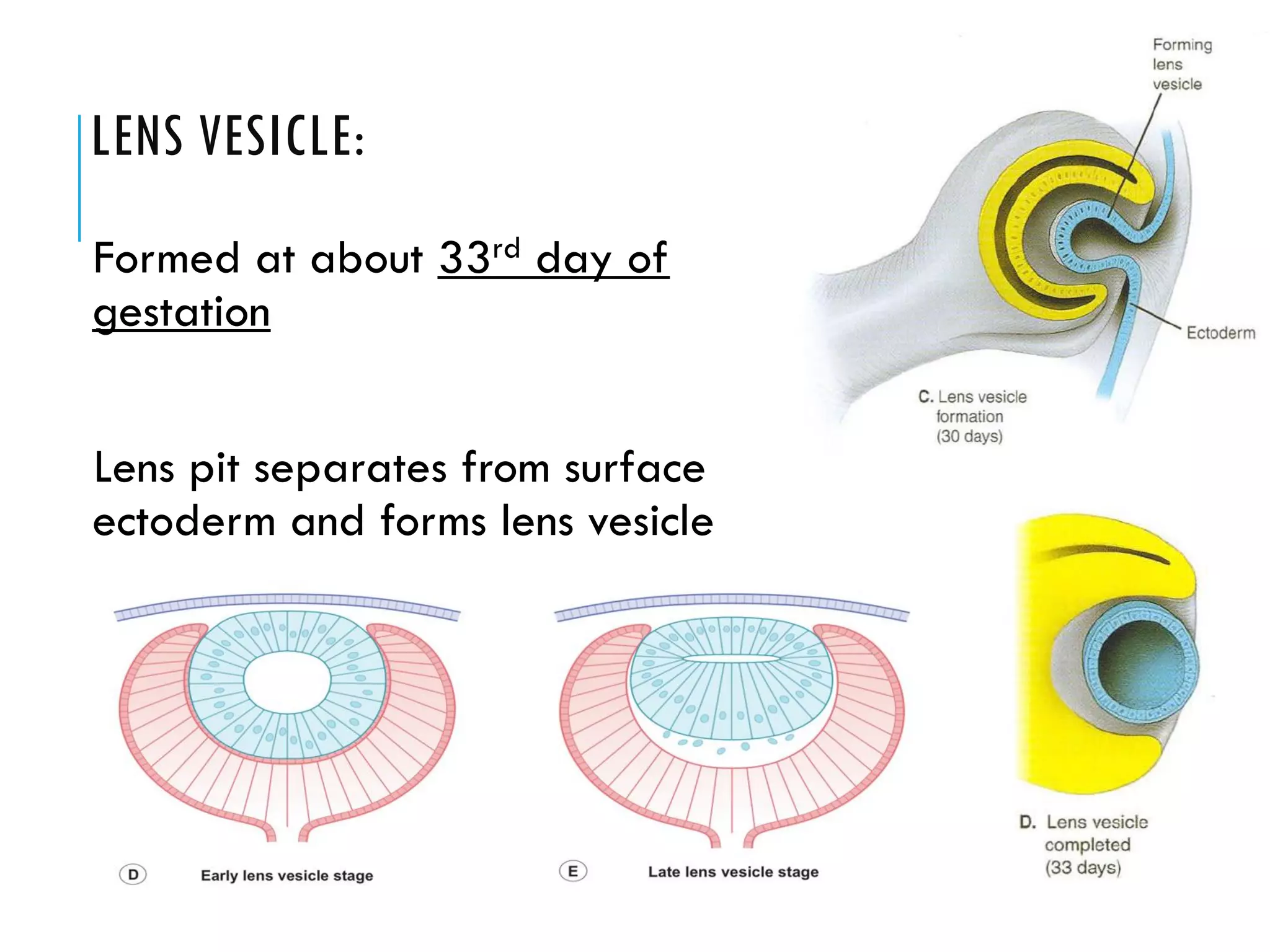

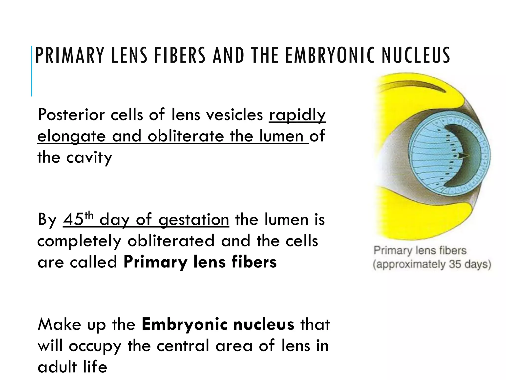
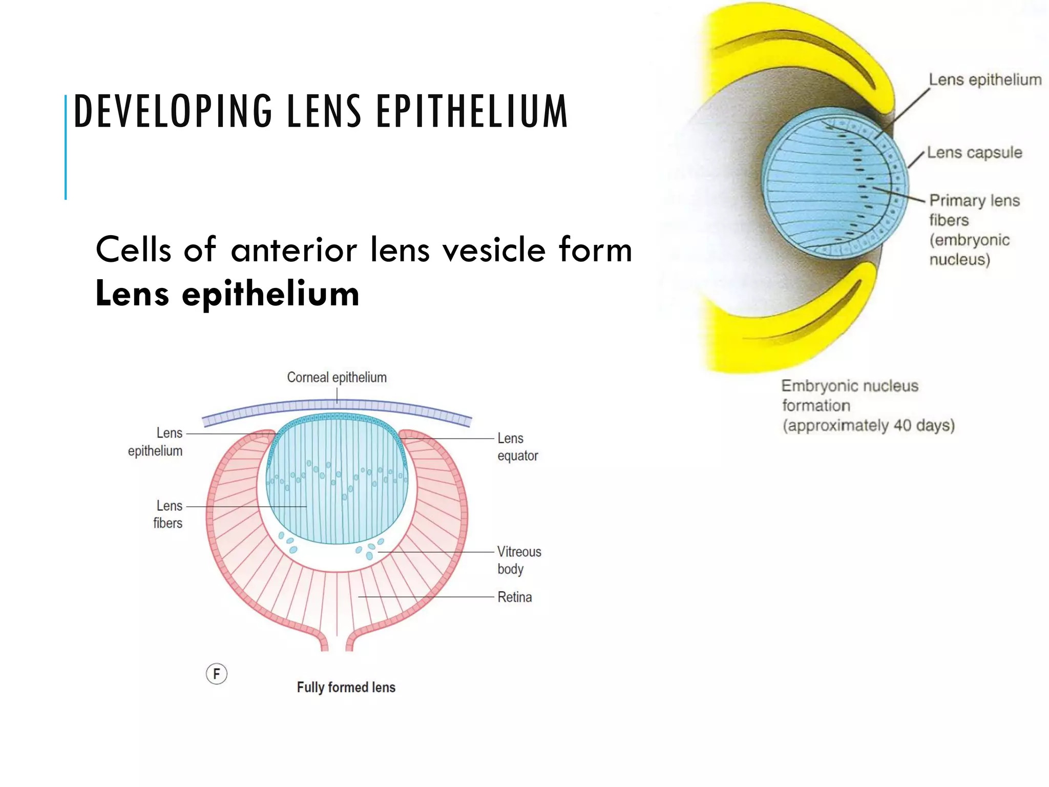

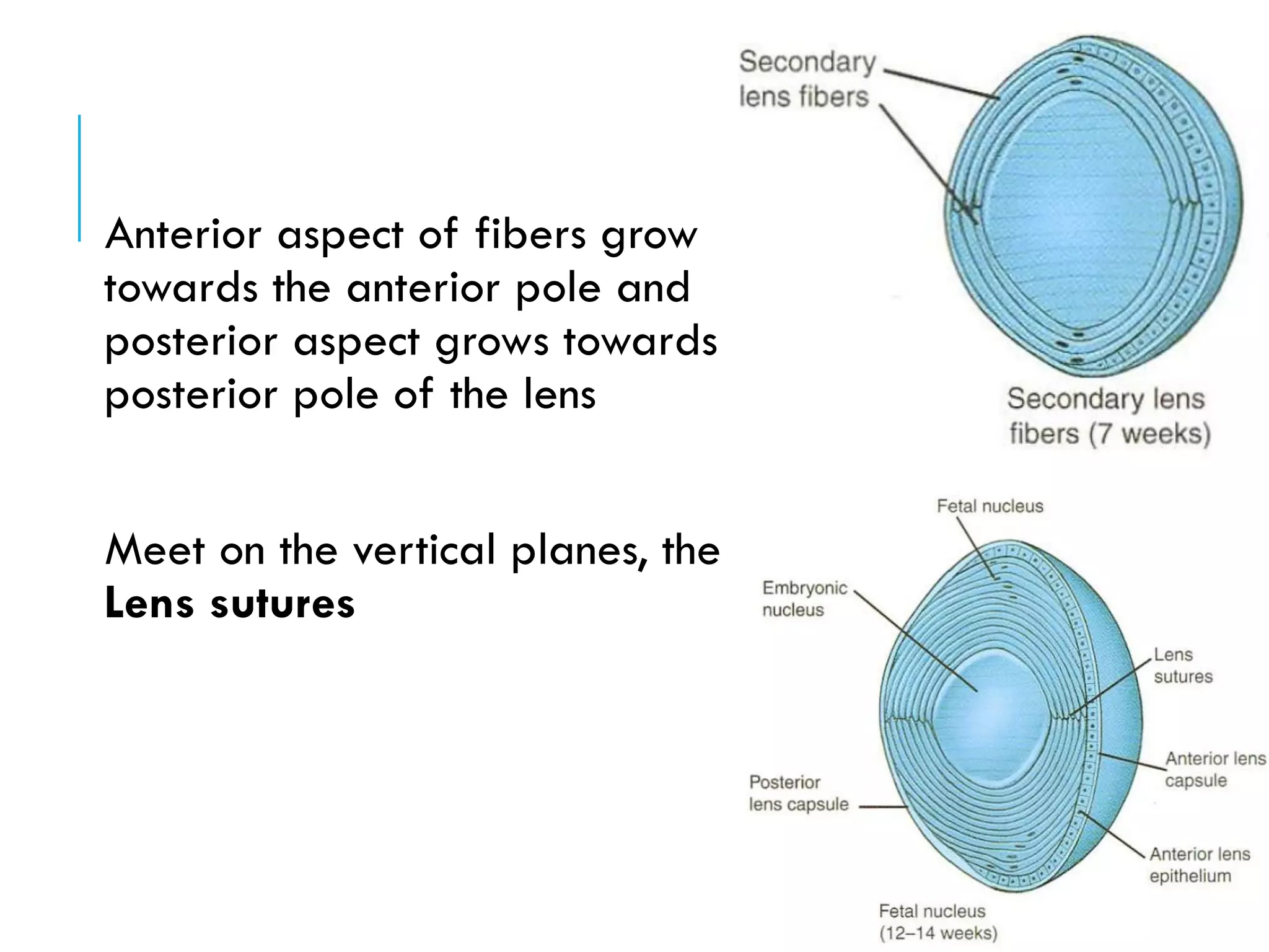
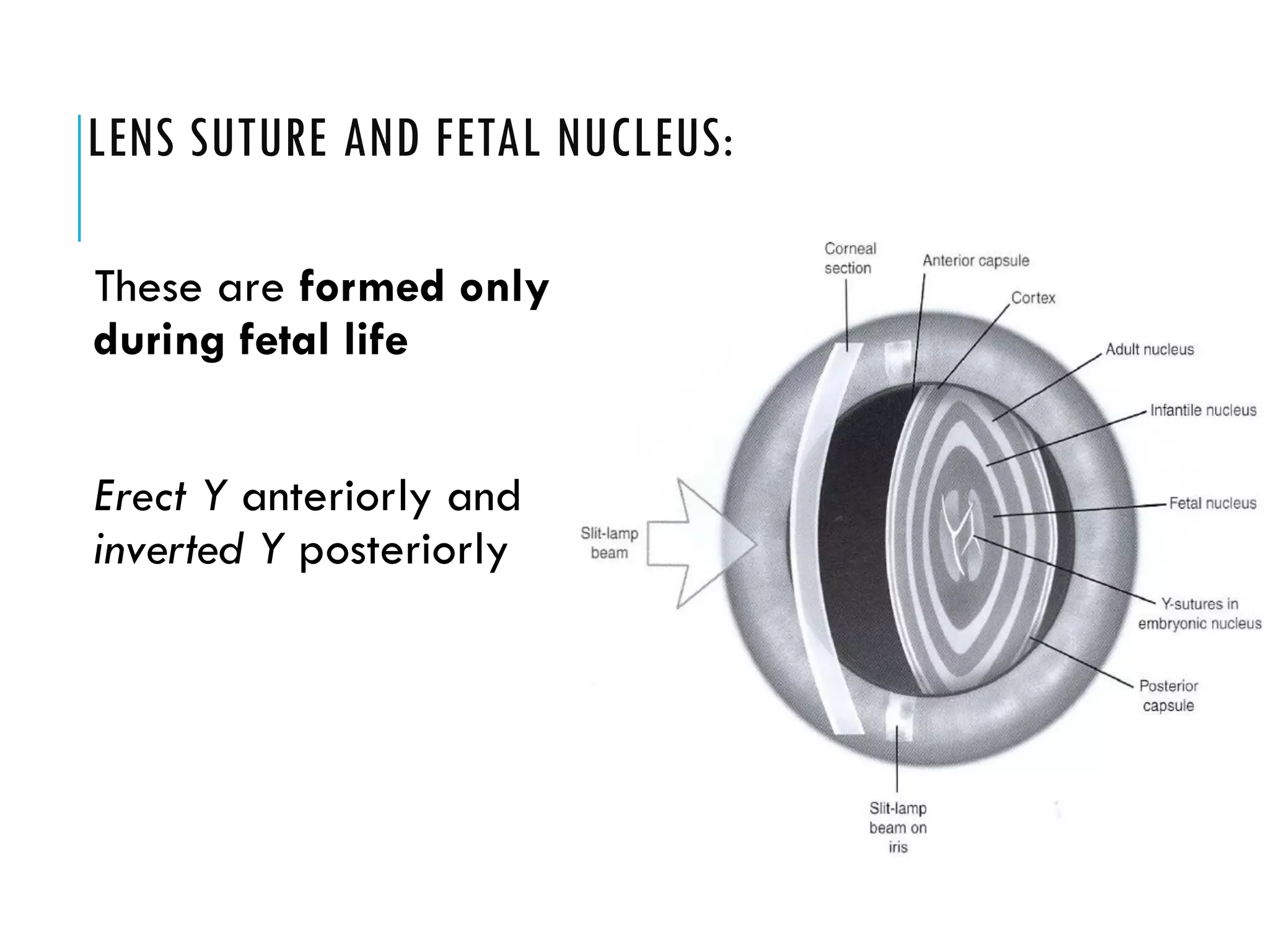


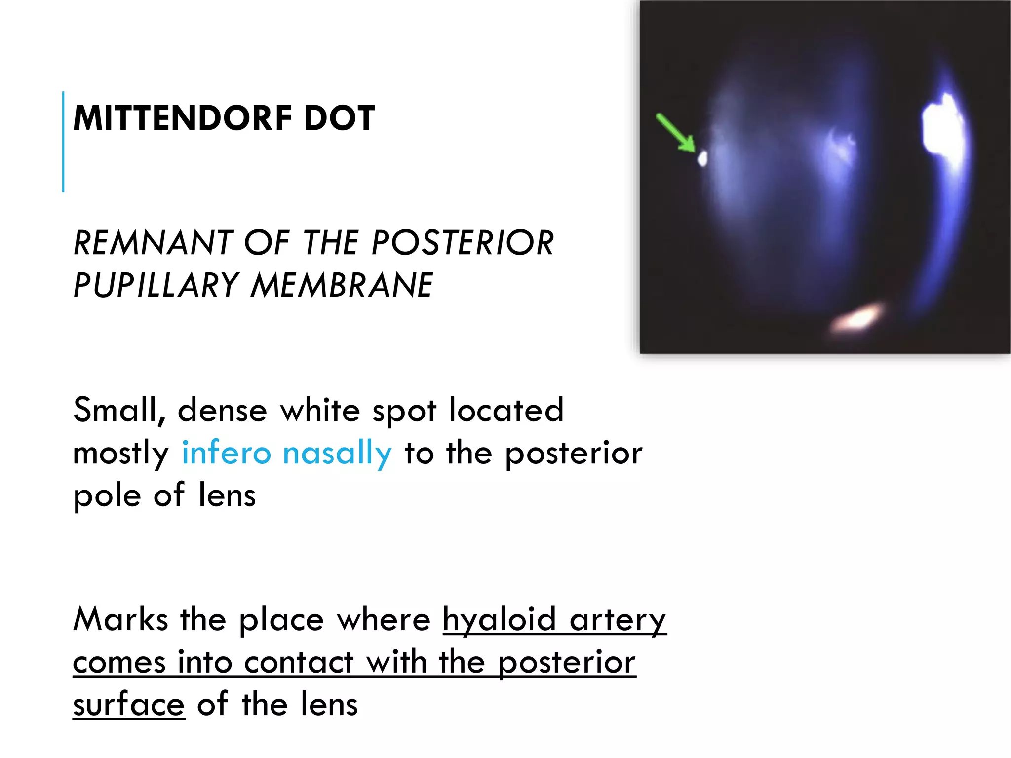


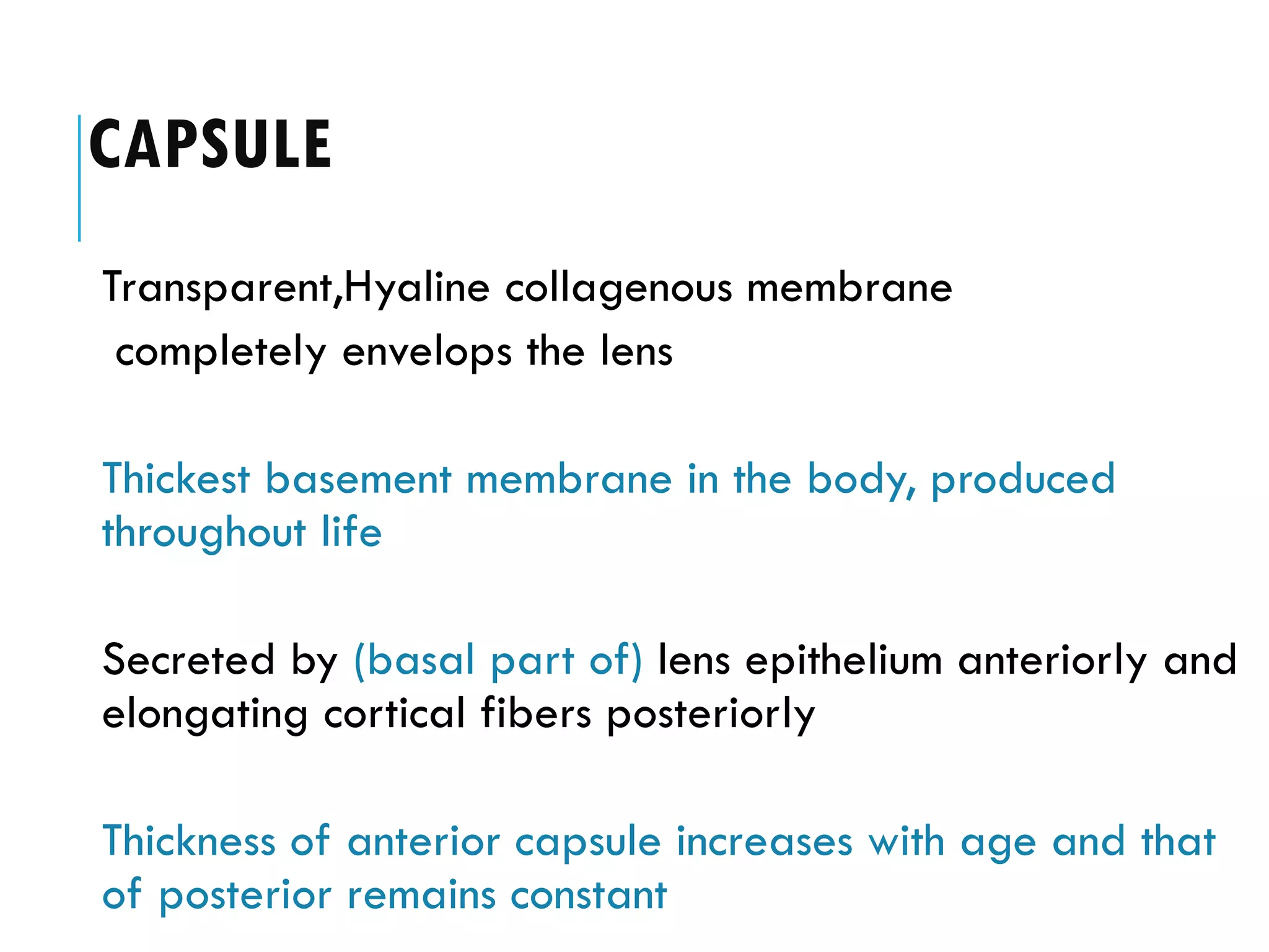


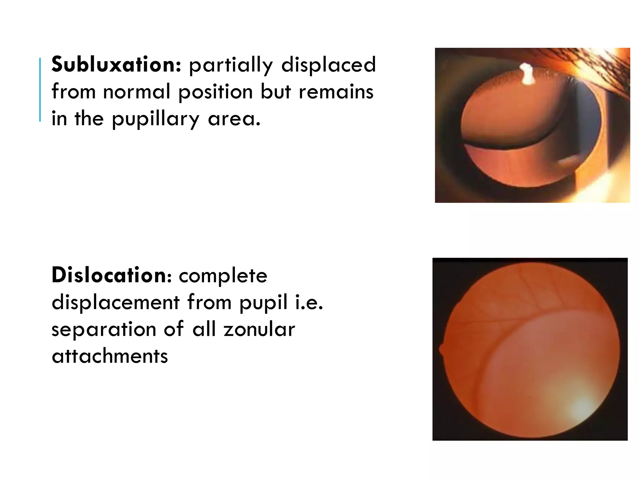


The document provides an extensive overview of the anatomy, physiology, and development of the lens in the eye, detailing its embryological origins, structural components, and functional properties related to accommodation and transparency. It covers various developmental anomalies, the significance of the lens capsule and zonular fibers, and the physiological mechanisms involved in maintaining lens clarity and refractive power. Additionally, the document discusses cataracts, their types, and implications for lens transparency, highlighting the importance of proper lens function throughout life.


