SlideShare a Scribd company logo
MEDICAL SURGICAL
NURSING (MSN)
The Cardiovascular system: General cardiovascular system,
Congenital heart defects, Disorders associated with the conducting
system, Congestive cardiac failure, Cardiac trauma, Myocardial
infarction & Angina
By C.Settley
Orientation to MSN
(Please see subject guide)
 Lecturer Ms C Settley Room 1
 023 347 0732/ 0844892450
 MSN Group
 settleyc@cput.ac.za /c.settley@gmail.com
 Work integrated learning
 Students must complete 80% of clinical hours as calculated per
year level will be allowed to enter for all the assessments
 Teaching methodology/strategies
 Emphasis is placed on self-directed learning!
Compiled by C.Settley 2018
Orientation to MSN
(Please see subject guide)
 Blackboard
 SDL
 Class attendance
 Compulsory prescribed books
 Recommended books
 Subject credits
 Purpose of this subject
Compiled by C.Settley 2018
Orientation to MSN
(Please see subject guide)
 Chapters with outcomes in subject guide
 Activities
 Assessment policy
 Guidelines for assignments
 Referencing, etc.
 Evaluation sheets
Compiled by C.Settley 2018
Orientation to MSN
(Please see subject guide)
Compiled by C.Settley 2018
ASSESSMENT WEIGHTING DATE
Summative 1 25% 16 February 2018
Deferred 1 03 April 2018
Summative 2 25% 23 March 2018
Deferred 2 16 April 2018
Summative 3 25% 09 April 2018
Deferred 3 ASAP
Summative 4 25% 18 May 2018
Deferred 4 04 June 2018
Outcomes
Apply knowledge regarding:
 Haematological studies
 Patho-physiology
 disease process
 clinical manifestations
 specific diagnostic and therapeutic interventions (tests and examinations)
Distinguish between the different health problems:
medical and surgical conditions of various body
systems
 Congenital defects
 Conducting system
 Congestive cardiac failure
 Coronary artery disease
 Angina
 Myocardial infarction
 Blood vessels disorders
 Peripheral arterial disease
 Deep vein thrombosis
 Cardiovascular disease in the elderly
Compiled by C.Settley
2018
 What is the cardio vascular system?
 What is it comprised of?
 What is its purpose? What does it circulate?
 How is the heart shaped?
 Where is it located?
 What are the 3 layers that the heart is made up, called?
 How many valves are in the heart?
Compiled by C.Settley 2018
Anatomy of the heart
Compiled by C.Settley 2018
Location of the heart The three layers of the heart wall
Compiled by C.Settley 2018
Compiled by C.Settley 2018Valves of the heart
Valves of the heart
 TRICUSPID VALVE: Closes off the upper right chamber (or
atrium) that holds blood coming in from the body.
 Opens to allow blood to flow from the top right chamber to
the lower right chamber (or from right atrium to right
ventricle).
 Prevents the back flow of blood from the ventricle to the
atrium when blood is pumped out of the ventricle.
Compiled by C.Settley 2018
Valves of the heart
 PULMONARY VALVE: Closes off the lower right chamber (or
right ventricle).
 Opens to allow blood to be pumped from the heart to the
lungs (through the pulmonary artery) where it will receive
oxygen.
Compiled by C.Settley 2018
Valves of the heart
 MITRAL VALVE: Closes off the upper left chamber (or left
atrium) collecting the oxygen-rich blood coming in from the
lungs.
 Opens to allow blood to pass from the upper left side to the
lower left side (or from the left atrium to the left ventricle).
Compiled by C.Settley 2018
Valves of the heart
 AORTIC VALVE: Closes off the lower left chamber that holds
the oxygen-rich blood before it is pumped out to the body.
 Opens to allow blood to leave the heart (from the left
ventricle to the aorta and on to the body).
Compiled by C.Settley 2018
Blood flow through the heart
Compiled by C.Settley 2018
Blood flow through the heart
Compiled by C.Settley 2018
Blood flow through the heart
 Watch video:
 Normal blood flow through the heart
Compiled by C.Settley 2018
Diagnostic tests- pg. 621
 Electrocardiography (ECG)
 A graphic representation of the electrical activity
of the heart muscle as it contracts and relaxes.
 A standard 12-lead electrocardiogram is used to
access the electrical activity of the heart and the
conduction of the cardiac impulse.
 Most cardiac conditions give rise to abnormal
recordings on the ECG.
 Video:
 ECG
Compiled by C.Settley 2018
Electrocardiography
(ECG) Compiled by C.Settley 2018
How to read an ECG
 HEART RATE
 Heart rate can be calculated using the following method
(if regular):
 Count the number of large squares present within one R-R
interval
 Divide 300 by this number to calculate the heart rate
 e.g. 4 large squares in an R-R interval: 300/4 = 75 beats
per minute
Compiled by C.Settley 2018
How to read an ECG
HEART RHYTHM
The heart rhythm can be regular or
irregular.
Irregular rhythms can be either:
Regularly irregular (i.e. a recurrent pattern
of irregularity)
Irregularly irregular (i.e. completely
disorganised)
Compiled by C.Settley 2018
How to read an ECG Compiled by C.Settley 2018
How to read an ECG
 Pre atrial contractions (pg636):
 Premature atrial contractions (PACs), also known as atrial
premature complexes (APC) or atrial premature beats (APB),
are a common cardiac dysrhythmia characterized
by premature heartbeats originating in the atria.
 While the sinoatrial node typically regulates the heartbeat
during normal sinus rhythm, PACs occur when another region of
the atria depolarizes before the sinoatrial node and thus triggers
a premature heartbeat.
Compiled by C.Settley 2018
How to read an ECG
Cardioversion is a medical procedure by which
an abnormally fast heart rate or other cardiac
arrhythmia is converted to a normal rhythm using
electricity or drugs
Compiled by C.Settley 2018
How to read an ECG
 Cardio pulmonary resuscitation and adrenalin
 Flatline:
Compiled by C.Settley 2018
Atrial flutter- pg636
 Irregular and fast heartbeat
 Cardio version may be required
 May be due to fluid overload, etc
Compiled by C.Settley 2018
Diagnostic tests- pg. 621
 Exercise ECG (Stress test)
 An exercise electrocardiogram (EKG or ECG) is a test that
checks for changes in your heart while you exercise.
 Sometimes ECG abnormalities can be seen only during
exercise or while symptoms are present. This test is
sometimes called a "stress test“.
 See box 32.2 INFORMATION REQUIRED
BEFORE A STRESS TEST IS DONE
Compiled by C.Settley 2018
Exercise ECG (Stress test)Compiled by C.Settley 2018
Diagnostic tests- pg. 621
 Chest radiography (CXR)
 An X-ray is an imaging test that uses small
amounts of radiation to produce pictures of the
organs, tissues, and bones of the body.
 When focused on the chest, it can help spot
abnormalities or diseases of the airways, blood
vessels, bones, heart, and lungs.
 Chest X-rays can also determine if patients have
fluid or air in their lungs.
 Heart size, etc. can be examined.
Compiled by C.Settley 2018
Chest radiography Compiled by C.Settley 2018
Diagnostic tests- pg. 621
 Echocardiography
An echocardiogram, often referred to as a
cardiac echo or simply an echo.
It is a sonogram of the heart.
Echocardiography uses standard two-
dimensional, three-dimensional, and
Doppler ultrasound to create images of the
heart.
Compiled by C.Settley 2018
Echocardiography Compiled by C.Settley 2018
Diagnostic tests- pg. 622
 Cardiac catherisation
Cardiac catheterisation is the insertion of a
catheter into a chamber or vessel of the
heart.
This is done both for diagnostic and
interventional purposes.
Compiled by C.Settley 2018
Cardiac catherisation Compiled by C.Settley 2018
Cardiac catherisation- pg 622
 VIDEO*:
 Cardiac catherisation
 Nursing responsibilities
 Consent
 NPO
 Cleansing and shaving of patient
 ECG as baseline to be taken
 Record any allergy history as the patient may react to x-ray dyes.
 Bedpan/urinal (empty bladder).
 Make patient comfortable as far as possible.
 Remove dentures, hearing aids, glasses, etc.
 Post procedure: monitor patient for dysrhythmias
and bleeding from catherisation site.
- Bedrest.
Compiled by C.Settley 2018
Diagnostic tests- pg. 622
 Angiography
 Angiography or arteriography is a medical imaging technique
used to visualize the inside, or lumen, of blood vessels and
organs of the body, with particular interest in the arteries,
veins and the heart chambers.
 VIDEO *
 Coronary angiogram
Compiled by C.Settley 2018
Diagnostic tests- pg. 622
 Doppler studies
A Doppler ultrasound is a test that uses
high-frequency sound waves to measure the
amount of blood flow through your arteries
and veins, usually those that supply blood to
your arms and legs.
Vascular flow studies, also known as blood
flow studies, can detect abnormal flow
within an artery or blood vessel.
Compiled by C.Settley 2018
Diagnostic tests- pg. 623
 Pulse oximetry
 Pulse oximetry is a non-invasive method for monitoring a
person's oxygen saturation (SO2).
Compiled by C.Settley 2018
Haematological tests in
cardiovascular conditions- pg. 623
TYPE OF TEST RATIONALE
SERUM ELECTROLYTES Potassium, sodium, calcium and
magnesium affect cardiac function.
Hypernatremia may be indicative of
dehydration while potassium deficit may
result in dysrhythmias.
BLOOD GASES Arterial gasses indicate the levels of
oxygen and carbon dioxide in the blood,
and are indicators of cardiac functioning.
SERUM ENZYMES Presence of enzymes are indicative of
damage to muscle tissue and indirect
indicator of damage to myocardium. The
following enzymes’ presence are however,
direct indicators of damage to the
myocardium: CPK, LDH, SGOT,
MB(CKMB), Trop T.
SERUM LIPIDS Presence of serum lipids indicates the
presence of atherosclerosis.
Compiled by C.Settley 2018
 Haematological tests in
cardiovascular conditions- pg. 623
 REFER TO HANDOUT
 Haematological tests can help diagnose anaemia, infection,
haemophilia, blood-clotting disorders, and leukaemia.
Compiled by C.Settley 2018
Classification of the cardiac
disorders- pg. 629
Causative factor Conditions
Congenital defects Septal defects
Defects affecting the valves
Defects affecting major arteries
Mixed defects
Disturbances of conduction Atrial dysrhythmias
Atrioventricular/junctional dysrhythmias
Ventricular dysrhythmias
Infection and inflammatory disorders Rheumatic fever
Infective endocarditis
Valvular heart disease
Myocarditis
Pericarditis
Structural disorders Mitral valve disease
Aortic valve disease
Tricuspid and pulmonic valve disease
cardiomyopathy
Congestive conditions
Trauma
Heart failure
Cardiac contusion
Cardiac tamponade
Pericardial rupture
Rupture of the heart wall, papillary muscles, heart valves
Compiled by C.Settley 2018
Risk factors for
cardiac disorders- pg. 629
 Lifestyle
 Unhealthy diet, high blood cholesterol and other fats
 Obesity & overweight
 Tabacco use
 Hypertension
 Diabetes Mellitus
 Stress
Compiled by C.Settley 2018
Congenital heart defects- pg. 630
 Congenital heart disease (CHD) is a problem with the
heart's structure and function that is present at birth.
 Congenital heart defects are the most common type of
birth defect.
 The defects can involve the walls of the heart, the valves
of the heart, and the arteries and veins near the heart.
 They can disrupt the normal flow of blood through the
heart.
 The blood flow can slow down, go in the wrong direction or
to the wrong place, or be blocked completely.
Compiled by C.Settley 2018
Congenital heart defects
Aetiology and risk factors- pg.
630
Viral infections during pregnancy. Especially the
1st trimester, eg Rubella.
Nutritional deficiencies during pregnancy.
Exposure to environmental factors such as
toxins and radiation.
Excessive alcohol consumption during
pregnancy.
Drugs or medication abuse.
Family history.
Compiled by C.Settley 2018
Congenital heart defects
Pathophysiology- pg 630
Defects form during early pregnancy
between the 6th and 12th week.
Heart of fetus develops from a single tube to
a heart with 4 chambers, veins and arteries
Compiled by C.Settley 2018
Congenital heart defects
Pathophysiology- pg. 630
 In congenital defects, part of the heart may thus not develop
(partially or not at all).
 Defects may include: a hole in the septum or the formation of
abnormal connections of the arteries and veins of the heart.
 This results in the mixing of deoxygenated and oxygenated
blood.
 Congenital defects are structural in nature and cause problems
because of their effect on blood flow through the heart.
 Sometimes the conducting system is involved.
 Defects range from simple to severe.
 VIDEO* (atrial defect blood flow)
Compiled by C.Settley 2018
Septal defects
Compiled by C.Settley 2018
Septal defects,
Heart valve defects &
major blood vessel defects- pg. 631
 Hole in the septum that divides
the R & L side of the heart.
 Small septal defects may close
with time or not, causing
defective flow.
 Larger holes can cause more
problems with functioning of the
heart.
 Major presentation: palpitations
and irregular pulse, poor colour
of blood.
Heart valve defects
Compiled by C.Settley 2018
Septal defects,
Heart valve defects &
major blood vessel defects- pg 631
 Heart valves allow blood to flow
in one direction.
 They open and close passively.
 Due to valve defects, these
valves may not open or close
properly (valve incompetence).
 Blood may flow backwards
(regurgitate).
 The heart now works harder and
it cannot get sufficient blood to
the lungs and body.
Septal defects,
Heart valve defects &
major blood vessel defects- pg. 631
 Major blood vessel defects
 Coarctation of the aorta
 Constriction of a segment of the aorta resulting in
obstruction of blood flow from the left ventricle.
Compiled by C.Settley 2018
Septal defects,
Heart valve defects &
major blood vessel defects- pg. 631
 Major blood vessel defects
 Transposition of the great arteries
 When the pulmonary artery arises from the left ventricle
and the aorta from the right ventricle sends
deoxygenated blood from the right atrium back to the
systemic circulation.
Compiled by C.Settley 2018
Septal defects,
Heart valve defects &
major blood vessel defects- pg. 631
 Major blood vessel defects
 Patent ductus arteriosus
 Before birth, the aorta and the pulmonary artery are
connected by a blood vessel called the ductus arteriosus
which is an essential part of the fetal circulation.
 Soon after birth, the vessel is supposed to close as part of
the normal changes occurring in the baby’s circulation.
 In some babies, it does not close, and the opening allows
oxygenated blood from the aorta to mix with deoxygenated
blood from the pulmonary artery.
 See other defects- pg. 632, Table 33.2 + figures
Compiled by C.Settley 2018
Clinical manifestations of
congenital defects- pg. 631
 Family history
 Child develops slow
 Tachypnea/cyanosed/dyspnea
 History of frequent chest infections
 The child, may squat when distressed- it increases blood
flow to the heart
 Clubbing of fingers and toes
 Enlarged liver or spleen
 Oedema of extremities
 ECG abnormalities
 Cardiac enlargement may be seen on x-ray and abnormal
heart sound may be heard
 Echocardiography and cardiac catherisation may be done
to confirm septal and valvular defects.
Compiled by C.Settley 2018
Management- pg. 631
Surgical correction of the defect which may involve
the use of catheters or open cardiac surgery.
The sooner the defect is corrected, the better in terms
of the child’s physical and mental health.
If the defect cannot be corrected, then conservative
management directed at preventing cardiac failure
and optimising pulmonary blood flow should be done.
Compiled by C.Settley 2018
What do you understand under
Decreased cardiac output?
 Inadequate blood pumped by the heart to meet the metabolic
demands of the body.
 Cardiac output is the amount of blood pumped by the heart
per minute.
 It is the product of the heart rate, which is the number of
beats per minute, and the stroke volume, which is amount
pumped per beat.
Compiled by C.Settley 2018
How would you assess a patient to evaluate
whether the patient has decreased cardiac
output?
 Abnormal heart sounds
 Angina
 Anxiety & restlessness
 Change in level of consciousness
 Breathing pattern
 Decreased activity tolerance/fatigue
 Decreased peripheral pulses; cold, clammy skin/poor capillary refill
 Decreased venous and arterial oxygen saturation
 Dysrhythmias
 Ejection fraction less than 40% (Ejection fraction is a measurement of the percentage
of blood leaving your heart each time it contracts. During each heartbeat pumping cycle,
the heart contracts and relaxes. When your heart contracts, it ejects blood from the two
pumping chambers (ventricles). When your heart relaxes, the ventricles refill with
blood.)
 Hypotension
 Weight gain, oedema, decreased urine output
Compiled by C.Settley 2018
Interventions for decreased
cardiac output?
Interventions Rationales
Record intake and output. If patient is acutely ill,
measure hourly urine output and note decreases in
output.
Reduced cardiac output results in reduced perfusion of
the kidneys, with a resulting decrease in urine output.
For patients with increased preload, limit fluids
and sodium as ordered.
Fluid restriction decreases extracellular fluid volume
and reduces demands on the heart.
Closely monitor fluid intake including IV lines. Maintain
fluid restriction if ordered.
In patients with decreased cardiac output, poorly
functioning ventricles may not tolerate increased fluid
volumes.
Auscultate heart sounds
The new onset of a gallop rhythm, tachycardia, and
fine crackles in lung bases can indicate onset of heart
failure. If patient develops pulmonary edema, there
will be coarse crackles on inspiration and severe
dyspnea.
Closely monitor for symptoms of heart failure and
decreased cardiac output, including diminished quality of
peripheral pulses, cold and clammy skin and extremities,
increased respiratory rate, presence of paroxysmal
nocturnal dyspnea or orthopnea, increased heart rate,
neck vein distention, decreased level of consciousness,
and presence of edema.
As these symptoms of heart failure progress, cardiac
output declines.
Compiled by C.Settley 2018
How would you assess a patient to evaluate
whether the patient has decreased cardiac
output?
Note chest pain. Identify location, radiation, severity,
quality, duration, associated manifestations such as
nausea, and precipitating and relieving factors.
Chest pain/discomfort is generally suggestive of an
inadequate blood supply to the heart, which can
compromise cardiac output. Patients with heart failure
can continue to have chest pain with angina or can
reinfarct.
If chest pain is present, have patient lie down, monitor
cardiac rhythm, give oxygen, run a strip, medicate for
pain, and notify the physician.
These actions can increase oxygen deliveryto
the coronary arteries and improve patient prognosis.
Place on cardiac monitor; monitor for dysrhythmias,
especially atrial fibrillation.
Atrial fibrillation is common in heart failure.
Examine laboratory data, especially arterial blood gases
and electrolytes, including potassium.
Patient may be receiving cardiac glycosides and the
potential for toxicity is greater
with hypokalemia; hypokalemia is common in heart
patients because of diuretic use.
Monitor laboratory tests such as complete blood
count, sodium level, and serum creatinine.
Routine blood work can provide insight into the
etiology of heart failure and extent of decompensation.
A low serum sodium level often is observed with
advanced heart failure and can be a poor prognostic
sign. Serum creatinine levels will elevate in patients
with severe heart failure because of decreased
perfusion to the kidneys. Creatinine may also elevate
because of ACE inhibitors.
Compiled by C.Settley 2018
How would you assess a patient to evaluate
whether the patient has decreased cardiac
output?
Administer medications as prescribed, noting side effects
and toxicity.
Depending on etiological factors, common medications
include digitalis therapy, diuretics, vasodilator
therapy, antidysrhythmics, angiotensin-converting
enzyme inhibitors, and inotropic agents.
Review results of EKG and chest Xray.
EKG can reveal previous MI, or evidence of left
ventricular hypertrophy, indicating aortic stenosis or
chronic systemic hypertension. Xray may provide
information on pulmonary edema, pleural effusions, or
enlarged cardiac silhouette found in dilated
cardiomyopathy or large pericardial effusion.
Compiled by C.Settley 2018
Describe interventions for activity
intolerance with possible rationales.
Assessment Rationales
Assess the physical activity level and mobility of the
patient.
 Take the resting pulse, blood pressure, and
respirations.
 Consider the rate, rhythm, and quality of the pulse.
 If the signs are normal, have the patient perform
the activity.
 Obtain the vital signs immediately after activity
 Have the patient rest for 3 minutes and then take
the vital signs again.
Provides baseline information for formulating nursing
goals during goal setting.
Discontinue the activity if the patient responds with:
 chest pain, vertigo, and/or dizziness
 decreased pulse rate, systemic blood pressure,
respiratory response
Reduce the duration and intensity of the activity if:
 Pulse takes longer than 3 to 4 minutes to return to
within 6-7 beats of the resting pulse.
 RR increase is excessive after the activity.
Investigate the patient’s perception of causes of activity
intolerance.
Causative factors may be temporary or permanent as
well as physical or psychological. Determining the cause
can help guide the nurse during the nursing intervention.
Assess the patient’s nutritional status. Adequate energy reserves are needed during activity.
Observe and monitor the patient’s sleep pattern and
the amount of sleep achieved over the past few days.
Sleep deprivation and difficulties during sleep can affect
the activity level of the patient – these needs to be
addressed before successful activity progression can be
achieved.
Compiled by C.Settley 2018
Describe interventions for activity
intolerance with possible rationales.
Determine the patient’s daily routine and over-the-
counter medication.
Fatigue can limit the patient’s ability to perform needed
activity. It can also be a medication side effect. Pay
attention to the patient’s use of beta-
blockers, calciumchannel blockers, tranquilizers,
antihistamines, relaxants, alcohol, and sedatives.
Assess the need for ambulation aids (e.g., cane, walker)
for ADLs.
Assistive devices enhance the mobility of the patient by
helping him overcome limitations.
Use portable pulse oximetry to assess for oxygen
desaturation during activity.
May determine the use of supplemental oxygen to help
compensate for the increased oxygen demands during
physical activity.
Assess the patient’s baseline cardiopulmonary status
(e.g., heart rate, orthostatic BP) before initiating
activity.
In normal adults, HR should not increase more than 20
to 30 beats/min above resting with routine activities.
Older patients are more susceptible to orthostatic
drops in BP with position changes.
Observe and document response to activity.
Close monitoring will serve as a guide for optimal
progression of activity.
Assess emotional response to limitations in physical
activity.
Depression over the inability to perform activities can
be a source of stress and frustration.
Compiled by C.Settley 2018
Describe interventions for activity
intolerance with possible rationales.
Establish guidelines and goals of activity with the patient
and/or SO.
Motivation and cooperation are enhanced if the patient
participates in goal setting.
Evaluate the need for additional help at home.
Coordinated efforts are more meaningful and effective
in assisting the patient in conserving energy.
Have the patient perform the activity more slowly, in a
longer time with more rest or pauses, or with assistance
if necessary.
Helps in increasing the tolerance for the activity.
Gradually increase activity with active range-of-motion
exercises in bed, increasing to sitting and then standing.
Gradual progression of the activity prevents
overexertion.
Dangle the legs from the bed side for 10 to 15 minutes. Prevents orthostatic hypotension.
Refrain from performing nonessential activities or
procedures.
Patient with limited activity tolerance need to prioritize
important taks first.
Assist with ADLs while avoiding patient dependency.
Assisting the patient with ADLs allows conservation of
energy. Carefully balance provision of
assistance; facilitating progressive endurance will
ultimately enhance the patient’s activity tolerance and
self-esteem.
Compiled by C.Settley 2018
Describe interventions for activity
intolerance with possible rationales.
Provide bedside commode as indicated.
Use of commode requires less energy expenditure
than using a bedpan or ambulating to the bathroom.
Encourage physical activity consistent with the
patient’s energy levels.
Helps promote a sense of autonomy while being
realistic about capabilities.
Instruct patient to plan activities for times when they
have the most energy.
Activities should be planned ahead to coincide with
the patient’s peak energy level. If the goal is too
low, negotiate.
Encourage verbalization of feelings regarding
limitations.
This helps the patient to cope. Acknowledgment that
living with activity intolerance is both physically and
emotionally difficult.
Gradually progress patient activity with the following:
 Range-of-motion (ROM) exercises in bed,
gradually increasing duration and frequency (then
intensity) to sitting and then standing.
 Deep-breathing exercises three or more times
daily.
 Sitting up in a chair 30 minutes three times daily.
 Walking in room 1 to 2 minutes TID.
 Walking down the hall 20 feet or walking through
the house, then slowly progressing walking
outside the house, saving energy for the return
trip.
Duration and frequency should be increased before
intensity.
Compiled by C.Settley 2018
Describe interventions for compromised family
coping with possible rationales.
Nursing Interventions Rationale
Observe for erratic behaviors (anger, tension, disorganization),
perception of crisis situation.
Information affecting the ability of the family to cope with
infant/child’s cardiac condition.
Encourage expression of feelings and provide factual
information about infant/child.
Reduces anxiety and enhances family’s understanding of the
condition.
Assess usual family coping methods and effectiveness.
Identifies need to develop new coping skills if existing
methods are ineffective in
changing behaviors exhibited.
Assess need for information and support.
Provides information about need for interventions to
relieve anxiety and concern.
Clarify any misinformation and answer questions
regarding disease process.
Prevents unnecessary anxiety resulting from
inaccurate knowledge or beliefs.
Assist in identifying and using
techniques to cope with and solve
problems and gain control over the situation.
Provides support for problem solving and management of the
situation.
Encourage to maintain the health of
family members and social contacts.
Chronic anxiety, fatigue, and isolation as result of infant care
will affect health and care capabilities of family.
Compiled by C.Settley 2018
Describe interventions for compromised family
coping with possible rationales.
Teach that overprotective behavior
may hinder growth and development
during infancy/ childhood.
Knowledge will enhance family understanding of the condition
and of adverse effects of behaviors.
Suggest and reinforce appropriate
coping behaviors, support family
decisions.
Promotes behavior change and adaptation to care for
infant/child.
Encourage parents to include ill infant/ child in family
activities rather than family revolving around needs of
infant/child.
Promotes normal growth and development of family
and infant/child.
Encourage to maintain consistent
behavior limits and modification
techniques.
Prevents behavioral problems and child control over family,
which interfere with child’s growth and family relationships.
Instruct parents in nutritional and
activity needs and/or limitations
and approaches that will assist
in establishing an effective pattern.
Assists in coping with effects and special needs of
infant/child with a cardiac defect.
Refer family for additional support
and counseling if indicated.
Referral supplies more assistance with coping than is
available from nursing personnel.
Compiled by C.Settley 2018
Disorders associated with
the conducting system- pg. 633
What is the conducting system?
 The cardiac conduction system is a group of
specialized cardiac muscle cells in the walls of the
heart that send signals to the heart muscle causing it
to contract.
 The main components of the cardiac conduction
system are the SA node, AV node, bundle of His,
bundle branches, and Purkinje
fibers.
Compiled by C.Settley 2018
Disorders associated with
the conducting system- pg. 633
- The SA node – sends out regular electrical impulses from the top chamber
(the atrium) causing it to contract and pump blood into the bottom chamber
(the ventricle).
- Also called the pacemaker, because it initiates each heartbeat.
- The electrical impulse then travels to the AV node. This causes the atria to
contract.
- From the AV node, the impulse passes down the ventricular septum to the
left and right bundle branches and the fibres of Purkinje to stimulate the
ventricles to contract.
- As a result of ventricular contraction, the blood is ejected into the aorta and
pulmonary circulation respectively.
- The normal rhythm of the heart is known as sinus rhythm as it is initiated
by the sinoatrial node.
Compiled by C.Settley 2018
Disorders associated with
the conducting system- pg. 633
The characteristics of normal sinus rhythm
are:
 The rhythm is regular.
 Rate is between 60-100 bpm.
 Each beat consists of a P - wave, QRS complex and a T – wave.
 Every P - wave is followed by a QRS complex
 The complexes are tall and narrow.
Compiled by C.Settley 2018
Disorders associated with
the conducting system- pg. 636
- Cardiac conducting system Video*
CLASSIFICATION OF DYSRYTHMIAS ACCORDING
TO:
- The rate of the dysrhythmia, whether fast or slow
- Whether life threatening or not
- The area of origin
Compiled by C.Settley 2018
Atrial dysthymias- pg. 636
 PAC
 Originates in the atria
 May occur in healthy individuals and are frequently due to
emotion, exertion or stimulants
 Mostly due to sympathetic over activity, hypoxia, stress,
smoking or anxiety
 Can also be associated with valvular heart disease, atrial
chamber enlargement and coronary artery disease
 May mark the onset of arterial fibrillation or heart failure
 P-waves are premature and differs from normal sinus P
waves
 Described as palpitations
 Underlying cause should be investigated
Compiled by C.Settley 2018
Atrial dysthymias- pg. 636
 Atrial flutter
 Irregular and fast
 A saw tooth pattern as baseline
 Common in rheumatic heart disease (Rheumatic fever is an inflammatory disease that
sometimes happens after an infection caused by a bacteria called group A streptococcus, like strep throat or scarlet fever.
Rheumatic fever happens when the infection is not completely treated with medicine (antibiotics). It may affect the heart,
joints, skin and brain).
 Fluid overload
 Cardioversion
Compiled by C.Settley 2018
Atrial dysthymias- pg. 636
 Atrial Fibrillation
 Atrial fibrillation is an irregular and often rapid heart rate that
can increase your risk of stroke, heart failure and other heart-
related complications.
 During atrial fibrillation, the heart's two upper chambers (the
atria) beat chaotically and irregularly — out of coordination with
the two lower chambers (the ventricles) of the heart.
 Atrial fibrillation symptoms often include heart palpitations,
shortness of breath and weakness.
Compiled by C.Settley 2018
Atrial dysthymias- pg. 636
 Atrial Fibrillation
 Episodes of atrial fibrillation can come and go, or you may
develop atrial fibrillation that doesn't go away and may require
treatment. Although atrial fibrillation itself usually isn't life-
threatening, it is a serious medical condition that sometimes
requires emergency treatment.
 It may lead to complications. Atrial fibrillation can lead to blood
clots forming in the heart that may circulate to other organs
and lead to blocked blood flow (ischemia).
 Treatments for atrial fibrillation may include medications and
other interventions to try to alter the heart's electrical system.
Compiled by C.Settley 2018
Atrial dysthymias- pg. 636
Atrial Fibrillation
 Some people with atrial fibrillation have no symptoms and are unaware of their
condition until it's discovered during a physical examination. Those who do have
atrial fibrillation symptoms may experience signs and symptoms such as:
 Palpitations, which are sensations of a racing, uncomfortable, irregular heartbeat
or a flip-flopping in your chest
 Weakness
 Reduced ability to exercise
 Fatigue
 Light-headedness
 Dizziness
 Confusion
 Shortness of breath
 Chest pain
Compiled by C.Settley 2018
Ventricular dysthymias - pg. 638
Ventricular tachycardia
 A condition in which the lower chambers of the heart (ventricles) beat very quickly.
 Heart rate above 100 beats per minute
 May be up to 180 beats per minute
 Treatment depends on haemodynamic status of the patient:
 A raised BP- Cardioversion will be required, anti arrhythmic agents (amiodarone)
and correction of electrolyte and acid-base balance must be implemented.
 If patient collapses then standard CPR must be commenced
Compiled by C.Settley 2018
Ventricular dysthymias - pg. 638
 Ventricular fibrillation
 Complete disorganisation of the cardiac rhythm
 Irregular heart waves on ECG
 Varies in size and shape
 No contraction therefore no cardiac output
 Patient would appear clinically dead as there would be no pulse
 Treatment: defibrillation and CPR
Compiled by C.Settley 2018
Ventricular dysthymias - pg. 638
 Ventricular asystole
 Ventricular asystole is indicative of cardiac arrest. It requires
immediate attempts at resuscitation, with a poor prognosis at that.
It is characterized by the absence of electrical activity for a length
of time with intermittent ventricular complexes of abnormal
configuration.
Compiled by C.Settley 2018
Disorders associated with
the conducting system- pg. 639
Nb:
 In emergencies due to arrhythmias, rapid and correct diagnosis is necessary for adequate
therapy.
 The clinical symptoms, the physical examination, and the 12-lead electrocardiogram are
important sources of information for making a correct diagnosis
 It is essential to get information about the underlying heart disease, understand the mechanism
of the present arrhythmia
 Amiodarone is currently regarded as the most effective antiarrhythmic drug available for the
treatment of patients with both supraventricular and ventricular tachy arrhythmias.
 Beta blockers. Beta blockers work by blocking the effect of hormones such as adrenaline on
the heart. They are commonly prescribed to people with heart failure or angina, or following
a heart attack. They are used to treat high blood pressure alongside another drug.
 The use of atropine in cardiovascular disorders is mainly in the management of patients with
bradycardia. Atropine increases the heart rate and improves the atrioventricular conduction by
blocking the parasympathetic influences on the heart.
Compiled by C.Settley 2018
Disorders associated with
the conducting system- pg. 635
Management of restoring a normal rhythm
 The application of electrical ‘shock’ therapy, defibrillation or cardioversion.
 The implantation of a temporary pacemaker (A pacemaker is a small
device that's placed in the chest or abdomen to help control abnormal heart
rhythms. This device uses electrical pulses to prompt the heart to beat at a
normal rate).
 Pacemakers can be temporary or permanent. Temporary pacemakers are
used to treat temporary heartbeat problems, such as a slow heartbeat
that's caused by a heart attack, heart surgery, or an overdose of medicine.
 They're used until a permanent pacemaker can be implanted or until the
temporary condition goes away.
 The use of medications.
 Cardiac ablation (Catheter ablation is a minimally-invasive procedure
used to remove or terminate a faulty electrical pathway from sections of
the hearts of those who are prone to developing cardiac arrhythmias)
 An implantable cardiac defibrillator.
 Surgery.
Compiled by C.Settley 2018
Congestive heart failure- pg. 643
• When the heart’s function as a pump is inadequate to
deliver oxygen rich blood to the body.
• Can be caused by diseases which weaken the heart
muscle
• Increased oxygen demand by the body beyond the
capability of the heart to deliver adequate oxygen
rich blood to the tissues.
• Aetiology
- Coronary artery disease
- Valvular disorders
- Hypertension
- Infections
- Arrhythmias
Compiled by C.Settley 2018
Congestive heart failure
Pathophysiology- pg. 643
 The normal heart is able to meet the body’s need for oxygen by
increasing its output in response to increased demand for
oxygen. BUT, in heart failure:
 The heart’s capacity to increase the force of contraction has been
exceeded and the heart cannot respond to the body’s demands.
 The heart does not function adequately due to a problem within the
heart itself, such as ischaemic heart diseases, cardiomyopathy and
constrictive pericarditis.
 If ↓ contractility of the heart muscle due to cardiac failure, the
cardiac output is compromised leading to ↓ stroke volume and
systemic arterial blood pressure.
Compiled by C.Settley 2018
Classification of
congestive cardiac heart failure
(CCF)- pg. 644
1. Systolic heart failure/ Diastolic heart failure
Compiled by C.Settley 2018
Classification of congestive cardiac heart failure
(CCF)- pg. 644
2. Right-sided heart failure/ Left sided heart failure
The failure of the pumping action of the right side of the heart causes swelling in the legs
and abdomen while that of the left side, causes congestion of the lungs.
Compiled by C.Settley 2018
Classification of congestive cardiac heart failure
(CCF)- pg. 644
3. Forward heart failure/ Backward heart failure
- Inability of the heart to pump sufficient blood to meet the
oxygen needs of the body during an exercise or rest leads
to forward heart failure;
- Whilst the inability of the heart to meet the oxygen needs
when the heart pressures are very high is backward
heart failure.
Compiled by C.Settley 2018
Clinical manifestations of
congestive cardiac heart failure (CCF)- pg. 644
 Respiratory symptoms
 Difficulty breathing, dyspnoea (Dyspnea: Difficult or labored breathing;
shortness of breath), orthopnoea (Orthopnea or orthopnoea is shortness of breath
(dyspnea) that occurs when lying flat, causing the person to have to sleep propped up in bed
or sitting in a chair).
 Disturbances in sleep
 Pleural effusion
 Gastrointestinal symptoms
 Enlarged liver & spleen
 Increased venous pressure
 Abdominal pain, digestive problems, anorexia, nausea and
vomiting & ascites (Ascites refers to abnormal accumulation fluid in the abdominal cavity).
Compiled by C.Settley 2018
Clinical manifestations of
congestive cardiac heart failure (CCF)- pg. 644
 Oedema
 Due to congestion and high pressure in the capillaries
 Prevents fluid from moving back into the blood vessels thus
collects in the tissues
 Retention of sodium and water
 Renal symptoms
 Renal impairment
 Poor perfusion
 Inadequate blood supply
Compiled by C.Settley 2018
Clinical manifestations of
congestive cardiac heart failure
(CCF)- pg. 644
 Neurological symptoms
 Cerebral hypoxia due to respiratory insufficiency
 Confusion
 Mental clouding
 Other symptoms
 Weakness and fatigue
 Cold extremities
 Distended neck veins due to congestion of circulation
 Physical examination
Compiled by C.Settley 2018
Diagnostic test results: CCF- PG. 646
 Chest x-ray
 Will show cardiac enlargement
 Congestion of the lungs
 Possible pleural effusion
 ECG
 Large complexes typical of cardiac strain and cardiac enlargement
 Blood gases
 Hypoxia
 Respiratory and metabolic acidosis
 Blood chemistry
 ↑ sodium levels
 Levels of urea
 These levels may be elevated due to renal insufficiency
 Liver enzymes may be raised (impaired liver function)
Compiled by C.Settley 2018
General nursing care plan for a patient with a
disorder of the cardiovascular system- pg.624
Compiled by C.Settley 2018
PROBLEM/NEED NURSING
DIAGNOSES
EXPECTED
OUTCOMES
NURSING
INTERVENTIONS &
RATIONALE
EVALUATION
Dyspnea, fatigue &
weakness
-Altered breathing
pattern related to
pulmonary congestion
and insufficient supply
to the lungs
Inadequate circulation,
poor oxygenation and
perfusion due to
reduces cardiac output
and impaired cardiac
function.
Relief from respiratory
distress
Rest and comfort
Adequate circulation
and tissue perfusion
O2, monitor
respiration, tissue
perfusion to improve
cardiac function and
circulation, enhances
metabolism to provide
energy and strength.
Semi fowlers position
Support with pillows,
armchair to improve
ventilation.
Monitor vital signs
Encourage deep
breathing and
coughing of
secretions. Remove
secretions
Administer medication
as prescribed.
Respiratory rate, blood
pressure within normal
ranges.
Perfusion and colour
of patient.
General nursing care plan for a patient with a
disorder of the cardiovascular system- pg.624
PROBLEM/NEED NURSING
DIAGNOSES
EXPECTED
OUTCOMES
NURSING
INTERVENTIONS &
RATIONALE
EVALUATION
Chest pain and
discomfort
Possible abdominal
distention
Congestion
Organ dysfunction due
to congestion and poor
circulation to gastro
intestinal organs
manifested by the
expression of pain,
dyspnea & tachycardia
Rest and comfort
Pain relief
Reduced fluid
retention
Fluid restrictions
Sodium restrictions
Note abdominal
distention increase
Monitor intake and
output
Semi fowlers position
Test urine daily for
proteins and blood +
weight
Abdominal girth is
reduced
Oedema is reduced
Clear urine
Weight
Oedema Fluid volume deficit
due to renal
impairment and
sodium retention
Optimum fluid and
electrolyte balance
Reduced oedema
comfort
Administer diuretics as
prescribed
Intake and output
monitor
Fluid restrictions, Salt
restrictions
Oedema test, Elevate
legs & Pressure care
Output=intake
Fluid balance
Compiled by C.Settley 2018
General nursing care plan for a patient with a
disorder of the cardiovascular system- pg.624
Compiled by C.Settley 2018
PROBLEM/NEED NURSING
DIAGNOSES
EXPECTED
OUTCOMES
NURSING
INTERVENTIONS &
RATIONALE
EVALUATION
Potential for anxiety Knowledge related to
condition, treatment
and medication of
lifestyle
Risk of complications
related to
cardiovascular
disorders
Health education
Compliance enhanced
Condition stabilised
Complication risk
reduced
Educate patient:
medication, complying
with treatment, self
management
Advise about intake
and output, oedema
Patient should be able
to manage condition
as far as possible at
home
Complications should
be detected early and
managed/referral
Nursing management- pg.624
 Bed rest. Balance.
 Diet.
 Oxygen therapy.
 Oedemous area care.
 Expression of feelings, concerns, etc.
 Health education.
Compiled by C.Settley 2018
Cardiac trauma- pg.647
TYPE OF TRAUMA SPECIFIC INJURIES
Blunt trauma, eg crushing of the chest
wall, blow to the anterior chest and
deceleration
• Pericardial rupture, rupture of the
heart wall
• Traumatic septal defects
• Injuries to the heart valves, papillary
muscles
• Cardiac contusion
Penetrating trauma, eg gunshots, stab
wounds
• Gunshots, stab wounds, injuries
from flying objects, intra-cardiac
catheters, pacemaker electrodes,
CVP lines
• Patients with structural damage
• Accompanied by massive
haemorrhage
Compiled by C.Settley 2018
Cardiac surgery- pg. 647
 Surgery may be performed to:
 Correct defects
 Repair or replace damaged heart valves
 Restore coronary circulation
 Repair cardiac structures
 Transplant the heart
 2 types: open and closed
 Open-heart surgery is any type of surgery where the chest is cut open and
surgery is performed on the muscles, valves, or arteries of the heart.
 Closed-heart surgery can be carried out without interrupting the flow of
blood through the heart and visualising internal structures.
Compiled by C.Settley 2018
Cardiac surgery:
preoperatively- pg.648
 Admission usually few days prior to surgery for work-up and
preparation.
 Cardiac function will be evaluated via studies: ECG, cardiac
catherisation, blood studies, echocardiography.
 Evaluation of respiratory function via pulmonary tests.
 Severe problems like dysrhythmias, congestive heart failure and
angina should be controlled before the patient goes to theatre.
 Psychological preparation.
 Skin preparation to guard against infections.
Compiled by C.Settley 2018
Cardiac surgery:
postoperatively- pg.648
 ICU care (for about 48 hours depending on condition).
 Ventilated.
 Vital signs, cardiac function should be monitored.
 Medication as prescribed.
 Fluids as prescribed to replace blood loss and electrolytes.
 Neurological status (complication).
 Chest drains monitored.
 Analgesics as prescribed.
 Slight pyrexia is normal as response to trauma (as the body
handles the operated body tissues). Monitor thoroughly for
infection.
 Early ambulation once tubes are removed depending on
progress.
 Encouragement.
Compiled by C.Settley 2018
Cardiac surgery:
complications- pg.648
 Dysrhythmias
 Haemorrhage.
 Renal shutdown.
 Cardiogenic shock.
 Cardiac tamponade.
 Electrolyte imbalance.
 Pneumothorax
 Emboli.
 Stress ulcers.
Compiled by C.Settley 2018
Coronary artery disease
- pg. 650
 Coronary artery disease (CAD) is the most common type of heart
disease.
 CAD happens when the arteries that supply blood to heart muscle
become hardened and narrowed.
 This is due to the build-up of cholesterol and other material, called
plaque, on their inner walls.
 This build-up is called atherosclerosis.
 As it grows, less blood can flow through the arteries.
 As a result, the heart muscle can't get the blood or oxygen it
needs. This can lead to chest pain (angina) or a heart attack.
Compiled by C.Settley 2018
Coronary artery disease
- pg. 650
 Most heart attacks happen when a blood clot suddenly cuts off the
hearts' blood supply, causing permanent heart damage.
 Over time, CAD can also weaken the heart muscle and contribute
to heart failure and arrhythmias.
 Heart failure means the heart can't pump blood well to the rest of
the body.
 ALSO KNOWN AS ISCHAEMIC CARDIAC DISEASE
Compiled by C.Settley 2018
Coronary artery disease-
pg. 650
Compiled by C.Settley 2018
Coronary artery disease-
pg. 650
Risk factors
Compiled by C.Settley 2018
 Direct cause not known. The following are specific risk
factors:
Increasing age
 Increases the chances of developing
arteriosclerosis & atherosclerosis
Gender
 Males are more prone to develop atherosclerosis
 Due to protective effect of oestrogen
 After menopause, the risk is equal
Coronary artery disease-
pg. 650
Risk factors
Compiled by C.Settley 2018
Heredity
 Family history
Stress factors
 Cardiac output increases
 It thus increases myocardial oxygen demand and
workload
 Mobilisation of fat from fat stores to provide
energy in response to stress may contribute to
developing the disease
Coronary artery disease-
pg. 650
Risk factors
Compiled by C.Settley 2018
Elevated serum cholesterol
 Increased amounts of cholesterol increases the risk
of developing the disease
Obesity
 Associated with higher levels of cholesterol,
hypertension & diabetes mellitus
 Increased cardiac workload by making the heart
work harder to pump blood through the tissues
Coronary artery disease-
pg. 650
Risk factors
Compiled by C.Settley 2018
Existing diseases
 HPT & DM increases the risk of developing the
disease
Diet
 High calorie diet
 Contribute to high blood lipid levels
 Abnormal metabolism
 Mobilisation of fats increases lip levels
 Hyperlipidaemia
 Gout
Coronary artery disease-
pg. 650
Risk factors
Compiled by C.Settley 2018
Smoking
 Nicotine, tar and carbon monoxide damages the
blood vessel
 Vasoconstriction
 Obstruction
Physical inactivity
Relationship between inactivity and
development of the disease
Coronary artery disease:
Pathophysiology- pg. 650
 Disruption in blood supply through arteries gets disrupted, or the
oxygen content of the blood is not adequate to meet the demands
of the body.
 In CAD, the arteries are blocked aby Atherosclerotic lesions
caused by the deposit of plaques on the arteries.
 These plaques protrude into the lumen of the artery, leading to
narrowing and obstruction of the blood flow.
 The vascular endothelium in the involved areas become necrotic,
narrow and rough, making them susceptible to clot formation.
Compiled by C.Settley 2018
Coronary artery disease:
Pathophysiology- pg. 650
 Thrombi forms on the surface of the plaque and the effects of
fibrin consolidate the thrombus, causing bleeding to the thrombus.
 This worsens by further deposits and enlargement takes place.
 The myocardial cells become ischaemic within ten seconds of
coronary occlusion, and the pumping function of the heart
deprives the ischaemic cells of the needed oxygen and glucose,
resulting in chest pain.
Compiled by C.Settley 2018
Coronary artery disease Compiled by C.Settley 2018
Coronary artery disease:
Subjective data- pg. 652
Chest pain
Frequency, duration, location, character
Aggravating & relieving factors
Associating factors
Dyspnoea
Exertion
Pallor
Diaphoresis
Dizziness
Nausea & vomiting
Compiled by C.Settley 2018
Coronary artery disease:
Subjective data-
pg. 652
Psychosocial information
 Lifestyle, e.g. Age & gender
Lifestyle & habits
 Smoking
 Alcohol intake
 Exercise
 Stress
 Occupation
Compiled by C.Settley 2018
Coronary artery disease:
Subjective data-
pg. 652
 Medication history
 Prescribed and over the counter medication
 Contraceptives, hormone therapy
 Past medical history
 As contribution
 Family medical history
 Familial tendency
 CVD
 DM
 HPT
 Hyperlipidaemia
Compiled by C.Settley 2018
Coronary artery disease:
Objective data-
pg. 652
 Physical assessment on examination with the following signs:
 Chest pain
 Pallor or cyanosis
 Distended neck veins
 Abnormal heart sounds
 Baseline:
 Pulse rate and rhythm
 Peripheral pulses
 Respiration rate, rhythm and depth
 Blood pressure
 Level of consciousness
 Urinary output
Compiled by C.Settley 2018
Coronary artery disease:
Diagnostic test results
- pg. 653
ECG
Exercise stress test
Coronary angiography
Echocardiogram
Compiled by C.Settley 2018
Coronary artery disease:
Diagnostic test results
- pg. 653
 Blood chemistry
 Creatine phosphokinase (CPK): raised levels indicate
that muscle have been damaged.
 Lactic dehydrogenase (LDH): muscle protein
 Creatine kinase - MB (CKMB): type of creatine that is
specific to myocarduim and raised levels indicate that
damage to the myocarduim has taken place
 Troponin (Trop T): protein found in muscle. Raised
levels indicates that myocardial damage has taken
place.
Compiled by C.Settley 2018
Angina pectoris-
pg. 653
A substernal pain
Radiates to the left arm, may radiate to other
areas such as the jaw, neck, back or epigastrium
region
Compiled by C.Settley 2018
Stable Unstable
Compiled by C.Settley 2018
Angina pectoris-
pg. 653
Types
Related to known
triggering factors.
Always relieved by
rest and/or a known
dose of nitrate.
Duration and
intensity is
predictable.
The condition never
varies much.
Unpredictable pattern.
Number of attacks,
intensity and duration,
has a tendency to
increase.
Strongly correlated with
myocardial infarction.
Unstable angina is
angina at rest or on
minimal exertion, and is
of recent onset.
Noctural Decubitus
Compiled by C.Settley 2018
Angina pectoris-
pg. 653
Types
Attacks occur at
night
Only during
REM sleep
Attacks occur
when the patient
is lying down
The pain is
relieved when the
patient stands
Intractable Variant
Compiled by C.Settley 2018
Angina pectoris-
pg. 653
Types
Severe
Incapacitating pain
Does not respond to
treatment
Indicative of
myocardial
infarction
Should not be
ignored
Urgent attention
As result of coronary
artery spasm
Unpredictable
During sleep
Pain usually relieved
by calcium channel
blockers
Angina:
Management-
pg. 653
Control of risk factors.
Dietary & lifestyle changes.
Follow-ups.
Nitrates (meds).
Educate patient: e.g. inform about
triggers such as eating a heavy meal.
Compiled by C.Settley 2018
Myocardial infarction (MI)-
pg. 654
 Heart attack: usually occurs
when a blood clot blocks
blood flow to the heart.
Without blood, tissue loses
oxygen and dies.
 Symptoms include tightness
or pain in the chest, neck,
back or arms, as well as
fatigue, light-headedness,
abnormal heartbeat and
anxiety. Women are more
likely to have atypical
symptoms than men.
Myocardial infarction (MI)
Clinical manifestations-
pg. 654
 Chest pain (retrosternal)
 Radiates to left arm- epigastrium or jaw
 Varies in intensity
 Lasts more than 30 minutes
 Associated with dyspnoea, sweating, nausea & vomiting
 Shock due to drop in cardiac output- drop in BP
 Clammy, cold, dizziness
 Tachycardia
 Distressed
 Breathless
 Fear and anxiety
Compiled by C.Settley 2018
Myocardial infarction (MI)
Medical management-
pg. 654
 Priority is to limit the extent of infarct
 Thrombolytic therapy (IV to dissolve the clot and re establish
coronary perfusion)
 Criteria: chest pain, S-T elevation in two contiguous ECG leads
 Complications of thrombolytic therapy: haemorrhage, pyrexia,
dysthymias, cardiac rupture
Compiled by C.Settley 2018
Myocardial infarction (MI)
Medical management-
pg. 654
To re establish coronary perfusion: (Coronary
perfusion pressure (CPP) refers to the pressure gradient that
drives coronary blood pressure, meaning the difference between
the diastolic aortic pressure and the right atrial end diastolic
pressure).
1. Percutaneous transluminal coronary angioplasty (PTCA)
 Treat narrowing of the coronary arteries of the heart found in coronary
artery disease.
2. Coronary artery bypass graft (CABG)
 Is a type of surgery that improves blood flow to the heart.
Compiled by C.Settley 2018
Myocardial infarction (MI)
Nursing management-
pg. 654
 Diagnosis
 Discomfort
 Decreased cardiac output
 Expected outcomes
 Reduced and controlled pain
 Optimised cardiac output
 Decreased anxiety
Compiled by C.Settley 2018
Myocardial infarction (MI)
Nursing management-
pg. 654-655
 Nursing interventions
 Severity of pain, radiation, duration, associating symptoms
 Vital signs monitoring (2-4 hourly)
 ECG, pulse oximetry, chest x-rays, blood tests
 O2 – 40%
 Nitrates to dilate coronary blood vessels and increase bloodflow
 Opioid analgesics- Morphine IV.
 Calcium channel blockers to relieve the coronary artery spasm
and increase blood flow
 Sedatives to promote rest.
 Βadrenergic blocking agents as prescribed to reduce contractility
of the myocardium (β1)
 Rest, quiet environment, calm activities
Compiled by C.Settley 2018
Myocardial infarction (MI)
Complications-
pg. 655-656
 Dysthymias
 Common in post myocardial infarction
 Cardiac monitoring important
 Cardiogenic shock
 Rupture
 Control dysthymias
 Monitor patient
 Stabilisation of fluids
 Thrombolytic therapy
Compiled by C.Settley 2018
Myocardial infarction (MI)
Complications-
pg. 655-656
 Cardiac failure
 Impairment in maintaining circulation
 Left sides heart failure occurs more often than right sided
heart failure as complication
 Digoxin contraindicated post myocardial infarction as it may
increase myocardial oxygen demand and lead to ischemia.
 Post infarction syndrome and extension of infarction
 Ongoing chest pain
 ECG monitoring
 Treat with nitrates, beta blockers & thrombolysis
Compiled by C.Settley 2018
Myocardial infarction (MI)
Complications-
pg. 655-656
 Cardiac rupture
 Necrotic tissue
 Fatal
 Rupture of intra cardiac structures
 Diagnosed by echocardiography
 Left ventricular aneurysm
 Takes weeks to develop
 A left ventricular aneurysm is a swelling of a weakened area
in the muscular wall of the left ventricle, the main pumping
chamber of the heart.
Compiled by C.Settley 2018
Myocardial infarction (MI)
Complications-
pg. 655-656
 Deep vein thrombosis and embolisms
 Due to increased immobility
 Treated with anti coagulants
 Pericarditis & Dressler’s syndrome
 Dressler's syndrome is a type of pericarditis — inflammation
of the sac surrounding the heart (pericardium).
 Health information
 Cardiac rehabilitation: diet, exercise, weight control, smoking
habits and suggestions on how to stop, stress management.
 Activities & self care
Compiled by C.Settley 2018
Reference list
 https://www.cardiachealth.org/heart-disease-tests/cardiac-
catheterization/
 http://alexisartscience.blogspot.co.za/2011/01/
 http://www.scielo.br/scielo.php?pid=S0102-
76382017000100057&script=sci_arttext
 https://topsy.one/hashtag.php?q=%23myocardial
 https://www.slideshare.net/10NEEL10/science-ppt-50633643
 https://www.mayoclinic.org/diseases-conditions/deep-vein-
thrombosis/symptoms-causes/syc-20352557
 https://embryology.med.unsw.edu.au/embryology/index.php/
Advanced_-_Cardiac_Conduction
 https://schoolworkhelper.net/coronary-artery-disease-
ischemic-heart-disease-overview-coronary-artery-disease-
ischemic-heart-disease-overview-pathophysiology/
Compiled by C.Settley 2018

More Related Content

What's hot

Nursing management of a patient with cardiomyopathy
Nursing management of a patient with cardiomyopathyNursing management of a patient with cardiomyopathy
Nursing management of a patient with cardiomyopathy
Suchithra Pv
 
Heart block
Heart blockHeart block
Heart failure
Heart failureHeart failure
Heart failureUNEP
 
Cardiovascular & Hematologic System
Cardiovascular & Hematologic SystemCardiovascular & Hematologic System
Cardiovascular & Hematologic System
Nurse ReviewDotOrg
 
Transposition of great arteries
Transposition of great arteriesTransposition of great arteries
Transposition of great arteriesPriya Dharshini
 
Congenital heart diseases
Congenital heart diseasesCongenital heart diseases
Congenital heart diseases
ABHIJIT BHOYAR
 
Atrial septal defect
Atrial septal defectAtrial septal defect
Atrial septal defect
Dr. Maimuna Sayeed
 
Holter monitoring
Holter monitoringHolter monitoring
Holter monitoring
Apoorva S Shetty
 
Heart block
 Heart block Heart block
Heart block
NetraGautam
 
Cardiac cycle
Cardiac cycleCardiac cycle
Cardiac cycle
Kashikant Yadav
 
Heart block
Heart blockHeart block
Heart block
Chinna Chadayan
 
Ventricular septal defect (vsd)
Ventricular septal defect (vsd)Ventricular septal defect (vsd)
Ventricular septal defect (vsd)
Miljie Tompong
 
Cardiac conduction system
Cardiac conduction systemCardiac conduction system
Cardiac conduction systemMichael Wrock
 
Cardiomyopathy
CardiomyopathyCardiomyopathy
Cardiomyopathy
Ratheeshkrishnakripa
 
Aortic Stenosis
Aortic StenosisAortic Stenosis
Aortic Stenosis
Abdullatif Al-Rashed
 
Coarctation of aorta
Coarctation of aortaCoarctation of aorta
Coarctation of aorta
Jerin Thunduparambil
 
CORONARY ARTERY DISEASE
CORONARY ARTERY DISEASECORONARY ARTERY DISEASE
CORONARY ARTERY DISEASE
salman habeeb
 
Coronary artery disease (cad)
Coronary artery disease (cad)Coronary artery disease (cad)
Coronary artery disease (cad)
Dr. Armaan Singh
 
Conduction Disorders
Conduction DisordersConduction Disorders
Conduction Disorders
Eneutron
 
Total Anomalous Pulmonary Venous Connection
Total Anomalous Pulmonary Venous ConnectionTotal Anomalous Pulmonary Venous Connection
Total Anomalous Pulmonary Venous ConnectionDang Thanh Tuan
 

What's hot (20)

Nursing management of a patient with cardiomyopathy
Nursing management of a patient with cardiomyopathyNursing management of a patient with cardiomyopathy
Nursing management of a patient with cardiomyopathy
 
Heart block
Heart blockHeart block
Heart block
 
Heart failure
Heart failureHeart failure
Heart failure
 
Cardiovascular & Hematologic System
Cardiovascular & Hematologic SystemCardiovascular & Hematologic System
Cardiovascular & Hematologic System
 
Transposition of great arteries
Transposition of great arteriesTransposition of great arteries
Transposition of great arteries
 
Congenital heart diseases
Congenital heart diseasesCongenital heart diseases
Congenital heart diseases
 
Atrial septal defect
Atrial septal defectAtrial septal defect
Atrial septal defect
 
Holter monitoring
Holter monitoringHolter monitoring
Holter monitoring
 
Heart block
 Heart block Heart block
Heart block
 
Cardiac cycle
Cardiac cycleCardiac cycle
Cardiac cycle
 
Heart block
Heart blockHeart block
Heart block
 
Ventricular septal defect (vsd)
Ventricular septal defect (vsd)Ventricular septal defect (vsd)
Ventricular septal defect (vsd)
 
Cardiac conduction system
Cardiac conduction systemCardiac conduction system
Cardiac conduction system
 
Cardiomyopathy
CardiomyopathyCardiomyopathy
Cardiomyopathy
 
Aortic Stenosis
Aortic StenosisAortic Stenosis
Aortic Stenosis
 
Coarctation of aorta
Coarctation of aortaCoarctation of aorta
Coarctation of aorta
 
CORONARY ARTERY DISEASE
CORONARY ARTERY DISEASECORONARY ARTERY DISEASE
CORONARY ARTERY DISEASE
 
Coronary artery disease (cad)
Coronary artery disease (cad)Coronary artery disease (cad)
Coronary artery disease (cad)
 
Conduction Disorders
Conduction DisordersConduction Disorders
Conduction Disorders
 
Total Anomalous Pulmonary Venous Connection
Total Anomalous Pulmonary Venous ConnectionTotal Anomalous Pulmonary Venous Connection
Total Anomalous Pulmonary Venous Connection
 

Similar to The cardiovascular system min

Heart chapter 2 r giger
Heart chapter 2 r gigerHeart chapter 2 r giger
Heart chapter 2 r giger
rgiger
 
Prevent Heart Attack - Simple Tips By Mr.Imtiyas Kondkari
Prevent Heart Attack - Simple Tips By Mr.Imtiyas KondkariPrevent Heart Attack - Simple Tips By Mr.Imtiyas Kondkari
Prevent Heart Attack - Simple Tips By Mr.Imtiyas Kondkari
Health Education Library for People
 
Cardiovascular system presen.05
Cardiovascular system  presen.05Cardiovascular system  presen.05
Cardiovascular system presen.05
biabellini
 
Diagnostic Procedures for Cardiovascular system
Diagnostic Procedures for Cardiovascular systemDiagnostic Procedures for Cardiovascular system
Diagnostic Procedures for Cardiovascular systemrangeles5
 
Surgical treatment of myocardial ischemia
Surgical treatment of myocardial ischemiaSurgical treatment of myocardial ischemia
Surgical treatment of myocardial ischemia
Ali Albarkaawi
 
Interventional+Procedures
Interventional+ProceduresInterventional+Procedures
Interventional+Proceduresdhavalshah4424
 
Cardiovascular assessment and diagnostic procedures
Cardiovascular assessment and diagnostic proceduresCardiovascular assessment and diagnostic procedures
Cardiovascular assessment and diagnostic procedures
ANILKUMAR BR
 
Cardiovascular assessment
Cardiovascular assessment Cardiovascular assessment
Cardiovascular assessment
kirtipandey19
 
Angina pectoris presentation
Angina pectoris presentationAngina pectoris presentation
Angina pectoris presentationTaher Haddad
 
Angina pectoris presentation
Angina pectoris presentationAngina pectoris presentation
Angina pectoris presentation
Taher Haddad
 
Angina pectoris presentation
Angina pectoris presentationAngina pectoris presentation
Angina pectoris presentationTaher Haddad
 
Congestive heart failure
Congestive heart failureCongestive heart failure
Congestive heart failure
Priya
 
Cardio
CardioCardio
Cardio
nbug123
 
Heart Slides
Heart SlidesHeart Slides
Heart Slides
Satnam
 
Introduction-to-the-Cardiovascular-System.pptx
Introduction-to-the-Cardiovascular-System.pptxIntroduction-to-the-Cardiovascular-System.pptx
Introduction-to-the-Cardiovascular-System.pptx
JupiterMars2
 
Cardiovascular System.ppt
Cardiovascular System.pptCardiovascular System.ppt
Cardiovascular System.pptShama
 
Scigmoid know your heart
Scigmoid know your heartScigmoid know your heart
Scigmoid know your heart
scigmoid
 
Chapter_36 dysrhythmias [Autosaved].pptx
Chapter_36 dysrhythmias [Autosaved].pptxChapter_36 dysrhythmias [Autosaved].pptx
Chapter_36 dysrhythmias [Autosaved].pptx
Ibrahimkargbo10
 

Similar to The cardiovascular system min (20)

Heart chapter 2 r giger
Heart chapter 2 r gigerHeart chapter 2 r giger
Heart chapter 2 r giger
 
Prevent Heart Attack - Simple Tips By Mr.Imtiyas Kondkari
Prevent Heart Attack - Simple Tips By Mr.Imtiyas KondkariPrevent Heart Attack - Simple Tips By Mr.Imtiyas Kondkari
Prevent Heart Attack - Simple Tips By Mr.Imtiyas Kondkari
 
Cardiovascular system presen.05
Cardiovascular system  presen.05Cardiovascular system  presen.05
Cardiovascular system presen.05
 
Diagnostic Procedures for Cardiovascular system
Diagnostic Procedures for Cardiovascular systemDiagnostic Procedures for Cardiovascular system
Diagnostic Procedures for Cardiovascular system
 
Surgical treatment of myocardial ischemia
Surgical treatment of myocardial ischemiaSurgical treatment of myocardial ischemia
Surgical treatment of myocardial ischemia
 
Interventional+Procedures
Interventional+ProceduresInterventional+Procedures
Interventional+Procedures
 
Cardiovascular assessment and diagnostic procedures
Cardiovascular assessment and diagnostic proceduresCardiovascular assessment and diagnostic procedures
Cardiovascular assessment and diagnostic procedures
 
Cardiovascular assessment
Cardiovascular assessment Cardiovascular assessment
Cardiovascular assessment
 
Angina pectoris presentation
Angina pectoris presentationAngina pectoris presentation
Angina pectoris presentation
 
Angina pectoris presentation
Angina pectoris presentationAngina pectoris presentation
Angina pectoris presentation
 
Angina pectoris presentation
Angina pectoris presentationAngina pectoris presentation
Angina pectoris presentation
 
Congestive heart failure
Congestive heart failureCongestive heart failure
Congestive heart failure
 
Cardio
CardioCardio
Cardio
 
Heart Slides
Heart SlidesHeart Slides
Heart Slides
 
Introduction-to-the-Cardiovascular-System.pptx
Introduction-to-the-Cardiovascular-System.pptxIntroduction-to-the-Cardiovascular-System.pptx
Introduction-to-the-Cardiovascular-System.pptx
 
Cardiovascular System.ppt
Cardiovascular System.pptCardiovascular System.ppt
Cardiovascular System.ppt
 
Scigmoid know your heart
Scigmoid know your heartScigmoid know your heart
Scigmoid know your heart
 
Chapter_36 dysrhythmias [Autosaved].pptx
Chapter_36 dysrhythmias [Autosaved].pptxChapter_36 dysrhythmias [Autosaved].pptx
Chapter_36 dysrhythmias [Autosaved].pptx
 
Coronary Artery Disease
Coronary Artery DiseaseCoronary Artery Disease
Coronary Artery Disease
 
Coronary Artery Disease
Coronary Artery DiseaseCoronary Artery Disease
Coronary Artery Disease
 

More from Chantal Settley

Preparation for Postnatal Care.pdf
Preparation for Postnatal Care.pdfPreparation for Postnatal Care.pdf
Preparation for Postnatal Care.pdf
Chantal Settley
 
Abortion and other Causes of Early Pregnancy Bleeding.pdf
Abortion and other Causes of Early Pregnancy Bleeding.pdfAbortion and other Causes of Early Pregnancy Bleeding.pdf
Abortion and other Causes of Early Pregnancy Bleeding.pdf
Chantal Settley
 
Regionalised perinatal care.pdf
Regionalised perinatal care.pdfRegionalised perinatal care.pdf
Regionalised perinatal care.pdf
Chantal Settley
 
Medical problems during pregnancy, labour and the puerperium.pdf
Medical problems during pregnancy, labour and the puerperium.pdfMedical problems during pregnancy, labour and the puerperium.pdf
Medical problems during pregnancy, labour and the puerperium.pdf
Chantal Settley
 
Family planning after pregnancy.pdf
Family planning after pregnancy.pdfFamily planning after pregnancy.pdf
Family planning after pregnancy.pdf
Chantal Settley
 
The puerperium.pdf
The puerperium.pdfThe puerperium.pdf
The puerperium.pdf
Chantal Settley
 
Third stage of labour.pdf
Third stage of labour.pdfThird stage of labour.pdf
Third stage of labour.pdf
Chantal Settley
 
2nd and 3rd stage of labour final.pdf
2nd and 3rd stage of labour final.pdf2nd and 3rd stage of labour final.pdf
2nd and 3rd stage of labour final.pdf
Chantal Settley
 
Monitoring the condition of the fetus during the first stage of labour.pdf
Monitoring the condition of the fetus during the first stage of labour.pdfMonitoring the condition of the fetus during the first stage of labour.pdf
Monitoring the condition of the fetus during the first stage of labour.pdf
Chantal Settley
 
stages of labour
stages of labourstages of labour
stages of labour
Chantal Settley
 
10.2 Preterm labour and preterm rupture of the membranes.pdf
10.2 Preterm labour and preterm rupture of the membranes.pdf10.2 Preterm labour and preterm rupture of the membranes.pdf
10.2 Preterm labour and preterm rupture of the membranes.pdf
Chantal Settley
 
10.1 Common Medical Disorders in Pregnancy.pdf
10.1 Common Medical Disorders in Pregnancy.pdf10.1 Common Medical Disorders in Pregnancy.pdf
10.1 Common Medical Disorders in Pregnancy.pdf
Chantal Settley
 
Antepartum Haemorrage.pdf
Antepartum Haemorrage.pdfAntepartum Haemorrage.pdf
Antepartum Haemorrage.pdf
Chantal Settley
 
Hypertensive disorders in pregnancy.pdf
Hypertensive disorders in pregnancy.pdfHypertensive disorders in pregnancy.pdf
Hypertensive disorders in pregnancy.pdf
Chantal Settley
 
Managing pregnant women with HIV Infection.pdf
Managing pregnant women with HIV Infection.pdfManaging pregnant women with HIV Infection.pdf
Managing pregnant women with HIV Infection.pdf
Chantal Settley
 
7.2 New Microsoft PowerPoint Presentation (2).pdf
7.2 New Microsoft PowerPoint Presentation (2).pdf7.2 New Microsoft PowerPoint Presentation (2).pdf
7.2 New Microsoft PowerPoint Presentation (2).pdf
Chantal Settley
 
6.4 Assessment of fetal growth and condition during pregnancy.pdf
6.4 Assessment of fetal growth and condition during pregnancy.pdf6.4 Assessment of fetal growth and condition during pregnancy.pdf
6.4 Assessment of fetal growth and condition during pregnancy.pdf
Chantal Settley
 
6.3 Antenatal assessment Subsequent visits.pdf
6.3 Antenatal assessment Subsequent visits.pdf6.3 Antenatal assessment Subsequent visits.pdf
6.3 Antenatal assessment Subsequent visits.pdf
Chantal Settley
 
6.2 Antenatal assessment Second visit.pdf
6.2 Antenatal assessment Second visit.pdf6.2 Antenatal assessment Second visit.pdf
6.2 Antenatal assessment Second visit.pdf
Chantal Settley
 
6.1 Antenatal assessment First visit.pdf
6.1 Antenatal assessment First visit.pdf6.1 Antenatal assessment First visit.pdf
6.1 Antenatal assessment First visit.pdf
Chantal Settley
 

More from Chantal Settley (20)

Preparation for Postnatal Care.pdf
Preparation for Postnatal Care.pdfPreparation for Postnatal Care.pdf
Preparation for Postnatal Care.pdf
 
Abortion and other Causes of Early Pregnancy Bleeding.pdf
Abortion and other Causes of Early Pregnancy Bleeding.pdfAbortion and other Causes of Early Pregnancy Bleeding.pdf
Abortion and other Causes of Early Pregnancy Bleeding.pdf
 
Regionalised perinatal care.pdf
Regionalised perinatal care.pdfRegionalised perinatal care.pdf
Regionalised perinatal care.pdf
 
Medical problems during pregnancy, labour and the puerperium.pdf
Medical problems during pregnancy, labour and the puerperium.pdfMedical problems during pregnancy, labour and the puerperium.pdf
Medical problems during pregnancy, labour and the puerperium.pdf
 
Family planning after pregnancy.pdf
Family planning after pregnancy.pdfFamily planning after pregnancy.pdf
Family planning after pregnancy.pdf
 
The puerperium.pdf
The puerperium.pdfThe puerperium.pdf
The puerperium.pdf
 
Third stage of labour.pdf
Third stage of labour.pdfThird stage of labour.pdf
Third stage of labour.pdf
 
2nd and 3rd stage of labour final.pdf
2nd and 3rd stage of labour final.pdf2nd and 3rd stage of labour final.pdf
2nd and 3rd stage of labour final.pdf
 
Monitoring the condition of the fetus during the first stage of labour.pdf
Monitoring the condition of the fetus during the first stage of labour.pdfMonitoring the condition of the fetus during the first stage of labour.pdf
Monitoring the condition of the fetus during the first stage of labour.pdf
 
stages of labour
stages of labourstages of labour
stages of labour
 
10.2 Preterm labour and preterm rupture of the membranes.pdf
10.2 Preterm labour and preterm rupture of the membranes.pdf10.2 Preterm labour and preterm rupture of the membranes.pdf
10.2 Preterm labour and preterm rupture of the membranes.pdf
 
10.1 Common Medical Disorders in Pregnancy.pdf
10.1 Common Medical Disorders in Pregnancy.pdf10.1 Common Medical Disorders in Pregnancy.pdf
10.1 Common Medical Disorders in Pregnancy.pdf
 
Antepartum Haemorrage.pdf
Antepartum Haemorrage.pdfAntepartum Haemorrage.pdf
Antepartum Haemorrage.pdf
 
Hypertensive disorders in pregnancy.pdf
Hypertensive disorders in pregnancy.pdfHypertensive disorders in pregnancy.pdf
Hypertensive disorders in pregnancy.pdf
 
Managing pregnant women with HIV Infection.pdf
Managing pregnant women with HIV Infection.pdfManaging pregnant women with HIV Infection.pdf
Managing pregnant women with HIV Infection.pdf
 
7.2 New Microsoft PowerPoint Presentation (2).pdf
7.2 New Microsoft PowerPoint Presentation (2).pdf7.2 New Microsoft PowerPoint Presentation (2).pdf
7.2 New Microsoft PowerPoint Presentation (2).pdf
 
6.4 Assessment of fetal growth and condition during pregnancy.pdf
6.4 Assessment of fetal growth and condition during pregnancy.pdf6.4 Assessment of fetal growth and condition during pregnancy.pdf
6.4 Assessment of fetal growth and condition during pregnancy.pdf
 
6.3 Antenatal assessment Subsequent visits.pdf
6.3 Antenatal assessment Subsequent visits.pdf6.3 Antenatal assessment Subsequent visits.pdf
6.3 Antenatal assessment Subsequent visits.pdf
 
6.2 Antenatal assessment Second visit.pdf
6.2 Antenatal assessment Second visit.pdf6.2 Antenatal assessment Second visit.pdf
6.2 Antenatal assessment Second visit.pdf
 
6.1 Antenatal assessment First visit.pdf
6.1 Antenatal assessment First visit.pdf6.1 Antenatal assessment First visit.pdf
6.1 Antenatal assessment First visit.pdf
 

Recently uploaded

Secret Tantric VIP Erotic Massage London
Secret Tantric VIP Erotic Massage LondonSecret Tantric VIP Erotic Massage London
Secret Tantric VIP Erotic Massage London
Secret Tantric - VIP Erotic Massage London
 
Performance Standards for Antimicrobial Susceptibility Testing
Performance Standards for Antimicrobial Susceptibility TestingPerformance Standards for Antimicrobial Susceptibility Testing
Performance Standards for Antimicrobial Susceptibility Testing
Nguyễn Thị Vân Anh
 
Overcome Your Phobias with Hypnotherapy.pptx
Overcome Your Phobias with Hypnotherapy.pptxOvercome Your Phobias with Hypnotherapy.pptx
Overcome Your Phobias with Hypnotherapy.pptx
renewlifehypnosis
 
Leading the Way in Nephrology: Dr. David Greene's Work with Stem Cells for Ki...
Leading the Way in Nephrology: Dr. David Greene's Work with Stem Cells for Ki...Leading the Way in Nephrology: Dr. David Greene's Work with Stem Cells for Ki...
Leading the Way in Nephrology: Dr. David Greene's Work with Stem Cells for Ki...
Dr. David Greene Arizona
 
the IUA Administrative Board and General Assembly meeting
the IUA Administrative Board and General Assembly meetingthe IUA Administrative Board and General Assembly meeting
the IUA Administrative Board and General Assembly meeting
ssuser787e5c1
 
Preventing Pickleball Injuries & Treatment
Preventing Pickleball Injuries & TreatmentPreventing Pickleball Injuries & Treatment
Preventing Pickleball Injuries & Treatment
LAB Sports Therapy
 
Navigating Challenges: Mental Health, Legislation, and the Prison System in B...
Navigating Challenges: Mental Health, Legislation, and the Prison System in B...Navigating Challenges: Mental Health, Legislation, and the Prison System in B...
Navigating Challenges: Mental Health, Legislation, and the Prison System in B...
Guillermo Rivera
 
Myopia Management & Control Strategies.pptx
Myopia Management & Control Strategies.pptxMyopia Management & Control Strategies.pptx
Myopia Management & Control Strategies.pptx
RitonDeb1
 
Deepfake Detection_Using Machine Learning .pptx
Deepfake Detection_Using Machine Learning .pptxDeepfake Detection_Using Machine Learning .pptx
Deepfake Detection_Using Machine Learning .pptx
mahalsuraj389
 
Roti bank chennai PPT [Autosaved].pptx1
Roti bank  chennai PPT [Autosaved].pptx1Roti bank  chennai PPT [Autosaved].pptx1
Roti bank chennai PPT [Autosaved].pptx1
roti bank
 
Dimensions of Healthcare Quality
Dimensions of Healthcare QualityDimensions of Healthcare Quality
Dimensions of Healthcare Quality
Naeemshahzad51
 
Antibiotic Stewardship by Anushri Srivastava.pptx
Antibiotic Stewardship by Anushri Srivastava.pptxAntibiotic Stewardship by Anushri Srivastava.pptx
Antibiotic Stewardship by Anushri Srivastava.pptx
AnushriSrivastav
 
Medical Technology Tackles New Health Care Demand - Research Report - March 2...
Medical Technology Tackles New Health Care Demand - Research Report - March 2...Medical Technology Tackles New Health Care Demand - Research Report - March 2...
Medical Technology Tackles New Health Care Demand - Research Report - March 2...
pchutichetpong
 
CANCER CANCER CANCER CANCER CANCER CANCER
CANCER  CANCER  CANCER  CANCER  CANCER CANCERCANCER  CANCER  CANCER  CANCER  CANCER CANCER
CANCER CANCER CANCER CANCER CANCER CANCER
KRISTELLEGAMBOA2
 
Haridwar ❤CALL Girls 🔝 89011★83002 🔝 ❤ℂall Girls IN Haridwar ESCORT SERVICE❤
Haridwar ❤CALL Girls 🔝 89011★83002 🔝 ❤ℂall Girls IN Haridwar ESCORT SERVICE❤Haridwar ❤CALL Girls 🔝 89011★83002 🔝 ❤ℂall Girls IN Haridwar ESCORT SERVICE❤
Haridwar ❤CALL Girls 🔝 89011★83002 🔝 ❤ℂall Girls IN Haridwar ESCORT SERVICE❤
ranishasharma67
 
Nursing Care of Client With Acute And Chronic Renal Failure.ppt
Nursing Care of Client With Acute And Chronic Renal Failure.pptNursing Care of Client With Acute And Chronic Renal Failure.ppt
Nursing Care of Client With Acute And Chronic Renal Failure.ppt
Rommel Luis III Israel
 
Contact Now 89011**83002 Dehradun ℂall Girls By Full Service ℂall Girl In De...
Contact Now  89011**83002 Dehradun ℂall Girls By Full Service ℂall Girl In De...Contact Now  89011**83002 Dehradun ℂall Girls By Full Service ℂall Girl In De...
Contact Now 89011**83002 Dehradun ℂall Girls By Full Service ℂall Girl In De...
aunty1x2
 
💘Ludhiana ℂall Girls 📞]][89011★83002][[ 📱 ❤ESCORTS service in Ludhiana💃💦Ludhi...
💘Ludhiana ℂall Girls 📞]][89011★83002][[ 📱 ❤ESCORTS service in Ludhiana💃💦Ludhi...💘Ludhiana ℂall Girls 📞]][89011★83002][[ 📱 ❤ESCORTS service in Ludhiana💃💦Ludhi...
💘Ludhiana ℂall Girls 📞]][89011★83002][[ 📱 ❤ESCORTS service in Ludhiana💃💦Ludhi...
ranishasharma67
 
ABDOMINAL COMPARTMENT SYSNDROME
ABDOMINAL COMPARTMENT SYSNDROMEABDOMINAL COMPARTMENT SYSNDROME
ABDOMINAL COMPARTMENT SYSNDROME
Rommel Luis III Israel
 
QA Paediatric dentistry department, Hospital Melaka 2020
QA Paediatric dentistry department, Hospital Melaka 2020QA Paediatric dentistry department, Hospital Melaka 2020
QA Paediatric dentistry department, Hospital Melaka 2020
Azreen Aj
 

Recently uploaded (20)

Secret Tantric VIP Erotic Massage London
Secret Tantric VIP Erotic Massage LondonSecret Tantric VIP Erotic Massage London
Secret Tantric VIP Erotic Massage London
 
Performance Standards for Antimicrobial Susceptibility Testing
Performance Standards for Antimicrobial Susceptibility TestingPerformance Standards for Antimicrobial Susceptibility Testing
Performance Standards for Antimicrobial Susceptibility Testing
 
Overcome Your Phobias with Hypnotherapy.pptx
Overcome Your Phobias with Hypnotherapy.pptxOvercome Your Phobias with Hypnotherapy.pptx
Overcome Your Phobias with Hypnotherapy.pptx
 
Leading the Way in Nephrology: Dr. David Greene's Work with Stem Cells for Ki...
Leading the Way in Nephrology: Dr. David Greene's Work with Stem Cells for Ki...Leading the Way in Nephrology: Dr. David Greene's Work with Stem Cells for Ki...
Leading the Way in Nephrology: Dr. David Greene's Work with Stem Cells for Ki...
 
the IUA Administrative Board and General Assembly meeting
the IUA Administrative Board and General Assembly meetingthe IUA Administrative Board and General Assembly meeting
the IUA Administrative Board and General Assembly meeting
 
Preventing Pickleball Injuries & Treatment
Preventing Pickleball Injuries & TreatmentPreventing Pickleball Injuries & Treatment
Preventing Pickleball Injuries & Treatment
 
Navigating Challenges: Mental Health, Legislation, and the Prison System in B...
Navigating Challenges: Mental Health, Legislation, and the Prison System in B...Navigating Challenges: Mental Health, Legislation, and the Prison System in B...
Navigating Challenges: Mental Health, Legislation, and the Prison System in B...
 
Myopia Management & Control Strategies.pptx
Myopia Management & Control Strategies.pptxMyopia Management & Control Strategies.pptx
Myopia Management & Control Strategies.pptx
 
Deepfake Detection_Using Machine Learning .pptx
Deepfake Detection_Using Machine Learning .pptxDeepfake Detection_Using Machine Learning .pptx
Deepfake Detection_Using Machine Learning .pptx
 
Roti bank chennai PPT [Autosaved].pptx1
Roti bank  chennai PPT [Autosaved].pptx1Roti bank  chennai PPT [Autosaved].pptx1
Roti bank chennai PPT [Autosaved].pptx1
 
Dimensions of Healthcare Quality
Dimensions of Healthcare QualityDimensions of Healthcare Quality
Dimensions of Healthcare Quality
 
Antibiotic Stewardship by Anushri Srivastava.pptx
Antibiotic Stewardship by Anushri Srivastava.pptxAntibiotic Stewardship by Anushri Srivastava.pptx
Antibiotic Stewardship by Anushri Srivastava.pptx
 
Medical Technology Tackles New Health Care Demand - Research Report - March 2...
Medical Technology Tackles New Health Care Demand - Research Report - March 2...Medical Technology Tackles New Health Care Demand - Research Report - March 2...
Medical Technology Tackles New Health Care Demand - Research Report - March 2...
 
CANCER CANCER CANCER CANCER CANCER CANCER
CANCER  CANCER  CANCER  CANCER  CANCER CANCERCANCER  CANCER  CANCER  CANCER  CANCER CANCER
CANCER CANCER CANCER CANCER CANCER CANCER
 
Haridwar ❤CALL Girls 🔝 89011★83002 🔝 ❤ℂall Girls IN Haridwar ESCORT SERVICE❤
Haridwar ❤CALL Girls 🔝 89011★83002 🔝 ❤ℂall Girls IN Haridwar ESCORT SERVICE❤Haridwar ❤CALL Girls 🔝 89011★83002 🔝 ❤ℂall Girls IN Haridwar ESCORT SERVICE❤
Haridwar ❤CALL Girls 🔝 89011★83002 🔝 ❤ℂall Girls IN Haridwar ESCORT SERVICE❤
 
Nursing Care of Client With Acute And Chronic Renal Failure.ppt
Nursing Care of Client With Acute And Chronic Renal Failure.pptNursing Care of Client With Acute And Chronic Renal Failure.ppt
Nursing Care of Client With Acute And Chronic Renal Failure.ppt
 
Contact Now 89011**83002 Dehradun ℂall Girls By Full Service ℂall Girl In De...
Contact Now  89011**83002 Dehradun ℂall Girls By Full Service ℂall Girl In De...Contact Now  89011**83002 Dehradun ℂall Girls By Full Service ℂall Girl In De...
Contact Now 89011**83002 Dehradun ℂall Girls By Full Service ℂall Girl In De...
 
💘Ludhiana ℂall Girls 📞]][89011★83002][[ 📱 ❤ESCORTS service in Ludhiana💃💦Ludhi...
💘Ludhiana ℂall Girls 📞]][89011★83002][[ 📱 ❤ESCORTS service in Ludhiana💃💦Ludhi...💘Ludhiana ℂall Girls 📞]][89011★83002][[ 📱 ❤ESCORTS service in Ludhiana💃💦Ludhi...
💘Ludhiana ℂall Girls 📞]][89011★83002][[ 📱 ❤ESCORTS service in Ludhiana💃💦Ludhi...
 
ABDOMINAL COMPARTMENT SYSNDROME
ABDOMINAL COMPARTMENT SYSNDROMEABDOMINAL COMPARTMENT SYSNDROME
ABDOMINAL COMPARTMENT SYSNDROME
 
QA Paediatric dentistry department, Hospital Melaka 2020
QA Paediatric dentistry department, Hospital Melaka 2020QA Paediatric dentistry department, Hospital Melaka 2020
QA Paediatric dentistry department, Hospital Melaka 2020
 

The cardiovascular system min

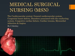
  • 1. MEDICAL SURGICAL NURSING (MSN) The Cardiovascular system: General cardiovascular system, Congenital heart defects, Disorders associated with the conducting system, Congestive cardiac failure, Cardiac trauma, Myocardial infarction & Angina By C.Settley
  • 2. Orientation to MSN (Please see subject guide)  Lecturer Ms C Settley Room 1  023 347 0732/ 0844892450  MSN Group  settleyc@cput.ac.za /c.settley@gmail.com  Work integrated learning  Students must complete 80% of clinical hours as calculated per year level will be allowed to enter for all the assessments  Teaching methodology/strategies  Emphasis is placed on self-directed learning! Compiled by C.Settley 2018
  • 3. Orientation to MSN (Please see subject guide)  Blackboard  SDL  Class attendance  Compulsory prescribed books  Recommended books  Subject credits  Purpose of this subject Compiled by C.Settley 2018
  • 4. Orientation to MSN (Please see subject guide)  Chapters with outcomes in subject guide  Activities  Assessment policy  Guidelines for assignments  Referencing, etc.  Evaluation sheets Compiled by C.Settley 2018
  • 5. Orientation to MSN (Please see subject guide) Compiled by C.Settley 2018 ASSESSMENT WEIGHTING DATE Summative 1 25% 16 February 2018 Deferred 1 03 April 2018 Summative 2 25% 23 March 2018 Deferred 2 16 April 2018 Summative 3 25% 09 April 2018 Deferred 3 ASAP Summative 4 25% 18 May 2018 Deferred 4 04 June 2018
  • 6. Outcomes Apply knowledge regarding:  Haematological studies  Patho-physiology  disease process  clinical manifestations  specific diagnostic and therapeutic interventions (tests and examinations) Distinguish between the different health problems: medical and surgical conditions of various body systems  Congenital defects  Conducting system  Congestive cardiac failure  Coronary artery disease  Angina  Myocardial infarction  Blood vessels disorders  Peripheral arterial disease  Deep vein thrombosis  Cardiovascular disease in the elderly Compiled by C.Settley 2018
  • 7.  What is the cardio vascular system?  What is it comprised of?  What is its purpose? What does it circulate?  How is the heart shaped?  Where is it located?  What are the 3 layers that the heart is made up, called?  How many valves are in the heart? Compiled by C.Settley 2018
  • 8. Anatomy of the heart Compiled by C.Settley 2018
  • 9. Location of the heart The three layers of the heart wall Compiled by C.Settley 2018
  • 10. Compiled by C.Settley 2018Valves of the heart
  • 11. Valves of the heart  TRICUSPID VALVE: Closes off the upper right chamber (or atrium) that holds blood coming in from the body.  Opens to allow blood to flow from the top right chamber to the lower right chamber (or from right atrium to right ventricle).  Prevents the back flow of blood from the ventricle to the atrium when blood is pumped out of the ventricle. Compiled by C.Settley 2018
  • 12. Valves of the heart  PULMONARY VALVE: Closes off the lower right chamber (or right ventricle).  Opens to allow blood to be pumped from the heart to the lungs (through the pulmonary artery) where it will receive oxygen. Compiled by C.Settley 2018
  • 13. Valves of the heart  MITRAL VALVE: Closes off the upper left chamber (or left atrium) collecting the oxygen-rich blood coming in from the lungs.  Opens to allow blood to pass from the upper left side to the lower left side (or from the left atrium to the left ventricle). Compiled by C.Settley 2018
  • 14. Valves of the heart  AORTIC VALVE: Closes off the lower left chamber that holds the oxygen-rich blood before it is pumped out to the body.  Opens to allow blood to leave the heart (from the left ventricle to the aorta and on to the body). Compiled by C.Settley 2018
  • 15. Blood flow through the heart Compiled by C.Settley 2018
  • 16. Blood flow through the heart Compiled by C.Settley 2018
  • 17. Blood flow through the heart  Watch video:  Normal blood flow through the heart Compiled by C.Settley 2018
  • 18. Diagnostic tests- pg. 621  Electrocardiography (ECG)  A graphic representation of the electrical activity of the heart muscle as it contracts and relaxes.  A standard 12-lead electrocardiogram is used to access the electrical activity of the heart and the conduction of the cardiac impulse.  Most cardiac conditions give rise to abnormal recordings on the ECG.  Video:  ECG Compiled by C.Settley 2018
  • 20. How to read an ECG  HEART RATE  Heart rate can be calculated using the following method (if regular):  Count the number of large squares present within one R-R interval  Divide 300 by this number to calculate the heart rate  e.g. 4 large squares in an R-R interval: 300/4 = 75 beats per minute Compiled by C.Settley 2018
  • 21. How to read an ECG HEART RHYTHM The heart rhythm can be regular or irregular. Irregular rhythms can be either: Regularly irregular (i.e. a recurrent pattern of irregularity) Irregularly irregular (i.e. completely disorganised) Compiled by C.Settley 2018
  • 22. How to read an ECG Compiled by C.Settley 2018
  • 23. How to read an ECG  Pre atrial contractions (pg636):  Premature atrial contractions (PACs), also known as atrial premature complexes (APC) or atrial premature beats (APB), are a common cardiac dysrhythmia characterized by premature heartbeats originating in the atria.  While the sinoatrial node typically regulates the heartbeat during normal sinus rhythm, PACs occur when another region of the atria depolarizes before the sinoatrial node and thus triggers a premature heartbeat. Compiled by C.Settley 2018
  • 24. How to read an ECG Cardioversion is a medical procedure by which an abnormally fast heart rate or other cardiac arrhythmia is converted to a normal rhythm using electricity or drugs Compiled by C.Settley 2018
  • 25. How to read an ECG  Cardio pulmonary resuscitation and adrenalin  Flatline: Compiled by C.Settley 2018
  • 26. Atrial flutter- pg636  Irregular and fast heartbeat  Cardio version may be required  May be due to fluid overload, etc Compiled by C.Settley 2018
  • 27. Diagnostic tests- pg. 621  Exercise ECG (Stress test)  An exercise electrocardiogram (EKG or ECG) is a test that checks for changes in your heart while you exercise.  Sometimes ECG abnormalities can be seen only during exercise or while symptoms are present. This test is sometimes called a "stress test“.  See box 32.2 INFORMATION REQUIRED BEFORE A STRESS TEST IS DONE Compiled by C.Settley 2018
  • 28. Exercise ECG (Stress test)Compiled by C.Settley 2018
  • 29. Diagnostic tests- pg. 621  Chest radiography (CXR)  An X-ray is an imaging test that uses small amounts of radiation to produce pictures of the organs, tissues, and bones of the body.  When focused on the chest, it can help spot abnormalities or diseases of the airways, blood vessels, bones, heart, and lungs.  Chest X-rays can also determine if patients have fluid or air in their lungs.  Heart size, etc. can be examined. Compiled by C.Settley 2018
  • 30. Chest radiography Compiled by C.Settley 2018
  • 31. Diagnostic tests- pg. 621  Echocardiography An echocardiogram, often referred to as a cardiac echo or simply an echo. It is a sonogram of the heart. Echocardiography uses standard two- dimensional, three-dimensional, and Doppler ultrasound to create images of the heart. Compiled by C.Settley 2018
  • 33. Diagnostic tests- pg. 622  Cardiac catherisation Cardiac catheterisation is the insertion of a catheter into a chamber or vessel of the heart. This is done both for diagnostic and interventional purposes. Compiled by C.Settley 2018
  • 34. Cardiac catherisation Compiled by C.Settley 2018
  • 35. Cardiac catherisation- pg 622  VIDEO*:  Cardiac catherisation  Nursing responsibilities  Consent  NPO  Cleansing and shaving of patient  ECG as baseline to be taken  Record any allergy history as the patient may react to x-ray dyes.  Bedpan/urinal (empty bladder).  Make patient comfortable as far as possible.  Remove dentures, hearing aids, glasses, etc.  Post procedure: monitor patient for dysrhythmias and bleeding from catherisation site. - Bedrest. Compiled by C.Settley 2018
  • 36. Diagnostic tests- pg. 622  Angiography  Angiography or arteriography is a medical imaging technique used to visualize the inside, or lumen, of blood vessels and organs of the body, with particular interest in the arteries, veins and the heart chambers.  VIDEO *  Coronary angiogram Compiled by C.Settley 2018
  • 37. Diagnostic tests- pg. 622  Doppler studies A Doppler ultrasound is a test that uses high-frequency sound waves to measure the amount of blood flow through your arteries and veins, usually those that supply blood to your arms and legs. Vascular flow studies, also known as blood flow studies, can detect abnormal flow within an artery or blood vessel. Compiled by C.Settley 2018
  • 38. Diagnostic tests- pg. 623  Pulse oximetry  Pulse oximetry is a non-invasive method for monitoring a person's oxygen saturation (SO2). Compiled by C.Settley 2018
  • 39. Haematological tests in cardiovascular conditions- pg. 623 TYPE OF TEST RATIONALE SERUM ELECTROLYTES Potassium, sodium, calcium and magnesium affect cardiac function. Hypernatremia may be indicative of dehydration while potassium deficit may result in dysrhythmias. BLOOD GASES Arterial gasses indicate the levels of oxygen and carbon dioxide in the blood, and are indicators of cardiac functioning. SERUM ENZYMES Presence of enzymes are indicative of damage to muscle tissue and indirect indicator of damage to myocardium. The following enzymes’ presence are however, direct indicators of damage to the myocardium: CPK, LDH, SGOT, MB(CKMB), Trop T. SERUM LIPIDS Presence of serum lipids indicates the presence of atherosclerosis. Compiled by C.Settley 2018
  • 40.  Haematological tests in cardiovascular conditions- pg. 623  REFER TO HANDOUT  Haematological tests can help diagnose anaemia, infection, haemophilia, blood-clotting disorders, and leukaemia. Compiled by C.Settley 2018
  • 41. Classification of the cardiac disorders- pg. 629 Causative factor Conditions Congenital defects Septal defects Defects affecting the valves Defects affecting major arteries Mixed defects Disturbances of conduction Atrial dysrhythmias Atrioventricular/junctional dysrhythmias Ventricular dysrhythmias Infection and inflammatory disorders Rheumatic fever Infective endocarditis Valvular heart disease Myocarditis Pericarditis Structural disorders Mitral valve disease Aortic valve disease Tricuspid and pulmonic valve disease cardiomyopathy Congestive conditions Trauma Heart failure Cardiac contusion Cardiac tamponade Pericardial rupture Rupture of the heart wall, papillary muscles, heart valves Compiled by C.Settley 2018
  • 42. Risk factors for cardiac disorders- pg. 629  Lifestyle  Unhealthy diet, high blood cholesterol and other fats  Obesity & overweight  Tabacco use  Hypertension  Diabetes Mellitus  Stress Compiled by C.Settley 2018
  • 43. Congenital heart defects- pg. 630  Congenital heart disease (CHD) is a problem with the heart's structure and function that is present at birth.  Congenital heart defects are the most common type of birth defect.  The defects can involve the walls of the heart, the valves of the heart, and the arteries and veins near the heart.  They can disrupt the normal flow of blood through the heart.  The blood flow can slow down, go in the wrong direction or to the wrong place, or be blocked completely. Compiled by C.Settley 2018
  • 44. Congenital heart defects Aetiology and risk factors- pg. 630 Viral infections during pregnancy. Especially the 1st trimester, eg Rubella. Nutritional deficiencies during pregnancy. Exposure to environmental factors such as toxins and radiation. Excessive alcohol consumption during pregnancy. Drugs or medication abuse. Family history. Compiled by C.Settley 2018
  • 45. Congenital heart defects Pathophysiology- pg 630 Defects form during early pregnancy between the 6th and 12th week. Heart of fetus develops from a single tube to a heart with 4 chambers, veins and arteries Compiled by C.Settley 2018
  • 46. Congenital heart defects Pathophysiology- pg. 630  In congenital defects, part of the heart may thus not develop (partially or not at all).  Defects may include: a hole in the septum or the formation of abnormal connections of the arteries and veins of the heart.  This results in the mixing of deoxygenated and oxygenated blood.  Congenital defects are structural in nature and cause problems because of their effect on blood flow through the heart.  Sometimes the conducting system is involved.  Defects range from simple to severe.  VIDEO* (atrial defect blood flow) Compiled by C.Settley 2018
  • 47. Septal defects Compiled by C.Settley 2018 Septal defects, Heart valve defects & major blood vessel defects- pg. 631  Hole in the septum that divides the R & L side of the heart.  Small septal defects may close with time or not, causing defective flow.  Larger holes can cause more problems with functioning of the heart.  Major presentation: palpitations and irregular pulse, poor colour of blood.
  • 48. Heart valve defects Compiled by C.Settley 2018 Septal defects, Heart valve defects & major blood vessel defects- pg 631  Heart valves allow blood to flow in one direction.  They open and close passively.  Due to valve defects, these valves may not open or close properly (valve incompetence).  Blood may flow backwards (regurgitate).  The heart now works harder and it cannot get sufficient blood to the lungs and body.
  • 49. Septal defects, Heart valve defects & major blood vessel defects- pg. 631  Major blood vessel defects  Coarctation of the aorta  Constriction of a segment of the aorta resulting in obstruction of blood flow from the left ventricle. Compiled by C.Settley 2018
  • 50. Septal defects, Heart valve defects & major blood vessel defects- pg. 631  Major blood vessel defects  Transposition of the great arteries  When the pulmonary artery arises from the left ventricle and the aorta from the right ventricle sends deoxygenated blood from the right atrium back to the systemic circulation. Compiled by C.Settley 2018
  • 51. Septal defects, Heart valve defects & major blood vessel defects- pg. 631  Major blood vessel defects  Patent ductus arteriosus  Before birth, the aorta and the pulmonary artery are connected by a blood vessel called the ductus arteriosus which is an essential part of the fetal circulation.  Soon after birth, the vessel is supposed to close as part of the normal changes occurring in the baby’s circulation.  In some babies, it does not close, and the opening allows oxygenated blood from the aorta to mix with deoxygenated blood from the pulmonary artery.  See other defects- pg. 632, Table 33.2 + figures Compiled by C.Settley 2018
  • 52. Clinical manifestations of congenital defects- pg. 631  Family history  Child develops slow  Tachypnea/cyanosed/dyspnea  History of frequent chest infections  The child, may squat when distressed- it increases blood flow to the heart  Clubbing of fingers and toes  Enlarged liver or spleen  Oedema of extremities  ECG abnormalities  Cardiac enlargement may be seen on x-ray and abnormal heart sound may be heard  Echocardiography and cardiac catherisation may be done to confirm septal and valvular defects. Compiled by C.Settley 2018
  • 53. Management- pg. 631 Surgical correction of the defect which may involve the use of catheters or open cardiac surgery. The sooner the defect is corrected, the better in terms of the child’s physical and mental health. If the defect cannot be corrected, then conservative management directed at preventing cardiac failure and optimising pulmonary blood flow should be done. Compiled by C.Settley 2018
  • 54. What do you understand under Decreased cardiac output?  Inadequate blood pumped by the heart to meet the metabolic demands of the body.  Cardiac output is the amount of blood pumped by the heart per minute.  It is the product of the heart rate, which is the number of beats per minute, and the stroke volume, which is amount pumped per beat. Compiled by C.Settley 2018
  • 55. How would you assess a patient to evaluate whether the patient has decreased cardiac output?  Abnormal heart sounds  Angina  Anxiety & restlessness  Change in level of consciousness  Breathing pattern  Decreased activity tolerance/fatigue  Decreased peripheral pulses; cold, clammy skin/poor capillary refill  Decreased venous and arterial oxygen saturation  Dysrhythmias  Ejection fraction less than 40% (Ejection fraction is a measurement of the percentage of blood leaving your heart each time it contracts. During each heartbeat pumping cycle, the heart contracts and relaxes. When your heart contracts, it ejects blood from the two pumping chambers (ventricles). When your heart relaxes, the ventricles refill with blood.)  Hypotension  Weight gain, oedema, decreased urine output Compiled by C.Settley 2018
  • 56. Interventions for decreased cardiac output? Interventions Rationales Record intake and output. If patient is acutely ill, measure hourly urine output and note decreases in output. Reduced cardiac output results in reduced perfusion of the kidneys, with a resulting decrease in urine output. For patients with increased preload, limit fluids and sodium as ordered. Fluid restriction decreases extracellular fluid volume and reduces demands on the heart. Closely monitor fluid intake including IV lines. Maintain fluid restriction if ordered. In patients with decreased cardiac output, poorly functioning ventricles may not tolerate increased fluid volumes. Auscultate heart sounds The new onset of a gallop rhythm, tachycardia, and fine crackles in lung bases can indicate onset of heart failure. If patient develops pulmonary edema, there will be coarse crackles on inspiration and severe dyspnea. Closely monitor for symptoms of heart failure and decreased cardiac output, including diminished quality of peripheral pulses, cold and clammy skin and extremities, increased respiratory rate, presence of paroxysmal nocturnal dyspnea or orthopnea, increased heart rate, neck vein distention, decreased level of consciousness, and presence of edema. As these symptoms of heart failure progress, cardiac output declines. Compiled by C.Settley 2018
  • 57. How would you assess a patient to evaluate whether the patient has decreased cardiac output? Note chest pain. Identify location, radiation, severity, quality, duration, associated manifestations such as nausea, and precipitating and relieving factors. Chest pain/discomfort is generally suggestive of an inadequate blood supply to the heart, which can compromise cardiac output. Patients with heart failure can continue to have chest pain with angina or can reinfarct. If chest pain is present, have patient lie down, monitor cardiac rhythm, give oxygen, run a strip, medicate for pain, and notify the physician. These actions can increase oxygen deliveryto the coronary arteries and improve patient prognosis. Place on cardiac monitor; monitor for dysrhythmias, especially atrial fibrillation. Atrial fibrillation is common in heart failure. Examine laboratory data, especially arterial blood gases and electrolytes, including potassium. Patient may be receiving cardiac glycosides and the potential for toxicity is greater with hypokalemia; hypokalemia is common in heart patients because of diuretic use. Monitor laboratory tests such as complete blood count, sodium level, and serum creatinine. Routine blood work can provide insight into the etiology of heart failure and extent of decompensation. A low serum sodium level often is observed with advanced heart failure and can be a poor prognostic sign. Serum creatinine levels will elevate in patients with severe heart failure because of decreased perfusion to the kidneys. Creatinine may also elevate because of ACE inhibitors. Compiled by C.Settley 2018
  • 58. How would you assess a patient to evaluate whether the patient has decreased cardiac output? Administer medications as prescribed, noting side effects and toxicity. Depending on etiological factors, common medications include digitalis therapy, diuretics, vasodilator therapy, antidysrhythmics, angiotensin-converting enzyme inhibitors, and inotropic agents. Review results of EKG and chest Xray. EKG can reveal previous MI, or evidence of left ventricular hypertrophy, indicating aortic stenosis or chronic systemic hypertension. Xray may provide information on pulmonary edema, pleural effusions, or enlarged cardiac silhouette found in dilated cardiomyopathy or large pericardial effusion. Compiled by C.Settley 2018
  • 59. Describe interventions for activity intolerance with possible rationales. Assessment Rationales Assess the physical activity level and mobility of the patient.  Take the resting pulse, blood pressure, and respirations.  Consider the rate, rhythm, and quality of the pulse.  If the signs are normal, have the patient perform the activity.  Obtain the vital signs immediately after activity  Have the patient rest for 3 minutes and then take the vital signs again. Provides baseline information for formulating nursing goals during goal setting. Discontinue the activity if the patient responds with:  chest pain, vertigo, and/or dizziness  decreased pulse rate, systemic blood pressure, respiratory response Reduce the duration and intensity of the activity if:  Pulse takes longer than 3 to 4 minutes to return to within 6-7 beats of the resting pulse.  RR increase is excessive after the activity. Investigate the patient’s perception of causes of activity intolerance. Causative factors may be temporary or permanent as well as physical or psychological. Determining the cause can help guide the nurse during the nursing intervention. Assess the patient’s nutritional status. Adequate energy reserves are needed during activity. Observe and monitor the patient’s sleep pattern and the amount of sleep achieved over the past few days. Sleep deprivation and difficulties during sleep can affect the activity level of the patient – these needs to be addressed before successful activity progression can be achieved. Compiled by C.Settley 2018
  • 60. Describe interventions for activity intolerance with possible rationales. Determine the patient’s daily routine and over-the- counter medication. Fatigue can limit the patient’s ability to perform needed activity. It can also be a medication side effect. Pay attention to the patient’s use of beta- blockers, calciumchannel blockers, tranquilizers, antihistamines, relaxants, alcohol, and sedatives. Assess the need for ambulation aids (e.g., cane, walker) for ADLs. Assistive devices enhance the mobility of the patient by helping him overcome limitations. Use portable pulse oximetry to assess for oxygen desaturation during activity. May determine the use of supplemental oxygen to help compensate for the increased oxygen demands during physical activity. Assess the patient’s baseline cardiopulmonary status (e.g., heart rate, orthostatic BP) before initiating activity. In normal adults, HR should not increase more than 20 to 30 beats/min above resting with routine activities. Older patients are more susceptible to orthostatic drops in BP with position changes. Observe and document response to activity. Close monitoring will serve as a guide for optimal progression of activity. Assess emotional response to limitations in physical activity. Depression over the inability to perform activities can be a source of stress and frustration. Compiled by C.Settley 2018
  • 61. Describe interventions for activity intolerance with possible rationales. Establish guidelines and goals of activity with the patient and/or SO. Motivation and cooperation are enhanced if the patient participates in goal setting. Evaluate the need for additional help at home. Coordinated efforts are more meaningful and effective in assisting the patient in conserving energy. Have the patient perform the activity more slowly, in a longer time with more rest or pauses, or with assistance if necessary. Helps in increasing the tolerance for the activity. Gradually increase activity with active range-of-motion exercises in bed, increasing to sitting and then standing. Gradual progression of the activity prevents overexertion. Dangle the legs from the bed side for 10 to 15 minutes. Prevents orthostatic hypotension. Refrain from performing nonessential activities or procedures. Patient with limited activity tolerance need to prioritize important taks first. Assist with ADLs while avoiding patient dependency. Assisting the patient with ADLs allows conservation of energy. Carefully balance provision of assistance; facilitating progressive endurance will ultimately enhance the patient’s activity tolerance and self-esteem. Compiled by C.Settley 2018
  • 62. Describe interventions for activity intolerance with possible rationales. Provide bedside commode as indicated. Use of commode requires less energy expenditure than using a bedpan or ambulating to the bathroom. Encourage physical activity consistent with the patient’s energy levels. Helps promote a sense of autonomy while being realistic about capabilities. Instruct patient to plan activities for times when they have the most energy. Activities should be planned ahead to coincide with the patient’s peak energy level. If the goal is too low, negotiate. Encourage verbalization of feelings regarding limitations. This helps the patient to cope. Acknowledgment that living with activity intolerance is both physically and emotionally difficult. Gradually progress patient activity with the following:  Range-of-motion (ROM) exercises in bed, gradually increasing duration and frequency (then intensity) to sitting and then standing.  Deep-breathing exercises three or more times daily.  Sitting up in a chair 30 minutes three times daily.  Walking in room 1 to 2 minutes TID.  Walking down the hall 20 feet or walking through the house, then slowly progressing walking outside the house, saving energy for the return trip. Duration and frequency should be increased before intensity. Compiled by C.Settley 2018
  • 63. Describe interventions for compromised family coping with possible rationales. Nursing Interventions Rationale Observe for erratic behaviors (anger, tension, disorganization), perception of crisis situation. Information affecting the ability of the family to cope with infant/child’s cardiac condition. Encourage expression of feelings and provide factual information about infant/child. Reduces anxiety and enhances family’s understanding of the condition. Assess usual family coping methods and effectiveness. Identifies need to develop new coping skills if existing methods are ineffective in changing behaviors exhibited. Assess need for information and support. Provides information about need for interventions to relieve anxiety and concern. Clarify any misinformation and answer questions regarding disease process. Prevents unnecessary anxiety resulting from inaccurate knowledge or beliefs. Assist in identifying and using techniques to cope with and solve problems and gain control over the situation. Provides support for problem solving and management of the situation. Encourage to maintain the health of family members and social contacts. Chronic anxiety, fatigue, and isolation as result of infant care will affect health and care capabilities of family. Compiled by C.Settley 2018
  • 64. Describe interventions for compromised family coping with possible rationales. Teach that overprotective behavior may hinder growth and development during infancy/ childhood. Knowledge will enhance family understanding of the condition and of adverse effects of behaviors. Suggest and reinforce appropriate coping behaviors, support family decisions. Promotes behavior change and adaptation to care for infant/child. Encourage parents to include ill infant/ child in family activities rather than family revolving around needs of infant/child. Promotes normal growth and development of family and infant/child. Encourage to maintain consistent behavior limits and modification techniques. Prevents behavioral problems and child control over family, which interfere with child’s growth and family relationships. Instruct parents in nutritional and activity needs and/or limitations and approaches that will assist in establishing an effective pattern. Assists in coping with effects and special needs of infant/child with a cardiac defect. Refer family for additional support and counseling if indicated. Referral supplies more assistance with coping than is available from nursing personnel. Compiled by C.Settley 2018
  • 65. Disorders associated with the conducting system- pg. 633 What is the conducting system?  The cardiac conduction system is a group of specialized cardiac muscle cells in the walls of the heart that send signals to the heart muscle causing it to contract.  The main components of the cardiac conduction system are the SA node, AV node, bundle of His, bundle branches, and Purkinje fibers. Compiled by C.Settley 2018
  • 66. Disorders associated with the conducting system- pg. 633 - The SA node – sends out regular electrical impulses from the top chamber (the atrium) causing it to contract and pump blood into the bottom chamber (the ventricle). - Also called the pacemaker, because it initiates each heartbeat. - The electrical impulse then travels to the AV node. This causes the atria to contract. - From the AV node, the impulse passes down the ventricular septum to the left and right bundle branches and the fibres of Purkinje to stimulate the ventricles to contract. - As a result of ventricular contraction, the blood is ejected into the aorta and pulmonary circulation respectively. - The normal rhythm of the heart is known as sinus rhythm as it is initiated by the sinoatrial node. Compiled by C.Settley 2018
  • 67. Disorders associated with the conducting system- pg. 633 The characteristics of normal sinus rhythm are:  The rhythm is regular.  Rate is between 60-100 bpm.  Each beat consists of a P - wave, QRS complex and a T – wave.  Every P - wave is followed by a QRS complex  The complexes are tall and narrow. Compiled by C.Settley 2018
  • 68. Disorders associated with the conducting system- pg. 636 - Cardiac conducting system Video* CLASSIFICATION OF DYSRYTHMIAS ACCORDING TO: - The rate of the dysrhythmia, whether fast or slow - Whether life threatening or not - The area of origin Compiled by C.Settley 2018
  • 69. Atrial dysthymias- pg. 636  PAC  Originates in the atria  May occur in healthy individuals and are frequently due to emotion, exertion or stimulants  Mostly due to sympathetic over activity, hypoxia, stress, smoking or anxiety  Can also be associated with valvular heart disease, atrial chamber enlargement and coronary artery disease  May mark the onset of arterial fibrillation or heart failure  P-waves are premature and differs from normal sinus P waves  Described as palpitations  Underlying cause should be investigated Compiled by C.Settley 2018
  • 70. Atrial dysthymias- pg. 636  Atrial flutter  Irregular and fast  A saw tooth pattern as baseline  Common in rheumatic heart disease (Rheumatic fever is an inflammatory disease that sometimes happens after an infection caused by a bacteria called group A streptococcus, like strep throat or scarlet fever. Rheumatic fever happens when the infection is not completely treated with medicine (antibiotics). It may affect the heart, joints, skin and brain).  Fluid overload  Cardioversion Compiled by C.Settley 2018
  • 71. Atrial dysthymias- pg. 636  Atrial Fibrillation  Atrial fibrillation is an irregular and often rapid heart rate that can increase your risk of stroke, heart failure and other heart- related complications.  During atrial fibrillation, the heart's two upper chambers (the atria) beat chaotically and irregularly — out of coordination with the two lower chambers (the ventricles) of the heart.  Atrial fibrillation symptoms often include heart palpitations, shortness of breath and weakness. Compiled by C.Settley 2018
  • 72. Atrial dysthymias- pg. 636  Atrial Fibrillation  Episodes of atrial fibrillation can come and go, or you may develop atrial fibrillation that doesn't go away and may require treatment. Although atrial fibrillation itself usually isn't life- threatening, it is a serious medical condition that sometimes requires emergency treatment.  It may lead to complications. Atrial fibrillation can lead to blood clots forming in the heart that may circulate to other organs and lead to blocked blood flow (ischemia).  Treatments for atrial fibrillation may include medications and other interventions to try to alter the heart's electrical system. Compiled by C.Settley 2018
  • 73. Atrial dysthymias- pg. 636 Atrial Fibrillation  Some people with atrial fibrillation have no symptoms and are unaware of their condition until it's discovered during a physical examination. Those who do have atrial fibrillation symptoms may experience signs and symptoms such as:  Palpitations, which are sensations of a racing, uncomfortable, irregular heartbeat or a flip-flopping in your chest  Weakness  Reduced ability to exercise  Fatigue  Light-headedness  Dizziness  Confusion  Shortness of breath  Chest pain Compiled by C.Settley 2018
  • 74. Ventricular dysthymias - pg. 638 Ventricular tachycardia  A condition in which the lower chambers of the heart (ventricles) beat very quickly.  Heart rate above 100 beats per minute  May be up to 180 beats per minute  Treatment depends on haemodynamic status of the patient:  A raised BP- Cardioversion will be required, anti arrhythmic agents (amiodarone) and correction of electrolyte and acid-base balance must be implemented.  If patient collapses then standard CPR must be commenced Compiled by C.Settley 2018
  • 75. Ventricular dysthymias - pg. 638  Ventricular fibrillation  Complete disorganisation of the cardiac rhythm  Irregular heart waves on ECG  Varies in size and shape  No contraction therefore no cardiac output  Patient would appear clinically dead as there would be no pulse  Treatment: defibrillation and CPR Compiled by C.Settley 2018
  • 76. Ventricular dysthymias - pg. 638  Ventricular asystole  Ventricular asystole is indicative of cardiac arrest. It requires immediate attempts at resuscitation, with a poor prognosis at that. It is characterized by the absence of electrical activity for a length of time with intermittent ventricular complexes of abnormal configuration. Compiled by C.Settley 2018
  • 77. Disorders associated with the conducting system- pg. 639 Nb:  In emergencies due to arrhythmias, rapid and correct diagnosis is necessary for adequate therapy.  The clinical symptoms, the physical examination, and the 12-lead electrocardiogram are important sources of information for making a correct diagnosis  It is essential to get information about the underlying heart disease, understand the mechanism of the present arrhythmia  Amiodarone is currently regarded as the most effective antiarrhythmic drug available for the treatment of patients with both supraventricular and ventricular tachy arrhythmias.  Beta blockers. Beta blockers work by blocking the effect of hormones such as adrenaline on the heart. They are commonly prescribed to people with heart failure or angina, or following a heart attack. They are used to treat high blood pressure alongside another drug.  The use of atropine in cardiovascular disorders is mainly in the management of patients with bradycardia. Atropine increases the heart rate and improves the atrioventricular conduction by blocking the parasympathetic influences on the heart. Compiled by C.Settley 2018
  • 78. Disorders associated with the conducting system- pg. 635 Management of restoring a normal rhythm  The application of electrical ‘shock’ therapy, defibrillation or cardioversion.  The implantation of a temporary pacemaker (A pacemaker is a small device that's placed in the chest or abdomen to help control abnormal heart rhythms. This device uses electrical pulses to prompt the heart to beat at a normal rate).  Pacemakers can be temporary or permanent. Temporary pacemakers are used to treat temporary heartbeat problems, such as a slow heartbeat that's caused by a heart attack, heart surgery, or an overdose of medicine.  They're used until a permanent pacemaker can be implanted or until the temporary condition goes away.  The use of medications.  Cardiac ablation (Catheter ablation is a minimally-invasive procedure used to remove or terminate a faulty electrical pathway from sections of the hearts of those who are prone to developing cardiac arrhythmias)  An implantable cardiac defibrillator.  Surgery. Compiled by C.Settley 2018
  • 79. Congestive heart failure- pg. 643 • When the heart’s function as a pump is inadequate to deliver oxygen rich blood to the body. • Can be caused by diseases which weaken the heart muscle • Increased oxygen demand by the body beyond the capability of the heart to deliver adequate oxygen rich blood to the tissues. • Aetiology - Coronary artery disease - Valvular disorders - Hypertension - Infections - Arrhythmias Compiled by C.Settley 2018
  • 80. Congestive heart failure Pathophysiology- pg. 643  The normal heart is able to meet the body’s need for oxygen by increasing its output in response to increased demand for oxygen. BUT, in heart failure:  The heart’s capacity to increase the force of contraction has been exceeded and the heart cannot respond to the body’s demands.  The heart does not function adequately due to a problem within the heart itself, such as ischaemic heart diseases, cardiomyopathy and constrictive pericarditis.  If ↓ contractility of the heart muscle due to cardiac failure, the cardiac output is compromised leading to ↓ stroke volume and systemic arterial blood pressure. Compiled by C.Settley 2018
  • 81. Classification of congestive cardiac heart failure (CCF)- pg. 644 1. Systolic heart failure/ Diastolic heart failure Compiled by C.Settley 2018
  • 82. Classification of congestive cardiac heart failure (CCF)- pg. 644 2. Right-sided heart failure/ Left sided heart failure The failure of the pumping action of the right side of the heart causes swelling in the legs and abdomen while that of the left side, causes congestion of the lungs. Compiled by C.Settley 2018
  • 83. Classification of congestive cardiac heart failure (CCF)- pg. 644 3. Forward heart failure/ Backward heart failure - Inability of the heart to pump sufficient blood to meet the oxygen needs of the body during an exercise or rest leads to forward heart failure; - Whilst the inability of the heart to meet the oxygen needs when the heart pressures are very high is backward heart failure. Compiled by C.Settley 2018
  • 84. Clinical manifestations of congestive cardiac heart failure (CCF)- pg. 644  Respiratory symptoms  Difficulty breathing, dyspnoea (Dyspnea: Difficult or labored breathing; shortness of breath), orthopnoea (Orthopnea or orthopnoea is shortness of breath (dyspnea) that occurs when lying flat, causing the person to have to sleep propped up in bed or sitting in a chair).  Disturbances in sleep  Pleural effusion  Gastrointestinal symptoms  Enlarged liver & spleen  Increased venous pressure  Abdominal pain, digestive problems, anorexia, nausea and vomiting & ascites (Ascites refers to abnormal accumulation fluid in the abdominal cavity). Compiled by C.Settley 2018
  • 85. Clinical manifestations of congestive cardiac heart failure (CCF)- pg. 644  Oedema  Due to congestion and high pressure in the capillaries  Prevents fluid from moving back into the blood vessels thus collects in the tissues  Retention of sodium and water  Renal symptoms  Renal impairment  Poor perfusion  Inadequate blood supply Compiled by C.Settley 2018
  • 86. Clinical manifestations of congestive cardiac heart failure (CCF)- pg. 644  Neurological symptoms  Cerebral hypoxia due to respiratory insufficiency  Confusion  Mental clouding  Other symptoms  Weakness and fatigue  Cold extremities  Distended neck veins due to congestion of circulation  Physical examination Compiled by C.Settley 2018
  • 87. Diagnostic test results: CCF- PG. 646  Chest x-ray  Will show cardiac enlargement  Congestion of the lungs  Possible pleural effusion  ECG  Large complexes typical of cardiac strain and cardiac enlargement  Blood gases  Hypoxia  Respiratory and metabolic acidosis  Blood chemistry  ↑ sodium levels  Levels of urea  These levels may be elevated due to renal insufficiency  Liver enzymes may be raised (impaired liver function) Compiled by C.Settley 2018
  • 88. General nursing care plan for a patient with a disorder of the cardiovascular system- pg.624 Compiled by C.Settley 2018 PROBLEM/NEED NURSING DIAGNOSES EXPECTED OUTCOMES NURSING INTERVENTIONS & RATIONALE EVALUATION Dyspnea, fatigue & weakness -Altered breathing pattern related to pulmonary congestion and insufficient supply to the lungs Inadequate circulation, poor oxygenation and perfusion due to reduces cardiac output and impaired cardiac function. Relief from respiratory distress Rest and comfort Adequate circulation and tissue perfusion O2, monitor respiration, tissue perfusion to improve cardiac function and circulation, enhances metabolism to provide energy and strength. Semi fowlers position Support with pillows, armchair to improve ventilation. Monitor vital signs Encourage deep breathing and coughing of secretions. Remove secretions Administer medication as prescribed. Respiratory rate, blood pressure within normal ranges. Perfusion and colour of patient.
  • 89. General nursing care plan for a patient with a disorder of the cardiovascular system- pg.624 PROBLEM/NEED NURSING DIAGNOSES EXPECTED OUTCOMES NURSING INTERVENTIONS & RATIONALE EVALUATION Chest pain and discomfort Possible abdominal distention Congestion Organ dysfunction due to congestion and poor circulation to gastro intestinal organs manifested by the expression of pain, dyspnea & tachycardia Rest and comfort Pain relief Reduced fluid retention Fluid restrictions Sodium restrictions Note abdominal distention increase Monitor intake and output Semi fowlers position Test urine daily for proteins and blood + weight Abdominal girth is reduced Oedema is reduced Clear urine Weight Oedema Fluid volume deficit due to renal impairment and sodium retention Optimum fluid and electrolyte balance Reduced oedema comfort Administer diuretics as prescribed Intake and output monitor Fluid restrictions, Salt restrictions Oedema test, Elevate legs & Pressure care Output=intake Fluid balance Compiled by C.Settley 2018
  • 90. General nursing care plan for a patient with a disorder of the cardiovascular system- pg.624 Compiled by C.Settley 2018 PROBLEM/NEED NURSING DIAGNOSES EXPECTED OUTCOMES NURSING INTERVENTIONS & RATIONALE EVALUATION Potential for anxiety Knowledge related to condition, treatment and medication of lifestyle Risk of complications related to cardiovascular disorders Health education Compliance enhanced Condition stabilised Complication risk reduced Educate patient: medication, complying with treatment, self management Advise about intake and output, oedema Patient should be able to manage condition as far as possible at home Complications should be detected early and managed/referral
  • 91. Nursing management- pg.624  Bed rest. Balance.  Diet.  Oxygen therapy.  Oedemous area care.  Expression of feelings, concerns, etc.  Health education. Compiled by C.Settley 2018
  • 92. Cardiac trauma- pg.647 TYPE OF TRAUMA SPECIFIC INJURIES Blunt trauma, eg crushing of the chest wall, blow to the anterior chest and deceleration • Pericardial rupture, rupture of the heart wall • Traumatic septal defects • Injuries to the heart valves, papillary muscles • Cardiac contusion Penetrating trauma, eg gunshots, stab wounds • Gunshots, stab wounds, injuries from flying objects, intra-cardiac catheters, pacemaker electrodes, CVP lines • Patients with structural damage • Accompanied by massive haemorrhage Compiled by C.Settley 2018
  • 93. Cardiac surgery- pg. 647  Surgery may be performed to:  Correct defects  Repair or replace damaged heart valves  Restore coronary circulation  Repair cardiac structures  Transplant the heart  2 types: open and closed  Open-heart surgery is any type of surgery where the chest is cut open and surgery is performed on the muscles, valves, or arteries of the heart.  Closed-heart surgery can be carried out without interrupting the flow of blood through the heart and visualising internal structures. Compiled by C.Settley 2018
  • 94. Cardiac surgery: preoperatively- pg.648  Admission usually few days prior to surgery for work-up and preparation.  Cardiac function will be evaluated via studies: ECG, cardiac catherisation, blood studies, echocardiography.  Evaluation of respiratory function via pulmonary tests.  Severe problems like dysrhythmias, congestive heart failure and angina should be controlled before the patient goes to theatre.  Psychological preparation.  Skin preparation to guard against infections. Compiled by C.Settley 2018
  • 95. Cardiac surgery: postoperatively- pg.648  ICU care (for about 48 hours depending on condition).  Ventilated.  Vital signs, cardiac function should be monitored.  Medication as prescribed.  Fluids as prescribed to replace blood loss and electrolytes.  Neurological status (complication).  Chest drains monitored.  Analgesics as prescribed.  Slight pyrexia is normal as response to trauma (as the body handles the operated body tissues). Monitor thoroughly for infection.  Early ambulation once tubes are removed depending on progress.  Encouragement. Compiled by C.Settley 2018
  • 96. Cardiac surgery: complications- pg.648  Dysrhythmias  Haemorrhage.  Renal shutdown.  Cardiogenic shock.  Cardiac tamponade.  Electrolyte imbalance.  Pneumothorax  Emboli.  Stress ulcers. Compiled by C.Settley 2018
  • 97. Coronary artery disease - pg. 650  Coronary artery disease (CAD) is the most common type of heart disease.  CAD happens when the arteries that supply blood to heart muscle become hardened and narrowed.  This is due to the build-up of cholesterol and other material, called plaque, on their inner walls.  This build-up is called atherosclerosis.  As it grows, less blood can flow through the arteries.  As a result, the heart muscle can't get the blood or oxygen it needs. This can lead to chest pain (angina) or a heart attack. Compiled by C.Settley 2018
  • 98. Coronary artery disease - pg. 650  Most heart attacks happen when a blood clot suddenly cuts off the hearts' blood supply, causing permanent heart damage.  Over time, CAD can also weaken the heart muscle and contribute to heart failure and arrhythmias.  Heart failure means the heart can't pump blood well to the rest of the body.  ALSO KNOWN AS ISCHAEMIC CARDIAC DISEASE Compiled by C.Settley 2018
  • 99. Coronary artery disease- pg. 650 Compiled by C.Settley 2018
  • 100. Coronary artery disease- pg. 650 Risk factors Compiled by C.Settley 2018  Direct cause not known. The following are specific risk factors: Increasing age  Increases the chances of developing arteriosclerosis & atherosclerosis Gender  Males are more prone to develop atherosclerosis  Due to protective effect of oestrogen  After menopause, the risk is equal
  • 101. Coronary artery disease- pg. 650 Risk factors Compiled by C.Settley 2018 Heredity  Family history Stress factors  Cardiac output increases  It thus increases myocardial oxygen demand and workload  Mobilisation of fat from fat stores to provide energy in response to stress may contribute to developing the disease
  • 102. Coronary artery disease- pg. 650 Risk factors Compiled by C.Settley 2018 Elevated serum cholesterol  Increased amounts of cholesterol increases the risk of developing the disease Obesity  Associated with higher levels of cholesterol, hypertension & diabetes mellitus  Increased cardiac workload by making the heart work harder to pump blood through the tissues
  • 103. Coronary artery disease- pg. 650 Risk factors Compiled by C.Settley 2018 Existing diseases  HPT & DM increases the risk of developing the disease Diet  High calorie diet  Contribute to high blood lipid levels  Abnormal metabolism  Mobilisation of fats increases lip levels  Hyperlipidaemia  Gout
  • 104. Coronary artery disease- pg. 650 Risk factors Compiled by C.Settley 2018 Smoking  Nicotine, tar and carbon monoxide damages the blood vessel  Vasoconstriction  Obstruction Physical inactivity Relationship between inactivity and development of the disease
  • 105. Coronary artery disease: Pathophysiology- pg. 650  Disruption in blood supply through arteries gets disrupted, or the oxygen content of the blood is not adequate to meet the demands of the body.  In CAD, the arteries are blocked aby Atherosclerotic lesions caused by the deposit of plaques on the arteries.  These plaques protrude into the lumen of the artery, leading to narrowing and obstruction of the blood flow.  The vascular endothelium in the involved areas become necrotic, narrow and rough, making them susceptible to clot formation. Compiled by C.Settley 2018
  • 106. Coronary artery disease: Pathophysiology- pg. 650  Thrombi forms on the surface of the plaque and the effects of fibrin consolidate the thrombus, causing bleeding to the thrombus.  This worsens by further deposits and enlargement takes place.  The myocardial cells become ischaemic within ten seconds of coronary occlusion, and the pumping function of the heart deprives the ischaemic cells of the needed oxygen and glucose, resulting in chest pain. Compiled by C.Settley 2018
  • 107. Coronary artery disease Compiled by C.Settley 2018
  • 108. Coronary artery disease Compiled by C.Settley 2018
  • 109. Coronary artery disease: Subjective data- pg. 652 Chest pain Frequency, duration, location, character Aggravating & relieving factors Associating factors Dyspnoea Exertion Pallor Diaphoresis Dizziness Nausea & vomiting Compiled by C.Settley 2018
  • 110. Coronary artery disease: Subjective data- pg. 652 Psychosocial information  Lifestyle, e.g. Age & gender Lifestyle & habits  Smoking  Alcohol intake  Exercise  Stress  Occupation Compiled by C.Settley 2018
  • 111. Coronary artery disease: Subjective data- pg. 652  Medication history  Prescribed and over the counter medication  Contraceptives, hormone therapy  Past medical history  As contribution  Family medical history  Familial tendency  CVD  DM  HPT  Hyperlipidaemia Compiled by C.Settley 2018
  • 112. Coronary artery disease: Objective data- pg. 652  Physical assessment on examination with the following signs:  Chest pain  Pallor or cyanosis  Distended neck veins  Abnormal heart sounds  Baseline:  Pulse rate and rhythm  Peripheral pulses  Respiration rate, rhythm and depth  Blood pressure  Level of consciousness  Urinary output Compiled by C.Settley 2018
  • 113. Coronary artery disease: Diagnostic test results - pg. 653 ECG Exercise stress test Coronary angiography Echocardiogram Compiled by C.Settley 2018
  • 114. Coronary artery disease: Diagnostic test results - pg. 653  Blood chemistry  Creatine phosphokinase (CPK): raised levels indicate that muscle have been damaged.  Lactic dehydrogenase (LDH): muscle protein  Creatine kinase - MB (CKMB): type of creatine that is specific to myocarduim and raised levels indicate that damage to the myocarduim has taken place  Troponin (Trop T): protein found in muscle. Raised levels indicates that myocardial damage has taken place. Compiled by C.Settley 2018
  • 115. Angina pectoris- pg. 653 A substernal pain Radiates to the left arm, may radiate to other areas such as the jaw, neck, back or epigastrium region Compiled by C.Settley 2018
  • 116. Stable Unstable Compiled by C.Settley 2018 Angina pectoris- pg. 653 Types Related to known triggering factors. Always relieved by rest and/or a known dose of nitrate. Duration and intensity is predictable. The condition never varies much. Unpredictable pattern. Number of attacks, intensity and duration, has a tendency to increase. Strongly correlated with myocardial infarction. Unstable angina is angina at rest or on minimal exertion, and is of recent onset.
  • 117. Noctural Decubitus Compiled by C.Settley 2018 Angina pectoris- pg. 653 Types Attacks occur at night Only during REM sleep Attacks occur when the patient is lying down The pain is relieved when the patient stands
  • 118. Intractable Variant Compiled by C.Settley 2018 Angina pectoris- pg. 653 Types Severe Incapacitating pain Does not respond to treatment Indicative of myocardial infarction Should not be ignored Urgent attention As result of coronary artery spasm Unpredictable During sleep Pain usually relieved by calcium channel blockers
  • 119. Angina: Management- pg. 653 Control of risk factors. Dietary & lifestyle changes. Follow-ups. Nitrates (meds). Educate patient: e.g. inform about triggers such as eating a heavy meal. Compiled by C.Settley 2018
  • 120. Compiled by C.Settley 2018 Myocardial infarction (MI)- pg. 654  Heart attack: usually occurs when a blood clot blocks blood flow to the heart. Without blood, tissue loses oxygen and dies.  Symptoms include tightness or pain in the chest, neck, back or arms, as well as fatigue, light-headedness, abnormal heartbeat and anxiety. Women are more likely to have atypical symptoms than men.
  • 121. Myocardial infarction (MI) Clinical manifestations- pg. 654  Chest pain (retrosternal)  Radiates to left arm- epigastrium or jaw  Varies in intensity  Lasts more than 30 minutes  Associated with dyspnoea, sweating, nausea & vomiting  Shock due to drop in cardiac output- drop in BP  Clammy, cold, dizziness  Tachycardia  Distressed  Breathless  Fear and anxiety Compiled by C.Settley 2018
  • 122. Myocardial infarction (MI) Medical management- pg. 654  Priority is to limit the extent of infarct  Thrombolytic therapy (IV to dissolve the clot and re establish coronary perfusion)  Criteria: chest pain, S-T elevation in two contiguous ECG leads  Complications of thrombolytic therapy: haemorrhage, pyrexia, dysthymias, cardiac rupture Compiled by C.Settley 2018
  • 123. Myocardial infarction (MI) Medical management- pg. 654 To re establish coronary perfusion: (Coronary perfusion pressure (CPP) refers to the pressure gradient that drives coronary blood pressure, meaning the difference between the diastolic aortic pressure and the right atrial end diastolic pressure). 1. Percutaneous transluminal coronary angioplasty (PTCA)  Treat narrowing of the coronary arteries of the heart found in coronary artery disease. 2. Coronary artery bypass graft (CABG)  Is a type of surgery that improves blood flow to the heart. Compiled by C.Settley 2018
  • 124. Myocardial infarction (MI) Nursing management- pg. 654  Diagnosis  Discomfort  Decreased cardiac output  Expected outcomes  Reduced and controlled pain  Optimised cardiac output  Decreased anxiety Compiled by C.Settley 2018
  • 125. Myocardial infarction (MI) Nursing management- pg. 654-655  Nursing interventions  Severity of pain, radiation, duration, associating symptoms  Vital signs monitoring (2-4 hourly)  ECG, pulse oximetry, chest x-rays, blood tests  O2 – 40%  Nitrates to dilate coronary blood vessels and increase bloodflow  Opioid analgesics- Morphine IV.  Calcium channel blockers to relieve the coronary artery spasm and increase blood flow  Sedatives to promote rest.  Βadrenergic blocking agents as prescribed to reduce contractility of the myocardium (β1)  Rest, quiet environment, calm activities Compiled by C.Settley 2018
  • 126. Myocardial infarction (MI) Complications- pg. 655-656  Dysthymias  Common in post myocardial infarction  Cardiac monitoring important  Cardiogenic shock  Rupture  Control dysthymias  Monitor patient  Stabilisation of fluids  Thrombolytic therapy Compiled by C.Settley 2018
  • 127. Myocardial infarction (MI) Complications- pg. 655-656  Cardiac failure  Impairment in maintaining circulation  Left sides heart failure occurs more often than right sided heart failure as complication  Digoxin contraindicated post myocardial infarction as it may increase myocardial oxygen demand and lead to ischemia.  Post infarction syndrome and extension of infarction  Ongoing chest pain  ECG monitoring  Treat with nitrates, beta blockers & thrombolysis Compiled by C.Settley 2018
  • 128. Myocardial infarction (MI) Complications- pg. 655-656  Cardiac rupture  Necrotic tissue  Fatal  Rupture of intra cardiac structures  Diagnosed by echocardiography  Left ventricular aneurysm  Takes weeks to develop  A left ventricular aneurysm is a swelling of a weakened area in the muscular wall of the left ventricle, the main pumping chamber of the heart. Compiled by C.Settley 2018
  • 129. Myocardial infarction (MI) Complications- pg. 655-656  Deep vein thrombosis and embolisms  Due to increased immobility  Treated with anti coagulants  Pericarditis & Dressler’s syndrome  Dressler's syndrome is a type of pericarditis — inflammation of the sac surrounding the heart (pericardium).  Health information  Cardiac rehabilitation: diet, exercise, weight control, smoking habits and suggestions on how to stop, stress management.  Activities & self care Compiled by C.Settley 2018
  • 130. Reference list  https://www.cardiachealth.org/heart-disease-tests/cardiac- catheterization/  http://alexisartscience.blogspot.co.za/2011/01/  http://www.scielo.br/scielo.php?pid=S0102- 76382017000100057&script=sci_arttext  https://topsy.one/hashtag.php?q=%23myocardial  https://www.slideshare.net/10NEEL10/science-ppt-50633643  https://www.mayoclinic.org/diseases-conditions/deep-vein- thrombosis/symptoms-causes/syc-20352557  https://embryology.med.unsw.edu.au/embryology/index.php/ Advanced_-_Cardiac_Conduction  https://schoolworkhelper.net/coronary-artery-disease- ischemic-heart-disease-overview-coronary-artery-disease- ischemic-heart-disease-overview-pathophysiology/ Compiled by C.Settley 2018