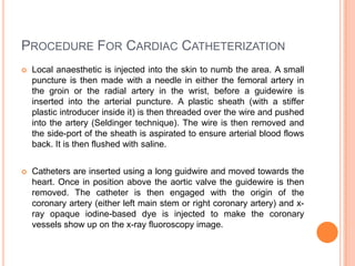
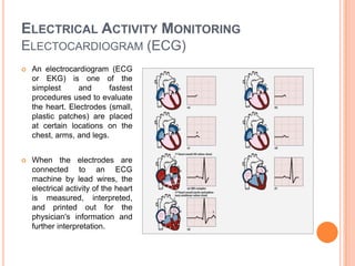
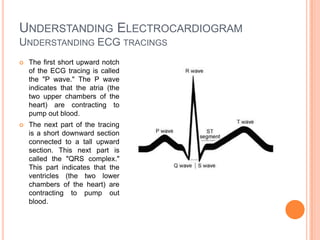
Cardiac catheterization is a procedure that inserts a catheter into the heart to evaluate heart function and disease. It can diagnose conditions like coronary artery disease and determine if treatments like angioplasty are needed. The catheter is inserted into an artery and guided to the heart where contrast dye is injected to image the heart and arteries. An electrocardiogram (ECG) records the heart's electrical activity through electrodes on the skin to check for issues like heart attacks or abnormal rhythms. A computed tomography (CT) scan uses x-rays to create clear pictures of the heart and assess conditions like tumors without invasive procedures.
continuous or intermittent monitoring of heart activity, generally by electrocardiography, with assessment of the patient's condition relative to their cardiac rhythm.
Hemodynamic monitoring- Hemodynamic monitoring refers to the measurement of pressure, flow and oxygenation within the cardiovascular system. Hemodynamic monitoring is amandatory process in all the critical care units to assess the patients progress. This presentation is aimed to create an insight on Hemodynamic monitoring.
pericardial effusion, cardiac tamponade and myocardial rupturegufuabdikadir96
consists of P.E,cardiac tamponade and myocardial rupture and describes their definitions, pathophysiologies, clinical manifestations, dx, medical-surgical mgt and nursing mgt
for more inquiries/feedback; gufuabdikadir96@gmail.com
indications, uses and types of cardiac catheterization, about intra cardiac pressure, about angiography and its technique, digital substraction angiography and its technique.
Pacemaker powerpoint presentation med surgNehaNupur8
pacemaker - artificial pump to the heart, this contained definition, components,working, types, indication, methods of pacaing, temporary and permanent pacemaker, signs of failure of pacemaker , medical and nursing management of patient with pacemaker.
Initial experience with the Glidesheath Slender for transradial coronary angiography and intervention: a feasibility study with prospective radial ultrasound follow-up
Your role and responsibility during an emergency in the cath lab. Communication is extremely important. This power point discusses how to do that. Effective teamwork and communication increases the success of resuscitation.
A moderately frequent illness called congestive heart failure occurs when the heart is unable to pump enough blood to meet the body's demands. It frequently happens as a result of a chronic illness or aging. The body makes an effort to make up for this by boosting blood salt levels and fluid retention.
Swelling, weight gain, and shortness of breath may result from this. Diabetes and high blood pressure are other conditions linked to congestive heart failure. Congestive heart failure, however, is most frequently brought on by coronary artery disease (CAD). This occurs when the arteries that carry blood to the heart start to constrict and narrow.
When calling a doctor is important to question Dr. Sumit shejol Cardiologist from Hrudaysparsh Clinic Suggests that if you recognize the majority of the symptoms of heart failure. Certain signs and symptoms, such as chest pain, acute breathlessness, an irregular heartbeat, extreme weakness, or fainting, demand rapid medical attention. Do not delay in seeking assistance, do not self-diagnose, and do not self-medicate if you feel any of that. Some of these symptoms may also be a sign of heart failure or another serious lung, heart, or cardiovascular disease. Your condition is stabilized as emergency room doctors try to identify the source of your symptoms. Call your doctor right away if you've already been given a heart failure diagnosis and you realize that your symptoms have gotten worse or a new symptom has appeared.
Congestive heart failure is a fatal condition with a high mortality rate. Congestive heart failure has a wide range of risk factors. Smoking, high blood pressure, diabetes, high cholesterol, being obese, and having experienced a heart attack in the past are some of them. It can also be brought on by a hereditary condition like cardiomyopathy. The condition can cause the heart muscle to expand and become excessively thick, which can result in heart failure. Congestive heart failure can be exacerbated by lifestyle choices including smoking, excessive alcohol intake, or tobacco use.
Denoising of ECG -- A discrete time approach using DWTIJERA Editor
This paper is about denoising of ECG signal using DWT transform. In this paper, ECG signals are denoised using DWT transform.Ecg signals are taken and noise at different frequencies are generated which are superimposed on this original ecg signal.High frequency noise is of 4000 hertz and power line interference is of 50 hertz.Decomposition of noisy signal is achieved through wavelet packet .wavelet packets are reconstructed and appropriate wavelet packets are combined to obtain a signal, very similar to original ecg signal.This technique results in the minimization of mean square error in the filtered signals.
Denoising of ECG -- A discrete time approach using DWT.IJERA Editor
This paper is about denoising of ECG signal using DWT transform. In this paper, ECG signals are denoised using DWT transform.Ecg signals are taken and noise at different frequencies are generated which are superimposed on this original ecg signal.High frequency noise is of 4000 hertz and power line interference is of 50 hertz.Decomposition of noisy signal is achieved through wavelet packet .wavelet packets are reconstructed and appropriate wavelet packets are combined to obtain a signal, very similar to original ecg signal.This technique results in the minimization of mean square error in the filtered signals.
ECG - Definition and Measurement techniques .pdfSathish M
Electrocardiogram
-Introduction
-Need for measurement
-How to be measured
-Principle of Measurement
-Lead Placements
-Reduce Art
ECG is used to record the electrical activity of the heart from different angles to both identify and locate pathology. Electrodes are placed on different parts of a patient’s limbs and chest to record the electrical activity.
#ECG
#Electrocardiogram
#Electrocardiograph
#Biomeical
#Cardiac
#Cardiac Monitoring
#12 lead system
#ECG Measurement
#Online
#Study material
#Concept
Title: Sense of Taste
Presenter: Dr. Faiza, Assistant Professor of Physiology
Qualifications:
MBBS (Best Graduate, AIMC Lahore)
FCPS Physiology
ICMT, CHPE, DHPE (STMU)
MPH (GC University, Faisalabad)
MBA (Virtual University of Pakistan)
Learning Objectives:
Describe the structure and function of taste buds.
Describe the relationship between the taste threshold and taste index of common substances.
Explain the chemical basis and signal transduction of taste perception for each type of primary taste sensation.
Recognize different abnormalities of taste perception and their causes.
Key Topics:
Significance of Taste Sensation:
Differentiation between pleasant and harmful food
Influence on behavior
Selection of food based on metabolic needs
Receptors of Taste:
Taste buds on the tongue
Influence of sense of smell, texture of food, and pain stimulation (e.g., by pepper)
Primary and Secondary Taste Sensations:
Primary taste sensations: Sweet, Sour, Salty, Bitter, Umami
Chemical basis and signal transduction mechanisms for each taste
Taste Threshold and Index:
Taste threshold values for Sweet (sucrose), Salty (NaCl), Sour (HCl), and Bitter (Quinine)
Taste index relationship: Inversely proportional to taste threshold
Taste Blindness:
Inability to taste certain substances, particularly thiourea compounds
Example: Phenylthiocarbamide
Structure and Function of Taste Buds:
Composition: Epithelial cells, Sustentacular/Supporting cells, Taste cells, Basal cells
Features: Taste pores, Taste hairs/microvilli, and Taste nerve fibers
Location of Taste Buds:
Found in papillae of the tongue (Fungiform, Circumvallate, Foliate)
Also present on the palate, tonsillar pillars, epiglottis, and proximal esophagus
Mechanism of Taste Stimulation:
Interaction of taste substances with receptors on microvilli
Signal transduction pathways for Umami, Sweet, Bitter, Sour, and Salty tastes
Taste Sensitivity and Adaptation:
Decrease in sensitivity with age
Rapid adaptation of taste sensation
Role of Saliva in Taste:
Dissolution of tastants to reach receptors
Washing away the stimulus
Taste Preferences and Aversions:
Mechanisms behind taste preference and aversion
Influence of receptors and neural pathways
Impact of Sensory Nerve Damage:
Degeneration of taste buds if the sensory nerve fiber is cut
Abnormalities of Taste Detection:
Conditions: Ageusia, Hypogeusia, Dysgeusia (parageusia)
Causes: Nerve damage, neurological disorders, infections, poor oral hygiene, adverse drug effects, deficiencies, aging, tobacco use, altered neurotransmitter levels
Neurotransmitters and Taste Threshold:
Effects of serotonin (5-HT) and norepinephrine (NE) on taste sensitivity
Supertasters:
25% of the population with heightened sensitivity to taste, especially bitterness
Increased number of fungiform papillae
micro teaching on communication m.sc nursing.pdfAnurag Sharma
Microteaching is a unique model of practice teaching. It is a viable instrument for the. desired change in the teaching behavior or the behavior potential which, in specified types of real. classroom situations, tends to facilitate the achievement of specified types of objectives.
Basavarajeeyam is an important text for ayurvedic physician belonging to andhra pradehs. It is a popular compendium in various parts of our country as well as in andhra pradesh. The content of the text was presented in sanskrit and telugu language (Bilingual). One of the most famous book in ayurvedic pharmaceutics and therapeutics. This book contains 25 chapters called as prakaranas. Many rasaoushadis were explained, pioneer of dhatu druti, nadi pareeksha, mutra pareeksha etc. Belongs to the period of 15-16 century. New diseases like upadamsha, phiranga rogas are explained.
Recomendações da OMS sobre cuidados maternos e neonatais para uma experiência pós-natal positiva.
Em consonância com os ODS – Objetivos do Desenvolvimento Sustentável e a Estratégia Global para a Saúde das Mulheres, Crianças e Adolescentes, e aplicando uma abordagem baseada nos direitos humanos, os esforços de cuidados pós-natais devem expandir-se para além da cobertura e da simples sobrevivência, de modo a incluir cuidados de qualidade.
Estas diretrizes visam melhorar a qualidade dos cuidados pós-natais essenciais e de rotina prestados às mulheres e aos recém-nascidos, com o objetivo final de melhorar a saúde e o bem-estar materno e neonatal.
Uma “experiência pós-natal positiva” é um resultado importante para todas as mulheres que dão à luz e para os seus recém-nascidos, estabelecendo as bases para a melhoria da saúde e do bem-estar a curto e longo prazo. Uma experiência pós-natal positiva é definida como aquela em que as mulheres, pessoas que gestam, os recém-nascidos, os casais, os pais, os cuidadores e as famílias recebem informação consistente, garantia e apoio de profissionais de saúde motivados; e onde um sistema de saúde flexível e com recursos reconheça as necessidades das mulheres e dos bebês e respeite o seu contexto cultural.
Estas diretrizes consolidadas apresentam algumas recomendações novas e já bem fundamentadas sobre cuidados pós-natais de rotina para mulheres e neonatos que recebem cuidados no pós-parto em unidades de saúde ou na comunidade, independentemente dos recursos disponíveis.
É fornecido um conjunto abrangente de recomendações para cuidados durante o período puerperal, com ênfase nos cuidados essenciais que todas as mulheres e recém-nascidos devem receber, e com a devida atenção à qualidade dos cuidados; isto é, a entrega e a experiência do cuidado recebido. Estas diretrizes atualizam e ampliam as recomendações da OMS de 2014 sobre cuidados pós-natais da mãe e do recém-nascido e complementam as atuais diretrizes da OMS sobre a gestão de complicações pós-natais.
O estabelecimento da amamentação e o manejo das principais intercorrências é contemplada.
Recomendamos muito.
Vamos discutir essas recomendações no nosso curso de pós-graduação em Aleitamento no Instituto Ciclos.
Esta publicação só está disponível em inglês até o momento.
Prof. Marcus Renato de Carvalho
www.agostodourado.com
Title: Sense of Smell
Presenter: Dr. Faiza, Assistant Professor of Physiology
Qualifications:
MBBS (Best Graduate, AIMC Lahore)
FCPS Physiology
ICMT, CHPE, DHPE (STMU)
MPH (GC University, Faisalabad)
MBA (Virtual University of Pakistan)
Learning Objectives:
Describe the primary categories of smells and the concept of odor blindness.
Explain the structure and location of the olfactory membrane and mucosa, including the types and roles of cells involved in olfaction.
Describe the pathway and mechanisms of olfactory signal transmission from the olfactory receptors to the brain.
Illustrate the biochemical cascade triggered by odorant binding to olfactory receptors, including the role of G-proteins and second messengers in generating an action potential.
Identify different types of olfactory disorders such as anosmia, hyposmia, hyperosmia, and dysosmia, including their potential causes.
Key Topics:
Olfactory Genes:
3% of the human genome accounts for olfactory genes.
400 genes for odorant receptors.
Olfactory Membrane:
Located in the superior part of the nasal cavity.
Medially: Folds downward along the superior septum.
Laterally: Folds over the superior turbinate and upper surface of the middle turbinate.
Total surface area: 5-10 square centimeters.
Olfactory Mucosa:
Olfactory Cells: Bipolar nerve cells derived from the CNS (100 million), with 4-25 olfactory cilia per cell.
Sustentacular Cells: Produce mucus and maintain ionic and molecular environment.
Basal Cells: Replace worn-out olfactory cells with an average lifespan of 1-2 months.
Bowman’s Gland: Secretes mucus.
Stimulation of Olfactory Cells:
Odorant dissolves in mucus and attaches to receptors on olfactory cilia.
Involves a cascade effect through G-proteins and second messengers, leading to depolarization and action potential generation in the olfactory nerve.
Quality of a Good Odorant:
Small (3-20 Carbon atoms), volatile, water-soluble, and lipid-soluble.
Facilitated by odorant-binding proteins in mucus.
Membrane Potential and Action Potential:
Resting membrane potential: -55mV.
Action potential frequency in the olfactory nerve increases with odorant strength.
Adaptation Towards the Sense of Smell:
Rapid adaptation within the first second, with further slow adaptation.
Psychological adaptation greater than receptor adaptation, involving feedback inhibition from the central nervous system.
Primary Sensations of Smell:
Camphoraceous, Musky, Floral, Pepperminty, Ethereal, Pungent, Putrid.
Odor Detection Threshold:
Examples: Hydrogen sulfide (0.0005 ppm), Methyl-mercaptan (0.002 ppm).
Some toxic substances are odorless at lethal concentrations.
Characteristics of Smell:
Odor blindness for single substances due to lack of appropriate receptor protein.
Behavioral and emotional influences of smell.
Transmission of Olfactory Signals:
From olfactory cells to glomeruli in the olfactory bulb, involving lateral inhibition.
Primitive, less old, and new olfactory systems with different path
Lung Cancer: Artificial Intelligence, Synergetics, Complex System Analysis, S...Oleg Kshivets
RESULTS: Overall life span (LS) was 2252.1±1742.5 days and cumulative 5-year survival (5YS) reached 73.2%, 10 years – 64.8%, 20 years – 42.5%. 513 LCP lived more than 5 years (LS=3124.6±1525.6 days), 148 LCP – more than 10 years (LS=5054.4±1504.1 days).199 LCP died because of LC (LS=562.7±374.5 days). 5YS of LCP after bi/lobectomies was significantly superior in comparison with LCP after pneumonectomies (78.1% vs.63.7%, P=0.00001 by log-rank test). AT significantly improved 5YS (66.3% vs. 34.8%) (P=0.00000 by log-rank test) only for LCP with N1-2. Cox modeling displayed that 5YS of LCP significantly depended on: phase transition (PT) early-invasive LC in terms of synergetics, PT N0—N12, cell ratio factors (ratio between cancer cells- CC and blood cells subpopulations), G1-3, histology, glucose, AT, blood cell circuit, prothrombin index, heparin tolerance, recalcification time (P=0.000-0.038). Neural networks, genetic algorithm selection and bootstrap simulation revealed relationships between 5YS and PT early-invasive LC (rank=1), PT N0—N12 (rank=2), thrombocytes/CC (3), erythrocytes/CC (4), eosinophils/CC (5), healthy cells/CC (6), lymphocytes/CC (7), segmented neutrophils/CC (8), stick neutrophils/CC (9), monocytes/CC (10); leucocytes/CC (11). Correct prediction of 5YS was 100% by neural networks computing (area under ROC curve=1.0; error=0.0).
CONCLUSIONS: 5YS of LCP after radical procedures significantly depended on: 1) PT early-invasive cancer; 2) PT N0--N12; 3) cell ratio factors; 4) blood cell circuit; 5) biochemical factors; 6) hemostasis system; 7) AT; 8) LC characteristics; 9) LC cell dynamics; 10) surgery type: lobectomy/pneumonectomy; 11) anthropometric data. Optimal diagnosis and treatment strategies for LC are: 1) screening and early detection of LC; 2) availability of experienced thoracic surgeons because of complexity of radical procedures; 3) aggressive en block surgery and adequate lymph node dissection for completeness; 4) precise prediction; 5) adjuvant chemoimmunoradiotherapy for LCP with unfavorable prognosis.
New Drug Discovery and Development .....NEHA GUPTA
The "New Drug Discovery and Development" process involves the identification, design, testing, and manufacturing of novel pharmaceutical compounds with the aim of introducing new and improved treatments for various medical conditions. This comprehensive endeavor encompasses various stages, including target identification, preclinical studies, clinical trials, regulatory approval, and post-market surveillance. It involves multidisciplinary collaboration among scientists, researchers, clinicians, regulatory experts, and pharmaceutical companies to bring innovative therapies to market and address unmet medical needs.
TEST BANK for Operations Management, 14th Edition by William J. Stevenson, Ve...kevinkariuki227
TEST BANK for Operations Management, 14th Edition by William J. Stevenson, Verified Chapters 1 - 19, Complete Newest Version.pdf
HOT NEW PRODUCT! BIG SALES FAST SHIPPING NOW FROM CHINA!! EU KU DB BK substit...GL Anaacs
Contact us if you are interested:
Email / Skype : kefaya1771@gmail.com
Threema: PXHY5PDH
New BATCH Ku !!! MUCH IN DEMAND FAST SALE EVERY BATCH HAPPY GOOD EFFECT BIG BATCH !
Contact me on Threema or skype to start big business!!
Hot-sale products:
NEW HOT EUTYLONE WHITE CRYSTAL!!
5cl-adba precursor (semi finished )
5cl-adba raw materials
ADBB precursor (semi finished )
ADBB raw materials
APVP powder
5fadb/4f-adb
Jwh018 / Jwh210
Eutylone crystal
Protonitazene (hydrochloride) CAS: 119276-01-6
Flubrotizolam CAS: 57801-95-3
Metonitazene CAS: 14680-51-4
Payment terms: Western Union,MoneyGram,Bitcoin or USDT.
Deliver Time: Usually 7-15days
Shipping method: FedEx, TNT, DHL,UPS etc.Our deliveries are 100% safe, fast, reliable and discreet.
Samples will be sent for your evaluation!If you are interested in, please contact me, let's talk details.
We specializes in exporting high quality Research chemical, medical intermediate, Pharmaceutical chemicals and so on. Products are exported to USA, Canada, France, Korea, Japan,Russia, Southeast Asia and other countries.
2. Cardiac Catheterization Cardiac catheterization (also called cardiac cath or coronary angiogram) is a procedure that allows your doctor to "see" how well your heart is functioning. The test involves inserting a long, narrow tube, called a catheter, into a blood vessel in your arm or leg, and guiding it to your heart with the aid of a special X-ray machine. Contrast dye is injected through the catheter so that X-ray movies of your valves, coronary arteries and heart chambers can be created.
3. Purpose of Cardiac Catheter Evaluate or confirm the presence of heart disease (such as coronary artery disease, heart valve disease or disease of the aorta). Evaluate heart muscle function. Determine the need for further treatment (for example, angioplasty or bypass surgery ) At many medical centers, cardiac catheterization is used to perform several interventional, or therapeutic, procedures to open blocked
4. Procedure For Cardiac Catheterization Local anaesthetic is injected into the skin to numb the area. A small puncture is then made with a needle in either the femoral artery in the groin or the radial artery in the wrist, before a guidewire is inserted into the arterial puncture. A plastic sheath (with a stiffer plastic introducer inside it) is then threaded over the wire and pushed into the artery (Seldinger technique). The wire is then removed and the side-port of the sheath is aspirated to ensure arterial blood flows back. It is then flushed with saline. Catheters are inserted using a long guidwire and moved towards the heart. Once in position above the aortic valve the guidewire is then removed. The catheter is then engaged with the origin of the coronary artery (either left main stem or right coronary artery) and x-ray opaque iodine-based dye is injected to make the coronary vessels show up on the x-ray fluoroscopy image.
5. Procedure For Cardiac Catheterization When the necessary procedures are complete, the catheter is removed. Firm pressure is applied to the site to prevent bleeding. This may be done by hand or with a mechanical device. Other closure techniques include an internal suture and plug. If the femoral artery was used, the patient will probably be asked to lie flat for several hours to prevent bleeding or the development of a hematoma. Cardiac interventions such as the insertion of a stent prolong both the procedure itself as well as the post-catheterization time spent in allowing the wound to clot. A cardiac catheterization is a general term for a group of procedures that are performed using this method, such as coronary angiography, as well as left ventrical angiography. Once the catheter is in place, it can be used to perform a number of procedures including angioplasty, angiography, balloon septostomy, and an Electrophysiology study.
6. Electrical Activity MonitoringElectocardiogram (ECG) An electrocardiogram (ECG or EKG) is one of the simplest and fastest procedures used to evaluate the heart. Electrodes (small, plastic patches) are placed at certain locations on the chest, arms, and legs. When the electrodes are connected to an ECG machine by lead wires, the electrical activity of the heart is measured, interpreted, and printed out for the physician's information and further interpretation.
7. Purpose of ECG Check the heart's electrical activity. Find the cause of unexplained chest pain, which could be caused by a heart attack, inflammation of the sac surrounding the heart (pericarditis), or angina. Find the cause of symptoms of heart disease, such as shortness of breath, dizziness, fainting, or rapid, irregular heartbeats (palpitations). Find out if the walls of the heart chambers are too thick (hypertrophied). Check how well medicines are working and whether they are causing side effects that affect the heart. Check how well mechanical devices that are implanted in the heart, such as pacemakers, are working to control a normal heartbeat. Check the health of the heart when other diseases or conditions are present, such as high blood pressure, high cholesterol, cigarette smoking, diabetes, or a family history of early heart disease.
8. Understanding ElectrocardiogramThe heart's electrical conduction system The heart is, in the simplest terms, a pump made up of muscle tissue. Like all pumps, the heart requires a source of energy in order to function. The heart's pumping energy comes from an inborn electrical conduction system. An electrical stimulus is generated by the sinus node (also called the sinoatrial node, or SA node), which is a small mass of specialized tissue located in the right atrium (right upper chamber) of the heart.
9. Understanding ElectrocardiogramThe heart's electrical conduction system The sinus node generates an electrical stimulus regularly at 60 to 100 times per minute under normal conditions. This electrical stimulus travels down through the conduction pathways (similar to the way electricity flows through power lines from the power plant to your house) and causes the heart's chambers to contract and pump out blood. The right and left atria (the two upper chambers of the heart) are stimulated first and contract a short period of time before the right and left ventricles (the two lower chambers of the heart). The electrical impulse travels from the sinus node to the atrioventricular (AV) node, where it stops for a very short period, then continues down the conduction pathways via the AV bundle of His" into the ventricles. The "bundle of His" divides into right and left pathways to provide electrical stimulation to both ventricles. This electrical activity of the heart is measured by an electrocardiogram. By placing electrodes at specific locations on the body (chest, arms, and legs), a graphic representation, or tracing, of the electrical activity can be obtained. Changes in an ECG from the normal tracing may indicate one or more of several heart-related conditions.
10. Understanding ElectrocardiogramUnderstanding ECG tracings The first short upward notch of the ECG tracing is called the "P wave." The P wave indicates that the atria (the two upper chambers of the heart) are contracting to pump out blood. The next part of the tracing is a short downward section connected to a tall upward section. This next part is called the "QRS complex." This part indicates that the ventricles (the two lower chambers of the heart) are contracting to pump out blood.
11. Understanding ElectrocardiogramUnderstanding ECG tracings The next short upward segment is called the "ST segment." The ST segment indicates the amount of time from the end of the contraction of the ventricles to the beginning of the rest period before the ventricles begin to contract for the next beat. The next upward curve is called the "T wave." The T wave indicates the resting period of the ventricles. When the physician views an ECG, he/she studies the size and length of each part of the ECG. Variations in size and length of the different parts of the tracing may be significant. The tracing for each lead of a 12-lead ECG will look different, but will have the same basic components as described above. Each lead of the 12-lead ECG is "looking" at a specific part of the heart, so variations in a lead may indicate a problem with the part of the heart associated with a particular lead.
12. Imaging ProceduresComputed Tomography (CT or CAT) Scan Cardiac computed tomography (to-MOG-rah-fee), or cardiac CT, is a painless test that uses an x-ray machine to take clear, detailed pictures of your heart. It's a common test for showing problems of the heart. During a cardiac CT scan, the x-ray machine will move around your body in a circle and take a picture of each part of your heart. Because an x-ray machine is used, cardiac CT scans involve radiation. However, the amount of radiation used is small. This test gives out a radiation dose similar to the amount of radiation you’re naturally exposed to over 3 years. There is a very small chance that cardiac CT will cause cancer.
13. Purpose of Computed Tomography to assess the chest and its organs for tumors and other lesions, injuries, intra-thoracic bleeding, infections, unexplained chest pain, obstructions, or other conditions, particularly when another type of examination, such as x-rays or physical examination, is not conclusive to evaluate the effects of treatment of thoracic tumors. Another use of chest CT is to provide guidance for biopsies and/or aspiration of tissue from the chest
14. Procedures for Computed Tomography In computed tomography, the x-ray beam moves in a circle around the body. This allows many different views of the same organ or structure. The x-ray information is sent to a computer that interprets the x-ray data and displays it in a two-dimensional (2D) form on a monitor. While many images are taken during a CT scan, in some cases the patient receives the same or less radiation exposure than with a single standard x-ray. CT scans may be done with or without "contrast." Contrast refers to a substance taken by mouth or injected into an intravenous (IV) line that causes the particular organ or tissue under study to be seen more clearly. Contrast examinations may require you to fast for a certain period of time before the procedure. Your physician will notify you of this prior to the procedure.
15. Procedures for Computed Tomography CT scans of the chest can provide more detailed information about organs and structures inside the chest than standard x-rays of the chest, thus providing more information related to injuries and/or diseases of the chest (thoracic) organs. CT scans of the chest may also be used to visualize placement of needles during biopsies of thoracic organs or tumors, or during aspiration (withdrawal) of fluid from the chest. CT scans of the chest are useful in monitoring tumors and other conditions of the chest before and after treatment.