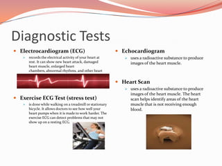
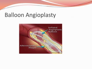
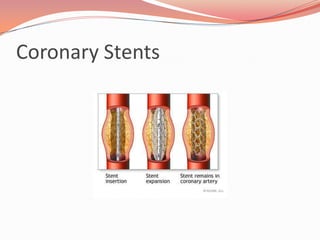
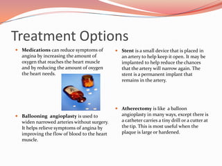
Coronary Artery DiseaseBy: Jessica Canterbury
About the HeartStrong muscle about the size of a fistIt lies in the center and tilted to the leftPumps blood, oxygen, and nutrients to al parts of the bodyThe septum divides the heart into the left and right sidesThe right side pumps blood only to the lungsThe left side pumps blood to the whole bodyThe left side is thicker
Blood Flow Through the HeartSuperior/Inferior vena cavaRight atriumTricuspid ValveRight ventriclePulmonary veinLungsPulmonary arteryLeft atriumMitral ValveLeft ventricleAorta (body)
What is Coronary Artery Disease?The walls inside your arteries are normally smooth and flexible, which allows blood to flow through easily. Over years, fatty deposits may build up on the inside of the artery’s wall.Therefore, vessels carrying blood and oxygen to your heart may be narrowed or blocked
Coronary Artery Disease (CAD) Risk FactorsRisk Factors You CANNOT changeRisk Factors you CAN changeYour age - risk goes up as you get olderGender - men are at greater risk than women and have attacks earlier in life. But women are more likely to die from a heart attackHeredity - Children of parents with heart disease are more likely to develop it themselvesRace - African American have higher risks, possibly due to high blood pressure, obesity, and diabetes Your family - most people with strong family history of heart disease have one or more other risk factorsExposure to tobacco smoke - secondhand smoke increases the risk for heart diseaseHigh blood pressure – makes the heart work harder, which causes the heart muscles to thick and weak. It also increases your risk for stroke, heart attack and kidney/heart failureHigh blood cholesterol – can be lowered with changes in diet, activity, and medicinePhysical inactivity –regular activity helps prevent heart and blood vessel diseaseDiabetes – seriously increases your risk of developing CADExcess body fat – increases the heart’s workload and raises blood pressure
Diagnostic TestsElectrocardiogram (ECG)records the electrical activity of your heart at rest. It can show new heart attack, damaged heart muscle, enlarged heart chambers, abnormal rhythms, and other heart conditionsExercise ECG Test (stress test)is done while walking on a treadmill or stationary bicycle. It allows doctors to see how well your heart pumps when it is made to work harder. The exercise ECG can detect problems that may not show up on a resting ECG.Echocardiogramuses a radioactive substance to produce images of the heart muscle.Heart Scanuses a radioactive substance to produce images of the heart muscle. The heart scan helps identify areas of the heart muscle that is not receiving enough blood.Preparing for the ProcedureNOTHING to eat or drink 6-8 hours before procedureCheck with your doctor several days before because you may be asked to take some medications for a few days beforeBring a list of the names and dosages of ALL the medications you are takingTell the nurse/doctor if you are allergic to any medications, x-ray dye (contrast), iodine/seafood, or if you have a history of bleeding problemsFor you comfort empty your bladder as much as possible before the procedure beginsYou may also be asked to remove any underclothing and jewelry before the procedure
What Happens During Catherization?Site is cleansed thoroughly (usually the groin)A small incision is made in the skinA guidewire is threaded into the arteryA sheath is slipped over the guidewire and into the artery (guidewire is then removed)The catheter is inserted into the artery and guided toward the heart, while staff watches its progress on a screenThe catheter maybe removed and replaced several times. It’s done to reach each of the heart chambers or coronary arteriesOnce the catheter is inside the heart, the doctor can measure the pressures in the left ventricle and take pictures of the coronary arteries and left ventricle
What Do Catherization Show?Measuring the PressuresCoronary AngiographyLeft Ventriculography
Cardiac Catherization
How Angioplasty Is Done?The sheath that was inserted in your artery during catherization can be used for inserting additional cathetersGuided catheter is inserted through the sheathThe catheter is directed toward the opening of the diseased artery while the staff watches its progressContrast is then injected into the artery to show exactly where the narrowed/blocked area isNext, a thinner balloon catheter is inserted through the guiding catheter. It has a small balloon at the tip that is used to open or widen the narrowed arteryThe balloon catheter is threaded into the artery and placed across the narrowed areaOnce the balloon is in place the balloon is inflated, this stretches the artery and flattens the plaqueThe balloon is then deflated and then removedThe larger opening in the artery allows more blood to flow through
Balloon Angioplasty
How Is a Stent ImplantedIf a stent is needed, it is usually implanted right after angioplasty, before the sheath is removedThe stent is usually mounted on a balloon catheterIt’s threaded into the coronary artery and placed at the site where the artery was widenedWhen the balloon inflated, the stent expands and presses against the inside wall of the arteryThe balloon is then deflated and removedThe stent remains in place, helping to keep the artery openOne or more stents may be used in the artery to span the length of the area where the narrowing wasIt’s a permanent implant that remains in the arteryWithin a few weeks, a thin layer of tissue will slowly grow over the stent and cover it
Coronary Stents
After the ProcedureYou will be taken do a recovery area or special care unitThe sheath is left in place for several hours, which allows doctors to check the treated coronary arteryIf no problems occur, the sheath is removed and a firm pressure is applied over the groin for about 20-30 minutes to keep site from bleedingAfter the sheath is removed, you will have to lie flat on your back for up to 8 hours, so that the site can begin to heal properly (DO NOT bend or lift leg)The nurse will check your pulse and blood pressure frequently, and will also check insertion site for bleedingIf you feel sudden pain at the site or if you notice bleeding let you nurse know IMMEDIATELY!!!Most patients go home within 24 hours after the procedureBefore leaving you will be given instructions about medications, physical activity, and follow up care.You should have someone to drive you home
Treatment OptionsMedicationscan reduce symptoms of angina by increasing the amount of oxygen that reaches the heart muscle and by reducing the amount of oxygen the heart needs.Ballooning  angioplasty is used to widen narrowed arteries without surgery. It helps relieve symptoms of angina by improving the flow of blood to the heart muscle.Stent is a small device that is placed in an artery to help keep it open. It may be implanted to help reduce the chances that the artery will narrow again. The stent is a permanent implant that remains in the artery.Atherectomy is like  a balloon angioplasty in many ways, except there is a catheter carries a tiny drill or a cutter at the tip. This is most useful when the plaque is large or hardened.
Ways to Prevent CADMaintain a healthy weightExercise or do physical activityEat a heart healthy diet low in fat and salt and high in fiberControl your blood pressure to keep in below 120/80Keep your cholesterol at a level that lowers your riskManage diabetes (if you have it)STOP smokingLearn how to manage stress
Life Style ChangesEat heart healthyLose weight and keep it offSTOP smokingExercise regularlyWatch blood pressureControl diabetes
The End!!
Coronary Artery Disease

Coronary Artery Disease

  • 1.
    Coronary Artery DiseaseBy:Jessica Canterbury
  • 2.
    About the HeartStrongmuscle about the size of a fistIt lies in the center and tilted to the leftPumps blood, oxygen, and nutrients to al parts of the bodyThe septum divides the heart into the left and right sidesThe right side pumps blood only to the lungsThe left side pumps blood to the whole bodyThe left side is thicker
  • 3.
    Blood Flow Throughthe HeartSuperior/Inferior vena cavaRight atriumTricuspid ValveRight ventriclePulmonary veinLungsPulmonary arteryLeft atriumMitral ValveLeft ventricleAorta (body)
  • 4.
    What is CoronaryArtery Disease?The walls inside your arteries are normally smooth and flexible, which allows blood to flow through easily. Over years, fatty deposits may build up on the inside of the artery’s wall.Therefore, vessels carrying blood and oxygen to your heart may be narrowed or blocked
  • 5.
    Coronary Artery Disease(CAD) Risk FactorsRisk Factors You CANNOT changeRisk Factors you CAN changeYour age - risk goes up as you get olderGender - men are at greater risk than women and have attacks earlier in life. But women are more likely to die from a heart attackHeredity - Children of parents with heart disease are more likely to develop it themselvesRace - African American have higher risks, possibly due to high blood pressure, obesity, and diabetes Your family - most people with strong family history of heart disease have one or more other risk factorsExposure to tobacco smoke - secondhand smoke increases the risk for heart diseaseHigh blood pressure – makes the heart work harder, which causes the heart muscles to thick and weak. It also increases your risk for stroke, heart attack and kidney/heart failureHigh blood cholesterol – can be lowered with changes in diet, activity, and medicinePhysical inactivity –regular activity helps prevent heart and blood vessel diseaseDiabetes – seriously increases your risk of developing CADExcess body fat – increases the heart’s workload and raises blood pressure
  • 6.
    Diagnostic TestsElectrocardiogram (ECG)recordsthe electrical activity of your heart at rest. It can show new heart attack, damaged heart muscle, enlarged heart chambers, abnormal rhythms, and other heart conditionsExercise ECG Test (stress test)is done while walking on a treadmill or stationary bicycle. It allows doctors to see how well your heart pumps when it is made to work harder. The exercise ECG can detect problems that may not show up on a resting ECG.Echocardiogramuses a radioactive substance to produce images of the heart muscle.Heart Scanuses a radioactive substance to produce images of the heart muscle. The heart scan helps identify areas of the heart muscle that is not receiving enough blood.Preparing for the ProcedureNOTHING to eat or drink 6-8 hours before procedureCheck with your doctor several days before because you may be asked to take some medications for a few days beforeBring a list of the names and dosages of ALL the medications you are takingTell the nurse/doctor if you are allergic to any medications, x-ray dye (contrast), iodine/seafood, or if you have a history of bleeding problemsFor you comfort empty your bladder as much as possible before the procedure beginsYou may also be asked to remove any underclothing and jewelry before the procedure
  • 7.
    What Happens DuringCatherization?Site is cleansed thoroughly (usually the groin)A small incision is made in the skinA guidewire is threaded into the arteryA sheath is slipped over the guidewire and into the artery (guidewire is then removed)The catheter is inserted into the artery and guided toward the heart, while staff watches its progress on a screenThe catheter maybe removed and replaced several times. It’s done to reach each of the heart chambers or coronary arteriesOnce the catheter is inside the heart, the doctor can measure the pressures in the left ventricle and take pictures of the coronary arteries and left ventricle
  • 8.
    What Do CatherizationShow?Measuring the PressuresCoronary AngiographyLeft Ventriculography
  • 9.
  • 10.
    How Angioplasty IsDone?The sheath that was inserted in your artery during catherization can be used for inserting additional cathetersGuided catheter is inserted through the sheathThe catheter is directed toward the opening of the diseased artery while the staff watches its progressContrast is then injected into the artery to show exactly where the narrowed/blocked area isNext, a thinner balloon catheter is inserted through the guiding catheter. It has a small balloon at the tip that is used to open or widen the narrowed arteryThe balloon catheter is threaded into the artery and placed across the narrowed areaOnce the balloon is in place the balloon is inflated, this stretches the artery and flattens the plaqueThe balloon is then deflated and then removedThe larger opening in the artery allows more blood to flow through
  • 11.
  • 12.
    How Is aStent ImplantedIf a stent is needed, it is usually implanted right after angioplasty, before the sheath is removedThe stent is usually mounted on a balloon catheterIt’s threaded into the coronary artery and placed at the site where the artery was widenedWhen the balloon inflated, the stent expands and presses against the inside wall of the arteryThe balloon is then deflated and removedThe stent remains in place, helping to keep the artery openOne or more stents may be used in the artery to span the length of the area where the narrowing wasIt’s a permanent implant that remains in the arteryWithin a few weeks, a thin layer of tissue will slowly grow over the stent and cover it
  • 13.
  • 14.
    After the ProcedureYouwill be taken do a recovery area or special care unitThe sheath is left in place for several hours, which allows doctors to check the treated coronary arteryIf no problems occur, the sheath is removed and a firm pressure is applied over the groin for about 20-30 minutes to keep site from bleedingAfter the sheath is removed, you will have to lie flat on your back for up to 8 hours, so that the site can begin to heal properly (DO NOT bend or lift leg)The nurse will check your pulse and blood pressure frequently, and will also check insertion site for bleedingIf you feel sudden pain at the site or if you notice bleeding let you nurse know IMMEDIATELY!!!Most patients go home within 24 hours after the procedureBefore leaving you will be given instructions about medications, physical activity, and follow up care.You should have someone to drive you home
  • 15.
    Treatment OptionsMedicationscan reducesymptoms of angina by increasing the amount of oxygen that reaches the heart muscle and by reducing the amount of oxygen the heart needs.Ballooning angioplasty is used to widen narrowed arteries without surgery. It helps relieve symptoms of angina by improving the flow of blood to the heart muscle.Stent is a small device that is placed in an artery to help keep it open. It may be implanted to help reduce the chances that the artery will narrow again. The stent is a permanent implant that remains in the artery.Atherectomy is like a balloon angioplasty in many ways, except there is a catheter carries a tiny drill or a cutter at the tip. This is most useful when the plaque is large or hardened.
  • 16.
    Ways to PreventCADMaintain a healthy weightExercise or do physical activityEat a heart healthy diet low in fat and salt and high in fiberControl your blood pressure to keep in below 120/80Keep your cholesterol at a level that lowers your riskManage diabetes (if you have it)STOP smokingLearn how to manage stress
  • 17.
    Life Style ChangesEatheart healthyLose weight and keep it offSTOP smokingExercise regularlyWatch blood pressureControl diabetes
  • 18.