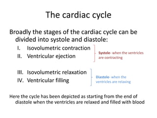
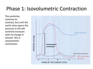
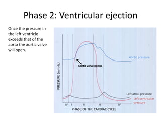
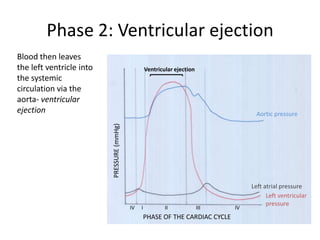
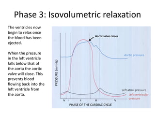
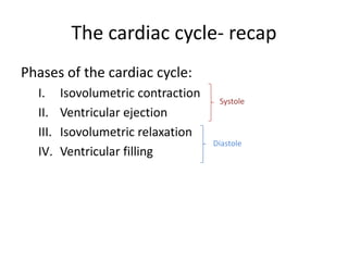
The cardiac cycle consists of 4 phases:
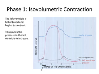
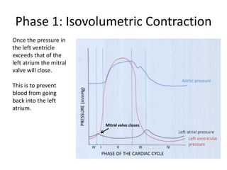
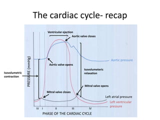
1) Isovolumetric contraction - the ventricles contract but volume remains constant until the aortic valve opens
2) Ventricular ejection - the aortic valve opens and blood is ejected from the ventricles into the aorta
3) Isovolumetric relaxation - the ventricles relax but volume is constant until the mitral valve opens
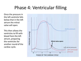
4) Ventricular filling - the mitral valve opens and the ventricles fill with blood from the atria in preparation for the next cycle.
Cardiac output (The Guyton and Hall Physiology)Maryam Fida
The volume of blood pumped by each ventricle per minute is called cardiac output
Cardiac output = Stroke Volume X Heart Rate
Normal value = 5 Liters /Minute
Cardiac output = Stroke Volume X Heart Rate
The factors which regulate stroke volume and Heart rate are basically regulating Cardiac output
Volume of blood ejected by each ventricle in single systole; Normal Value = 70 ml/beat
Stroke Volume = End diastolic Volume – End Systolic Volume
So stroke volume is mainly controlled by
EDV
ESV
VENOUS RETURN: What ever blood volume returns to the heart, same is pumped forward through the Frank’s Starlings Law. According to this law 13- 15 liters of blood volume can be pumped out without cardiac stimulation.
DURATION OF DIASTOLE OR FILLING TIME: ventricular filling occurs during diastole, so there must be adequate ventricular filling time.
DISTENSIBILITY OF THE VENTRICLES: Normally ventricles are distensible to accommodate adequate blood volume. Infarction decreases the distensibility which decreases the EDV.
ATRIAL CONTRACTION: There must be adequate atrial contraction to have adequate EDV. If atrial function is not adequate then EDV will decrease.
E.S.V is basically CONTROLLED BY MYOCARDIAL CONTRACTION
FORCE OF MYOCARDIAL CONTRACTION: It depends upon the initial length of muscle fibers according to frank’s starlings law.
PRELOAD: The effect of EDV on initial length is called preload. So EDV also effects the ESV.
AFTER LOAD: Force of contraction is also dependant upon the resistance against which the ventricles have to pump
CONDITION OF THE MYOCARDIUM : It also effects the force of contraction.
AUTONOMIC NERVES : Sympathetic stimulation increases and parasympathetic stimulation decreases force of contraction
HORMONES: Catecholamines, thyroxine, glucagon, digitalis, calcium, increased temp, caffeine, theophyline increase the force.
Force decreases by hypoxia, acidosis, barniturates, procainamide and quinidine decrease the force of contraction.
HEART RATE
REGULATION OF HEART RATE
VASOMOTOR CENTER – CARDIAC CENTER
MOTOR (EFFERENT) NERVE FIBERS TO HEART
FACTORS AFFECTING VASOMOTOR CENTER
for all medical & health care students
Describe events in cardiac cycle.
Describe atrial, ventricular and aortic pressure changes during cardiac cycle.
Describe the changes in ventricular volume & stroke volume during cardiac cycle.
Relate ECG changes to the phases of cardiac cycle.
Describe the functions of cardiac valves and relate their state to the production of heart sounds during cardiac cycle.
med_students0
CARDIAC CYCLE, ECG AND HEART SOUNDS.pptxthiru murugan
CARDIAC CYCLE, ECG AND HEART SOUNDS: BY Wincy Thirumurugan..
“Cardiac cycle refers to the series of events that take place when the heart beats.”
Each cycle is initiated by spontaneous contraction in the SA node and then transmit through the A-V bundle and branches into the ventricles results completion of one cycle.
EVENTS OR PHASES OF CARDIAC CYCLE: Diastolic phase (Diastole) in this phase the heart chamber are in the state of relaxation and fills with blood that receives from the veins [IVC, SVC,PULMONARY VEINS]
Systolic phase (Systole) in this the heart chambers are contracting and pumps the blood towards the periphery via the arteries. [ Pulmonary artery and aorta]
PHASES OF THE CARDIAC CYCLE
The different phases of the cardiac cycle involve:
Atrial diastole - Atrial relaxation
Atrial systole -Atrial contraction
Isovolumic relaxation -ventricular relaxation in the early phase but blood will not move and the Atrio ventricular valves will be closed
Ventricular filling - ventricular relaxation, the Atrio ventricular valves will be open allows filling blood in the ventricles
Isovolumic contraction of ventricle – ventricular systole in the early phase but no movement of the blood. The semilunar valves will be closed.
Ventricular ejection -ventricular contraction and send blood out of the ventricles through opened semilunar valves.
6. Ventricular Filling Stage: second phase. Rapid Filling, Slow Filling & Last Rapid Filling Duration of Cardiac Cycle:
In a normal person, a heartbeat is 72 beats/minute.
An Electrocardiogram (ECG) is a medical test that detects cardiac (heart) abnormalities by measuring the electrical activity generated by the heart as it. The machine that records the patient’s ECG is called an electrocardiograph.
contracts.
PLACEMENT OF ECG LEADS
ECG WAVES:
The P wave is caused by spread of depolarization through the atria, After the onset of the P wave, The QRS waves Occurs as a result of electrical depolarization of the ventricles, the ventricular T wave represents the stage of repolarization of the ventricles, The 'U' wave is a wave comes after the T wave of ventricular repolarization and may not always be observed.
HEART SOUNDS: First Heart Sound (S1)
The first heart sound results from the closing of the mitral and tricuspid valves. Second Heart Sound (S2): The second heart sound is produced by the closure of the aortic and pulmonic valves. Third Heart Sound (S3):
The third heart sound, also known as the “ventricular gallop,” occurs just after S2 when the mitral valve opens, allowing passive filling of the left ventricle. The S3 sound is actually produced by the large amount of blood striking a very compliant LV.
[Compliance heart means how easily the chamber of heart or the lumen of blood vessels expands when it is filling with the blood]
Fourth Heart Sound (S4):
The fourth heart sound, also known as the “atrial gallop,” occurs just before S1 when the atria contract to force blood into the LV.
Sa nodal action potential, conducting system of heart and spread of cardiac i...Maryam Fida
SA NODE, AV NODE and Purkinje System are specialized cells of the heart having unstable phase IV.
SA Node has no role of Voltage gated sodium channels(although they are present in SA Node) and
so the depolarization in it occurs through voltage gated slow calcium channels
The membrane of SA Node is Inherently leaky to Sodium and Calcium Ions.
It is the Pre Potential Slope or spontaneous slow depolarization which accounts for the Pace maker activity of SA node i.e. Automaticity
It is caused by the inherent leakiness of SA Nodal membrane to Sodium and Calcium leading to influx of Na+ , causing a slow rise in the RMP in the positive direction.
Thus, the “resting” potential gradually rises between each two heartbeats.
When the potential reaches a threshold voltage of about -40 millivolts, the Sodium-Calcium channels become “activated,” thus causing the action potential.
It is the upstroke of action potential
When the membrane potential reaches the thresh hold level i.e. -40 mV, voltage gated slow calcium channels open up leading to influx of calcium causing depolarization
Voltage gated sodium channels has no role in SA nodal depolarization because at the level of -55 mV, the fast sodium channels mainly have already become “inactivated,” which means that they have become blocked.
The cause of this is that any time the membrane potential remains less negative than about -55 mV for more than a few milliseconds, the inactivation gates on the inside of the cell membrane that close the fast sodium channels become closed and remain so. Therefore, only the slow sodium-calcium channels can open (i.e., can become “activated”) and thereby cause the action potential.
Cardiac cycle (The Guyton and Hall physiology)Maryam Fida
Sequence of events from the beginning of one systole to the beginning of next consecutive systole.
One heart beat consists of one systole and one diastole.
Each cardiac cycle is initiated by the cardiac impulse which originates from the SA node.
During each cardiac cycle, certain events occur in the heart and these include pressure changes, volume changes, production of heart sounds, closure and opening of heart valves and electrical changes in the heart.
Ventricular Systole refers to the phase of the cardiac cycle where the left and right ventricles contract at the same time and pump blood into the aorta and pulmonary trunk, respectively
Useful for medical and biology students who want to study the cardiac cycle in a short time with big benefits !!
CVS physiology - Wigger Diagram - ECG of cardiac cycle - Heart sounds
HOT NEW PRODUCT! BIG SALES FAST SHIPPING NOW FROM CHINA!! EU KU DB BK substit...GL Anaacs
Contact us if you are interested:
Email / Skype : kefaya1771@gmail.com
Threema: PXHY5PDH
New BATCH Ku !!! MUCH IN DEMAND FAST SALE EVERY BATCH HAPPY GOOD EFFECT BIG BATCH !
Contact me on Threema or skype to start big business!!
Hot-sale products:
NEW HOT EUTYLONE WHITE CRYSTAL!!
5cl-adba precursor (semi finished )
5cl-adba raw materials
ADBB precursor (semi finished )
ADBB raw materials
APVP powder
5fadb/4f-adb
Jwh018 / Jwh210
Eutylone crystal
Protonitazene (hydrochloride) CAS: 119276-01-6
Flubrotizolam CAS: 57801-95-3
Metonitazene CAS: 14680-51-4
Payment terms: Western Union,MoneyGram,Bitcoin or USDT.
Deliver Time: Usually 7-15days
Shipping method: FedEx, TNT, DHL,UPS etc.Our deliveries are 100% safe, fast, reliable and discreet.
Samples will be sent for your evaluation!If you are interested in, please contact me, let's talk details.
We specializes in exporting high quality Research chemical, medical intermediate, Pharmaceutical chemicals and so on. Products are exported to USA, Canada, France, Korea, Japan,Russia, Southeast Asia and other countries.
Acute scrotum is a general term referring to an emergency condition affecting the contents or the wall of the scrotum.
There are a number of conditions that present acutely, predominantly with pain and/or swelling
A careful and detailed history and examination, and in some cases, investigations allow differentiation between these diagnoses. A prompt diagnosis is essential as the patient may require urgent surgical intervention
Testicular torsion refers to twisting of the spermatic cord, causing ischaemia of the testicle.
Testicular torsion results from inadequate fixation of the testis to the tunica vaginalis producing ischemia from reduced arterial inflow and venous outflow obstruction.
The prevalence of testicular torsion in adult patients hospitalized with acute scrotal pain is approximately 25 to 50 percent
- Video recording of this lecture in English language: https://youtu.be/lK81BzxMqdo
- Video recording of this lecture in Arabic language: https://youtu.be/Ve4P0COk9OI
- Link to download the book free: https://nephrotube.blogspot.com/p/nephrotube-nephrology-books.html
- Link to NephroTube website: www.NephroTube.com
- Link to NephroTube social media accounts: https://nephrotube.blogspot.com/p/join-nephrotube-on-social-media.html
ARTIFICIAL INTELLIGENCE IN HEALTHCARE.pdfAnujkumaranit
Artificial intelligence (AI) refers to the simulation of human intelligence processes by machines, especially computer systems. It encompasses tasks such as learning, reasoning, problem-solving, perception, and language understanding. AI technologies are revolutionizing various fields, from healthcare to finance, by enabling machines to perform tasks that typically require human intelligence.
Explore natural remedies for syphilis treatment in Singapore. Discover alternative therapies, herbal remedies, and lifestyle changes that may complement conventional treatments. Learn about holistic approaches to managing syphilis symptoms and supporting overall health.
Tom Selleck Health: A Comprehensive Look at the Iconic Actor’s Wellness Journeygreendigital
Tom Selleck, an enduring figure in Hollywood. has captivated audiences for decades with his rugged charm, iconic moustache. and memorable roles in television and film. From his breakout role as Thomas Magnum in Magnum P.I. to his current portrayal of Frank Reagan in Blue Bloods. Selleck's career has spanned over 50 years. But beyond his professional achievements. fans have often been curious about Tom Selleck Health. especially as he has aged in the public eye.
Follow us on: Pinterest
Introduction
Many have been interested in Tom Selleck health. not only because of his enduring presence on screen but also because of the challenges. and lifestyle choices he has faced and made over the years. This article delves into the various aspects of Tom Selleck health. exploring his fitness regimen, diet, mental health. and the challenges he has encountered as he ages. We'll look at how he maintains his well-being. the health issues he has faced, and his approach to ageing .
Early Life and Career
Childhood and Athletic Beginnings
Tom Selleck was born on January 29, 1945, in Detroit, Michigan, and grew up in Sherman Oaks, California. From an early age, he was involved in sports, particularly basketball. which played a significant role in his physical development. His athletic pursuits continued into college. where he attended the University of Southern California (USC) on a basketball scholarship. This early involvement in sports laid a strong foundation for his physical health and disciplined lifestyle.
Transition to Acting
Selleck's transition from an athlete to an actor came with its physical demands. His first significant role in "Magnum P.I." required him to perform various stunts and maintain a fit appearance. This role, which he played from 1980 to 1988. necessitated a rigorous fitness routine to meet the show's demands. setting the stage for his long-term commitment to health and wellness.
Fitness Regimen
Workout Routine
Tom Selleck health and fitness regimen has evolved. adapting to his changing roles and age. During his "Magnum, P.I." days. Selleck's workouts were intense and focused on building and maintaining muscle mass. His routine included weightlifting, cardiovascular exercises. and specific training for the stunts he performed on the show.
Selleck adjusted his fitness routine as he aged to suit his body's needs. Today, his workouts focus on maintaining flexibility, strength, and cardiovascular health. He incorporates low-impact exercises such as swimming, walking, and light weightlifting. This balanced approach helps him stay fit without putting undue strain on his joints and muscles.
Importance of Flexibility and Mobility
In recent years, Selleck has emphasized the importance of flexibility and mobility in his fitness regimen. Understanding the natural decline in muscle mass and joint flexibility with age. he includes stretching and yoga in his routine. These practices help prevent injuries, improve posture, and maintain mobilit
263778731218 Abortion Clinic /Pills In Harare ,sisternakatoto
263778731218 Abortion Clinic /Pills In Harare ,ABORTION WOMEN’S CLINIC +27730423979 IN women clinic we believe that every woman should be able to make choices in her pregnancy. Our job is to provide compassionate care, safety,affordable and confidential services. That’s why we have won the trust from all generations of women all over the world. we use non surgical method(Abortion pills) to terminate…Dr.LISA +27730423979women Clinic is committed to providing the highest quality of obstetrical and gynecological care to women of all ages. Our dedicated staff aim to treat each patient and her health concerns with compassion and respect.Our dedicated group ABORTION WOMEN’S CLINIC +27730423979 IN women clinic we believe that every woman should be able to make choices in her pregnancy. Our job is to provide compassionate care, safety,affordable and confidential services. That’s why we have won the trust from all generations of women all over the world. we use non surgical method(Abortion pills) to terminate…Dr.LISA +27730423979women Clinic is committed to providing the highest quality of obstetrical and gynecological care to women of all ages. Our dedicated staff aim to treat each patient and her health concerns with compassion and respect.Our dedicated group of receptionists, nurses, and physicians have worked together as a teamof receptionists, nurses, and physicians have worked together as a team wwww.lisywomensclinic.co.za/
Title: Sense of Taste
Presenter: Dr. Faiza, Assistant Professor of Physiology
Qualifications:
MBBS (Best Graduate, AIMC Lahore)
FCPS Physiology
ICMT, CHPE, DHPE (STMU)
MPH (GC University, Faisalabad)
MBA (Virtual University of Pakistan)
Learning Objectives:
Describe the structure and function of taste buds.
Describe the relationship between the taste threshold and taste index of common substances.
Explain the chemical basis and signal transduction of taste perception for each type of primary taste sensation.
Recognize different abnormalities of taste perception and their causes.
Key Topics:
Significance of Taste Sensation:
Differentiation between pleasant and harmful food
Influence on behavior
Selection of food based on metabolic needs
Receptors of Taste:
Taste buds on the tongue
Influence of sense of smell, texture of food, and pain stimulation (e.g., by pepper)
Primary and Secondary Taste Sensations:
Primary taste sensations: Sweet, Sour, Salty, Bitter, Umami
Chemical basis and signal transduction mechanisms for each taste
Taste Threshold and Index:
Taste threshold values for Sweet (sucrose), Salty (NaCl), Sour (HCl), and Bitter (Quinine)
Taste index relationship: Inversely proportional to taste threshold
Taste Blindness:
Inability to taste certain substances, particularly thiourea compounds
Example: Phenylthiocarbamide
Structure and Function of Taste Buds:
Composition: Epithelial cells, Sustentacular/Supporting cells, Taste cells, Basal cells
Features: Taste pores, Taste hairs/microvilli, and Taste nerve fibers
Location of Taste Buds:
Found in papillae of the tongue (Fungiform, Circumvallate, Foliate)
Also present on the palate, tonsillar pillars, epiglottis, and proximal esophagus
Mechanism of Taste Stimulation:
Interaction of taste substances with receptors on microvilli
Signal transduction pathways for Umami, Sweet, Bitter, Sour, and Salty tastes
Taste Sensitivity and Adaptation:
Decrease in sensitivity with age
Rapid adaptation of taste sensation
Role of Saliva in Taste:
Dissolution of tastants to reach receptors
Washing away the stimulus
Taste Preferences and Aversions:
Mechanisms behind taste preference and aversion
Influence of receptors and neural pathways
Impact of Sensory Nerve Damage:
Degeneration of taste buds if the sensory nerve fiber is cut
Abnormalities of Taste Detection:
Conditions: Ageusia, Hypogeusia, Dysgeusia (parageusia)
Causes: Nerve damage, neurological disorders, infections, poor oral hygiene, adverse drug effects, deficiencies, aging, tobacco use, altered neurotransmitter levels
Neurotransmitters and Taste Threshold:
Effects of serotonin (5-HT) and norepinephrine (NE) on taste sensitivity
Supertasters:
25% of the population with heightened sensitivity to taste, especially bitterness
Increased number of fungiform papillae
Knee anatomy and clinical tests 2024.pdfvimalpl1234
This includes all relevant anatomy and clinical tests compiled from standard textbooks, Campbell,netter etc..It is comprehensive and best suited for orthopaedicians and orthopaedic residents.
New Drug Discovery and Development .....NEHA GUPTA
The "New Drug Discovery and Development" process involves the identification, design, testing, and manufacturing of novel pharmaceutical compounds with the aim of introducing new and improved treatments for various medical conditions. This comprehensive endeavor encompasses various stages, including target identification, preclinical studies, clinical trials, regulatory approval, and post-market surveillance. It involves multidisciplinary collaboration among scientists, researchers, clinicians, regulatory experts, and pharmaceutical companies to bring innovative therapies to market and address unmet medical needs.
2. The cardiac cycle
This presentation goes through the stages of the
cardiac cycle that occur in one full contraction
of the heart.
3. The cardiac cycle
There are four stages of the cardiac cycle:
I. Isovolumetric contraction
II. Ventricular ejection
III. Isovolumetric relaxation
IV. Ventricular filling
4. The cardiac cycle
Broadly the stages of the cardiac cycle can be
divided into systole and diastole:
I. Isovolumetric contraction
Systole- when the ventricles
II. Ventricular ejection are contracting
III. Isovolumetric relaxation
Diastole- when the
IV. Ventricular filling ventricles are relaxing
Here the cycle has been depicted as starting from the end of
diastole when the ventricles are relaxed and filled with blood
5. Phase 1: Isovolumetric Contraction
The left ventricle is
full of blood and
begins to contract.
This causes the
pressure in the left Aortic pressure
ventricle to increase.
PRESSURE (mmHg)
Left atrial pressure
Left ventricular
pressure
Left ventricular
IV I II III IV
pressure
PHASE OF THE CARDIAC CYCLE
6. Phase 1: Isovolumetric Contraction
Once the pressure in
the left ventricle
exceeds that of the
left atrium the mitral
valve will close.
Aortic pressure
This is to prevent
PRESSURE (mmHg)
blood from going
back into the left
atrium.
Mitral valve closes
Left atrial pressure
Left ventricular
pressure
Left ventricular
IV I II III IV
pressure
PHASE OF THE CARDIAC CYCLE
7. Phase 1: Isovolumetric Contraction
The ventricles
continue to
contract, but until the
aortic valve opens the
pressure in the left
ventricle increases Aortic pressure
with no change in
PRESSURE (mmHg)
volume- this is
isovolumetric
contraction. Isovolumetric
contraction
Left atrial pressure
Left ventricular
pressure
Left ventricular
IV I II III IV
pressure
PHASE OF THE CARDIAC CYCLE
8. Phase 2: Ventricular ejection
Once the pressure in
the left ventricle
exceeds that of the
aorta the aortic valve
will open.
PRESSURE (mmHg) Aortic pressure
Aortic valve opens
Left atrial pressure
Left ventricular
pressure
Left ventricular
IV I II III IV
pressure
PHASE OF THE CARDIAC CYCLE
9. Phase 2: Ventricular ejection
Blood then leaves
the left ventricle into Ventricular ejection
the systemic
circulation via the
aorta- ventricular
ejection Aortic pressure
PRESSURE (mmHg)
Left atrial pressure
Left ventricular
pressure
Left ventricular
IV I II III IV
pressure
PHASE OF THE CARDIAC CYCLE
10. Phase 3: Isovolumetric relaxation
The ventricles now
begin to relax once
Aortic valve closes
the blood has been
ejected.
When the pressure Aortic pressure
in the left ventricle
PRESSURE (mmHg)
falls below that of
the aorta the aortic
valve will close. This
prevents blood
flowing back into the
left ventricle from Left atrial pressure
the aorta. Left ventricular
pressure
Left ventricular
IV I II III IV
pressure
PHASE OF THE CARDIAC CYCLE
11. Phase 3: Isovolumetric relaxation
As the ventricles
relax the pressure
will fall, but there
will be no change in
volume until the
mitral valve opens- Aortic pressure
this is isovolumetric
PRESSURE (mmHg)
relaxation Isovolumeteric
relaxation
Left atrial pressure
Left ventricular
pressure
Left ventricular
IV I II III IV
pressure
PHASE OF THE CARDIAC CYCLE
12. Phase 4: Ventricular filling
Once the pressure in
the left ventricle falls
below that in the left
atrium the mitral
valve will open.
Aortic pressure
This then allows the
PRESSURE (mmHg)
ventricles to fill with
blood from the left
atrium, preparing
the ventricles for Mitral valve opens
another round of the
cardiac cycle. Left atrial pressure
Left ventricular
pressure
Left ventricular
IV I II III IV
pressure
PHASE OF THE CARDIAC CYCLE
13. The cardiac cycle- recap
Phases of the cardiac cycle:
I. Isovolumetric contraction
Systole
II. Ventricular ejection
III. Isovolumetric relaxation
Diastole
IV. Ventricular filling
14. The cardiac cycle- recap
Ventricular ejection
Aortic valve closes
PRESSURE (mmHg)
Aortic pressure
Aortic valve opens
Isovolumeteric
Isovolumetric relaxation
contraction
Mitral valve opens
Mitral valve closes
Left atrial pressure
Left ventricular
pressure
IV I II III IV
PHASE OF THE CARDIAC CYCLE