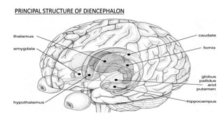
.
NEROPSYCHOLOGY
FOREBRAIN
The forebrain surrounds the rostral end of the neural tube. Its two major
components are the telencephalon and the diencephalon.
 Telencephalon
 The telencephalon includes most of the two symmetrical cerebral
hemispheres that make up the cerebrum.
 The cerebral hemispheres are covered by the cerebral cortex and contain
the limbic system and the basal ganglia.
 The latter two sets of structures are primarily in the subcortical regions of
the brain—those located deep within it, beneath the cerebral cortex.
CEREBRAL CORTEX
Cerebral Cortex
 Cerebral Cortex surrounds the cerebral hemispheres like the bark of
a tree.
 In humans the cerebral cortex is greatly convoluted; consisting of
 sulci (small grooves)
 fissures (large grooves)
 gyri (bulges between adjacent sulci or fissures)
 In fact, two-thirds of the surface of the cortex is hidden in the grooves;
thus, the presence of these convolutions triples the area of the cerebral
cortex.
 The total surface area is approximately 2360 cm2 (2.5 ft2), and the
thickness is approximately 3 mm.
 The cerebral cortex consists mostly of glia and the cell bodies, dendrites,
and interconnecting axons of neurons.
Cerebral Cortex
Since cell bodies predominate,
giving the cerebral cortex a grayish
tan appearance, it is referred to as
Gray matter.
 Beneath the cerebral cortex run
millions of axons that connect the
neurons of the cerebral cortex with
those located elsewhere in the
brain.
The large concentration of myelin
gives this tissue an opaque white
appearance—hence the term white
matter.
ROLE IN SENSORY FUNCTIONS
 Three areas of the cerebral cortex receive information from the
sensory organs.
 The primary visual cortex, which receives visual information, is located at the
back of the brain, on the inner surfaces of the cerebral hemispheres—
primarily on the upper and lower banks of the calcarine fissure.
The primary auditory cortex, which receives auditory information, is located
on the lower surface of a deep fissure in the side of the brain—the lateral
fissure.
The primary somatosensory cortex, a vertical strip of cortex just caudal to the
central sulcus, receives information from the body senses. Different regions
of the primary somatosensory cortex receive information from different
regions of the body.
 With the exception of olfaction and gustation (taste), sensory information from
the body or the environment is sent to primary sensory cortex of the
contralateral hemisphere.
 Thus, the primary somatosensory cortex of the left hemisphere learns what the
right hand is holding, the left primary visual cortex learns what is happening
toward the person’s right, and so on.
 The region of the cerebral cortex that is most directly involved in the control of
movement is the primary motor cortex, located just in front of the primary
somatosensory cortex.
 Neurons in different parts of the primary motor cortex are connected to
muscles in different parts of the body.
• The rostral region is involved
in movement-related
activities, such as planning
and executing behaviors. The
caudal region is involved in
perceiving and learning.
• The cerebral cortex is
divided into four areas, or
lobes, named for the bones
of the skull that cover them:
• the frontal lobe
• parietal lobe
• temporal lobe
• occipital lobe.
LOBES OF THE BRAIN
 The brain contains two of each lobe, one in each hemisphere. The frontal lobe
(the “front”) includes everything in front of the central sulcus.
 The parietal lobe (the “wall”) is located on the side of the cerebral
hemisphere, just behind the central sulcus, caudal to the frontal lobe.
 The temporal lobe (the “temple”) juts forward from the base of the brain,
ventral to the frontal and parietal lobes. The occipital lobe (from the Latin
ob,“in back of,” and caput, “head”) lies at the very back of the brain, caudal to
the parietal and temporal lobes.
Two cerebral hemispheres cooperate with each other, they do not perform identical
functions. Some functions are lateralized—located primarily on one side of the brain.
LEFT HEMISPHERE
Participates in the analysis of information—
the extraction of the elements that make up
the whole of an experience.
This ability makes the left hemisphere
particularly good at recognizing serial
events—events whose elements occur one
after the other—and controlling sequences
of behavior.
 The serial functions that are performed by
the left hemisphere include verbal activities,
such as talking, understanding the speech of
other people, reading, and writing.
RIGHT HEMISPHERE
 Specialized for synthesis .
 Putting isolated elements together to
perceive things as a whole. For example, our
ability to draw sketches (especially of three-
dimensional objects), read maps, and
construct complex objects out of smaller
elements depends heavily on circuits of
neurons that are located in the right
hemisphere.
CORPUS CALLOSUM
We are not aware of the fact that each hemisphere perceives the world
differently. Although the two cerebral hemispheres perform somewhat
different functions, our perceptions and our memories are unified. This
unity is accomplished by the corpus callosum, a large band of axons that
connects corresponding parts of the cerebral cortex of the left and right
hemispheres:
The left and right temporal lobes are connected, the left and right parietal
lobes are connected, and so on. Because of the corpus callosum, each
region of the association cortex knows what is happening in the
corresponding region of the opposite side of the brain.
The corpus callosum also makes a few asymmetrical connections that link
different regions of the two hemispheres.
SUBCORTICAL FOREBRAIN
(DIENCEPHALON)
SUBCORTICAL FOREBRAIN
(DIENCEPHALON)
•The second major division of the forebrain, the diencephalon,
is situated between the telencephalon and the mesencephalon;
it surrounds the third ventricle.
•The diencephalon (“interbrain”) is the region of the vertebrate neural tube that gives rise
to posterior forebrain structures.
•It is made up of three distinct components: the thalamus, the hypothalamus, and the
epithalamus.
PRINCIPAL STRUCTURE OF DIENCEPHALON
THALAMUS
• The thalamus (from the Greek thalamos,“inner chamber”) makes up the dorsal part of the
diencephalon.
• It resembles two avocados joined side by side, one in the left hemisphere and one in the right.
• It is a small structure in the center of the brain that acts as a relay center for sensory and motor
information.
• The thalamus is divided into several nuclei. Some thalamic nuclei receive sensory information from the
sensory systems.
• For example, the lateral geniculate nucleus receives information from the eye and sends axons to the
primary visual cortex, and the medial geniculate nucleus receives information from the inner ear and
sends axons to the primary auditory cortex.
• The thalamus plays a major role in regulating arousal, awareness level, and activity.
• Damage to the thalamus may produce resting tremor, chorea or rapid, jerky involuntary movements,
and dystonia or uncontrolled movement.
HYPOTHALAMUS
• The group of nuclei of the diencephalon situated beneath the thalamus.
• It controls the autonomic nervous system and the endocrine system and organizes behaviors related to
survival of the species—the so-called four F ’s: fighting, feeding, fleeing, and mating.
• The hypothalamus produces and secretes a wide variety of neurohormones that lead to the release or
inhibition of pituitary gland hormone
• In response to message from the hypothalamus, the pituitary synthesizes hormones that the blood
carries to organs throughout the body
• The posterior pituitary is composed of tissue derived from the hypothalamus, whereas the anterior
pituitary is derived from epithelial tissue
• For example, gonadotropin-releasing hormone causes the anterior pituitary gland to secrete the
gonadotropic hormones, which play a role in reproductive physiology and behavior
• Damage to any hypothalamic nucleus leads to abnormalities in motivated behaviours such as feeding,
drinking,temperature regulation, sexual behaviour,fighting.or activity level
EPITHALAMUS
• The Epithalamus is a dorsal posterior segment of the diencephalon
• It includes the habenula and their interconnecting fibers (the habenular
commissure), the stria medullaris, and the pineal gland
• The habenular commissure is a band of nerve fibers situated in front of the pineal
gland that connects the habenular nuclei on both sides of the diencephalon
• The stria medullaris, also known as stria medullaris thalami, is a fiber bundle
containing afferent fibers from the septal nuclei, lateral preoptic hypothalamic region,
and anterior thalamic nuclei to the habenula
EPITHALAMIC FUNCTIONS
• A main function of the epithalamus is the secretion of melatonin by the pineal gland.
• It also helps in regulation of motor pathways and emotions.
• The epithalamus is connected with both the limbic system and the basal ganglia.
BASAL GANGLIA
• Basal ganglia,a group of subcortical structures lateral to the thalamus,include three
major structures:the caudate nucleus, the putamen,and the globus pallidus.
• Basal ganglia are important for attention, language,planning,and other cognitive
functions.
• Damage to basal ganglia impairs movement,as in condition such as Parkinson’s
disease and Huntigton’s disease.
HIPPOCAMPUS
• The Hippocampus (from the Latin word meaning “seahorse”,a shape suggested by the
hippocampus) is a large structure between the thalamus and the cerebral cortex,mostly
towards the posterior of the forebrain
• The Hippocampus is the part of the brain that plays an important role in
forming,organizing and storing memory and also associate with spatial navigation
• People with hippocampus damage have trouble in storing new memories,but they do
not lose all the memories they had before the damage occurred
• For eg; The hippocampus seems to be involved in some mental illnesses.In
schizophrenia and some severe dipressions,where the hippocampus shrinks
BRAIN STEM
BRAIN STEM
• On entering the skull, the spinal cord
becomes the brain stem, which is formed of
three principal structures:
1. the medulla oblongata (“oblong
marrow”)
2. the pons (“bridge”)
3. and the midbrain
• The cerebellum sits astride this region and
forms massive connections with it
• It connects the cerebrum with the spinal cord.
ANATOMICAL RELATIONS:
• The brainstem is located in posterior
side of the brain -
• Above, the midbrain is continuous
with the cerebral hemisphere.
• Below, the medulla is continuous
with the spinal cord.
• Posteriorly, the pons and medulla
are separated from the cerebellum
by the fourth ventricle.
• Motor and sensory neurons travel through the brainstem allowing for
the relay of signals between the brain and the spinal cord.
• The brainstem coordinates motor control signals sent from the ​brain
to the body.
• Most cranial nerves are found in the brainstem.
• This brain region also controls life supporting autonomic functions of
the peripheral nervous system.
FUNCTIONS
• The midbrain : Visual information is relayed to this part of the brain and
some forms of visual reflex, for instance, blinking, pupillary constriction,
and certain eye movements, are organized here.
• Similarly, auditory reflexes, for example, starting at a sudden noise,
originate in this region.
• Autonomic functions : salivation, control of blood pressure, respiration,
and movements in the alimentary canal are influenced through the ANS
from this region.
• Reticular formation : Cells in this formation communicate with cells
throughout the spinal cord, cerebellum, and the higher regions of the
brain.
• A particular feature is the ascending reticular activating system (ARAS).
This system receives inputs from sensory pathways on their upward
journey, and projects onto the cerebral cortex through a variety of
intermediate centers.
• The result is a generalized arousal of the whole forebrain, and is an
important component in wakefulness, in the maintenance of
consciousness, and in attention.
• The cranial nerves : They consist of a set of twelve pairs of nerves, some sensory,
some motor, and some combining both functions, which almost entirely project to
the head and neck.
• Apart from sub serving sensation in this region, and providing motor control, the
cranial nerves serve the special senses through the olfactory nerves (smell), the
optic nerves (vision), the auditory nerves (hearing and equilibrium), and the facial,
glossopharyngeal, and vagus nerves (taste).
• Eye movements are also directly controlled by the cranial nerves.
• The brain stem is concerned with the special senses, with vital processes, and with
other visceral and somatic functions, all of which may be modified by impulses
entering by the cranial nerves, down from the cerebellum or from the forebrain.
MIDBRAIN
.
• The midbrain—also called the mesencephalon—is the smallest portion of the brain. It is
underneath the cerebral cortex, just above the brain stem, and is located toward the
centre of the brain
• Major structures –
1. TECTUM – The tectum is located in the dorsal portion of the midbrain. Its principle
structures are the superior colliculi and the inferior colliculi, which appears as four
bumps on the dorsal surface of the brain stem.
• The superior colliculus receives input from the retina and the visual cortex and
participates in a variety of visual reflexes, particularly the tracking of objects in the
visual field
• The inferior colliculus is a part of the midbrain that serves as a main auditory (sound)
center for the body.
2. TEGMENTUM : The tegmentum lies beneath the tectum. It includes the the
reticular formation, the periaqueductal gray matter, the red nucleus and substantia
nigra.
a) RETICULAR FORMATION – It is a large structure characterized by a diffuse ,
interconnected network of neurons. It occupies the core the brain stem, from the
lower border of the medulla to upper border of the midbrain.
• It receives sensory information by means of various pathways and projects axon to
the cerebral cortex, thalamus and spinal cord
• It plays a role in sleep and arousal, attention, muscle tonus, movement and various
vital reflexes
b)THE PERI-AQUEDUCTAL GRAY MATTER:
• It is so called because it consist of mostly of cell-body of neurons
• It contains neural circuits that control sequence of movements that constitute
species -typical behavior such as fighting
• It appears to function primarily in pain suppression as result of its naturally high
concentrations of endorphins
c) THE RED NUCLEUS :
• It is a large structure located centrally within tegmentum that is involved in the
coordination of sensorimotor information
• A bundle of axons that arises from red nucleus constitutes one of the major fiber
system that bring motor information from cerebral cortex and cerebellum to the
spinal cord
.
d) THE SUBSTANTIA NIGRA : “ black substance”
Darkly pigmented cluster of neurons with cells (containing melanin – the sleep
hormone) that make the neurotransmitter dopamine
Therefore, controls voluntary movement, controls the brain Reward system & regulates
sleep wake cycle
3)CEREBRAL PEDUNCLES -
• The back of the midbrain contains a pair of large nerve fibre bundles that connect the
rest of the brainstem to the forebrain
• These cerebral peduncles are the main highway for signals that need to be
transported from the cortex to other parts of the central nervous system (CNS), and
are especially important for body coordination
DYSFUNCTION :
• Neurodegeneration of nerve cells in the substantia nigra results in a drop off of
dopamine production. Significant loss in dopamine levels (60-80%) may result in
the development of Parkinson's disease
• Parinaud's syndrome - Inability to move the eyes up and down. Results from
injury to the dorsal midbrain
• Weber's syndrome, caused by midbrain infarction is characterized by spastic
half sided slight paralysis or weakness in the body , loss of facial muscles with
the exception of the upper part of the facial muscles and a half sided failure of
the tongue
• Benedikt syndrome, caused by a lesion (infarction, hemorrhage,
tumor, or tuberculosis) in the tegmentum of the midbrain and
cerebellum.
• People with this syndrome have a palsy or a waddling walk through coordination
disorder (ataxia).
- involuntary vibrations which occur when performing a deliberate operation
(intention tremor)
- abnormal involuntary movements (hyperkinesias)
HINDBRAIN
Introduction
• Hindbrain, as per Greek roots is commonly called as Rhomb encephalon (literally, meaning
parallelogram-brain). It is the posterior part of the brain.
Rhomb
encephalon
Myelencephalon
(after brain)
Pons Cerebellum
Myelencephalon
(marrow-brain)
Medulla
oblongata
Met encephalon
•Cerebellum
• The cerebellum (“little brain”), with its two hemispheres, resembles a miniature version of the
cerebrum.
• It is covered by the cerebellar cortex and has a set of deep cerebellar nuclei. These nuclei receive
projections from the cerebellar cortex and themselves send projections out of the cerebellum to
other parts of the brain.
• Each hemisphere of the cerebellum is attached to the dorsal surface of the Pons by bundles of
axons: the superior, middle, and inferior cerebellar peduncles (“little feet”).
Functions of cerebellum
• The cerebellum’s main role is to coordinate muscular activity, both in postural and locomotors
mechanisms, all of which is carried out at a subconscious level.
• Most positions of the body are achieved by the antagonistic activity of opposed groups of muscles.
These muscles, if not properly coordinated, are sufficiently powerful to break bones or at least to tear
other muscles out of position. It is therefore vital that these groups of muscles be carefully
coordinated.
• Cerebellum, thus play an important role in motor control as we can achieve this feat of control so
effortlessly, and not only avoid self-injury but also execute the most delicate, accurate, smooth, and
graceful movements.
• The cerebellum achieves this control by receiving information from the skin and from muscles,
tendons, joints, and the semicircular canals (the organs of positional sense and balance near the ear),
as well as from the visual and auditory systems.
• In turn, it discharges out to the cerebral cortex and spinal cord, executing control over the timing of
the execution of motor events. It thus ensures smooth, controlled, and well organized movement.
• The cerebellum integrates this information and modifies the motor outflow, exerting a coordinating
and smoothing effect on the movements.
Dysfunctions to cerebellum
• People with cerebellar damage are clumsy and lose their balance, but the functions of the
cerebellum extend far beyond balance and coordination. (A virtuoso pianist or other performing
musician owes much to his or her cerebellum).
• People with damage to the cerebellum have trouble shifting their attention back and forth
between auditory and visual stimuli.
• They have much difficulty with timing, including sensory timing. For example, they are poor at
judging whether one rhythm is faster than another.
Pons
• The Pons, a large bulge in the brain stem, lies between the mesencephalon and medulla oblongata,
immediately ventral to the cerebellum.
• Pons means “bridge,” but it does not really look like one. the name reflects the fact that many axons
in the Pons cross from one side of the brain to the other.
• This is in fact the location where axons from each half of the brain cross to the opposite side of the
spinal cord so that the left hemisphere controls the muscles of the right side of the body and the right
hemisphere controls the left side.
• The Pons contains, in its core, a portion of the reticular formation, including some nuclei that appear
to be important in sleep and arousal. It also contains a large nucleus that relays information from the
cerebral cortex to the cerebellum.
• The medulla and Pons also contain the reticular formation and the raphe system.
• The reticular formation has descending and ascending portions. The descending portion is one of
several brain areas that controls the motor areas of the spinal cord.
• The ascending portion sends output to much of the cerebral cortex, selectively increasing arousal and
attention in one area or another.
• The raphe system also sends axons to much of the forebrain, modifying the brain’s readiness to
respond to stimuli.
Myelencephalon
•Medulla
• The medulla, or medulla oblongata, is just above the spinal cord and could be regarded as
an enlarged extension of the spinal cord but located in the skull.
• The medulla controls some vital reflexes—including breathing, heart rate, vomiting,
salivation, coughing, and sneezing—through the cranial nerves, which control sensations
from the head, muscle movements in the head, and much of the parasympathetic output to
the organs.
• Some of the cranial nerves include both sensory and motor components; others have just
one or the other.
• Just as the lower parts of the body are connected to the spinal cord via sensory and motor nerves,
the receptors and muscles of the head and organs connect to the brain by 12 pairs of cranial nerves
(one of each pair on the right of the brain and one on the left)
• Each cranial nerve originates in a nucleus (cluster of neurons) that integrates the sensory
information, regulates the motor output, or both.
• The cranial nerve nuclei for nerves V through XII are in the medulla and Pons. Those for cranial nerves
I through IV are in the midbrain and forebrain
• Damage to the medulla is frequently fatal, and large doses of opiates are life-threatening because they
suppress activity of the medulla.
THE END
THANK YOU….!!!!

Neuropsychology.pptx

  • 1.
  • 2.
  • 3.
    The forebrain surroundsthe rostral end of the neural tube. Its two major components are the telencephalon and the diencephalon.  Telencephalon  The telencephalon includes most of the two symmetrical cerebral hemispheres that make up the cerebrum.  The cerebral hemispheres are covered by the cerebral cortex and contain the limbic system and the basal ganglia.  The latter two sets of structures are primarily in the subcortical regions of the brain—those located deep within it, beneath the cerebral cortex.
  • 4.
  • 5.
    Cerebral Cortex  CerebralCortex surrounds the cerebral hemispheres like the bark of a tree.  In humans the cerebral cortex is greatly convoluted; consisting of  sulci (small grooves)  fissures (large grooves)  gyri (bulges between adjacent sulci or fissures)  In fact, two-thirds of the surface of the cortex is hidden in the grooves; thus, the presence of these convolutions triples the area of the cerebral cortex.  The total surface area is approximately 2360 cm2 (2.5 ft2), and the thickness is approximately 3 mm.  The cerebral cortex consists mostly of glia and the cell bodies, dendrites, and interconnecting axons of neurons.
  • 6.
    Cerebral Cortex Since cellbodies predominate, giving the cerebral cortex a grayish tan appearance, it is referred to as Gray matter.  Beneath the cerebral cortex run millions of axons that connect the neurons of the cerebral cortex with those located elsewhere in the brain. The large concentration of myelin gives this tissue an opaque white appearance—hence the term white matter.
  • 7.
    ROLE IN SENSORYFUNCTIONS  Three areas of the cerebral cortex receive information from the sensory organs.  The primary visual cortex, which receives visual information, is located at the back of the brain, on the inner surfaces of the cerebral hemispheres— primarily on the upper and lower banks of the calcarine fissure. The primary auditory cortex, which receives auditory information, is located on the lower surface of a deep fissure in the side of the brain—the lateral fissure. The primary somatosensory cortex, a vertical strip of cortex just caudal to the central sulcus, receives information from the body senses. Different regions of the primary somatosensory cortex receive information from different regions of the body.
  • 8.
     With theexception of olfaction and gustation (taste), sensory information from the body or the environment is sent to primary sensory cortex of the contralateral hemisphere.  Thus, the primary somatosensory cortex of the left hemisphere learns what the right hand is holding, the left primary visual cortex learns what is happening toward the person’s right, and so on.  The region of the cerebral cortex that is most directly involved in the control of movement is the primary motor cortex, located just in front of the primary somatosensory cortex.  Neurons in different parts of the primary motor cortex are connected to muscles in different parts of the body.
  • 9.
    • The rostralregion is involved in movement-related activities, such as planning and executing behaviors. The caudal region is involved in perceiving and learning. • The cerebral cortex is divided into four areas, or lobes, named for the bones of the skull that cover them: • the frontal lobe • parietal lobe • temporal lobe • occipital lobe.
  • 10.
    LOBES OF THEBRAIN  The brain contains two of each lobe, one in each hemisphere. The frontal lobe (the “front”) includes everything in front of the central sulcus.  The parietal lobe (the “wall”) is located on the side of the cerebral hemisphere, just behind the central sulcus, caudal to the frontal lobe.  The temporal lobe (the “temple”) juts forward from the base of the brain, ventral to the frontal and parietal lobes. The occipital lobe (from the Latin ob,“in back of,” and caput, “head”) lies at the very back of the brain, caudal to the parietal and temporal lobes.
  • 11.
    Two cerebral hemispherescooperate with each other, they do not perform identical functions. Some functions are lateralized—located primarily on one side of the brain. LEFT HEMISPHERE Participates in the analysis of information— the extraction of the elements that make up the whole of an experience. This ability makes the left hemisphere particularly good at recognizing serial events—events whose elements occur one after the other—and controlling sequences of behavior.  The serial functions that are performed by the left hemisphere include verbal activities, such as talking, understanding the speech of other people, reading, and writing. RIGHT HEMISPHERE  Specialized for synthesis .  Putting isolated elements together to perceive things as a whole. For example, our ability to draw sketches (especially of three- dimensional objects), read maps, and construct complex objects out of smaller elements depends heavily on circuits of neurons that are located in the right hemisphere.
  • 12.
    CORPUS CALLOSUM We arenot aware of the fact that each hemisphere perceives the world differently. Although the two cerebral hemispheres perform somewhat different functions, our perceptions and our memories are unified. This unity is accomplished by the corpus callosum, a large band of axons that connects corresponding parts of the cerebral cortex of the left and right hemispheres: The left and right temporal lobes are connected, the left and right parietal lobes are connected, and so on. Because of the corpus callosum, each region of the association cortex knows what is happening in the corresponding region of the opposite side of the brain. The corpus callosum also makes a few asymmetrical connections that link different regions of the two hemispheres.
  • 13.
  • 14.
    SUBCORTICAL FOREBRAIN (DIENCEPHALON) •The secondmajor division of the forebrain, the diencephalon, is situated between the telencephalon and the mesencephalon; it surrounds the third ventricle. •The diencephalon (“interbrain”) is the region of the vertebrate neural tube that gives rise to posterior forebrain structures. •It is made up of three distinct components: the thalamus, the hypothalamus, and the epithalamus.
  • 15.
  • 16.
    THALAMUS • The thalamus(from the Greek thalamos,“inner chamber”) makes up the dorsal part of the diencephalon. • It resembles two avocados joined side by side, one in the left hemisphere and one in the right. • It is a small structure in the center of the brain that acts as a relay center for sensory and motor information. • The thalamus is divided into several nuclei. Some thalamic nuclei receive sensory information from the sensory systems. • For example, the lateral geniculate nucleus receives information from the eye and sends axons to the primary visual cortex, and the medial geniculate nucleus receives information from the inner ear and sends axons to the primary auditory cortex. • The thalamus plays a major role in regulating arousal, awareness level, and activity. • Damage to the thalamus may produce resting tremor, chorea or rapid, jerky involuntary movements, and dystonia or uncontrolled movement.
  • 17.
    HYPOTHALAMUS • The groupof nuclei of the diencephalon situated beneath the thalamus. • It controls the autonomic nervous system and the endocrine system and organizes behaviors related to survival of the species—the so-called four F ’s: fighting, feeding, fleeing, and mating. • The hypothalamus produces and secretes a wide variety of neurohormones that lead to the release or inhibition of pituitary gland hormone • In response to message from the hypothalamus, the pituitary synthesizes hormones that the blood carries to organs throughout the body • The posterior pituitary is composed of tissue derived from the hypothalamus, whereas the anterior pituitary is derived from epithelial tissue • For example, gonadotropin-releasing hormone causes the anterior pituitary gland to secrete the gonadotropic hormones, which play a role in reproductive physiology and behavior • Damage to any hypothalamic nucleus leads to abnormalities in motivated behaviours such as feeding, drinking,temperature regulation, sexual behaviour,fighting.or activity level
  • 18.
    EPITHALAMUS • The Epithalamusis a dorsal posterior segment of the diencephalon • It includes the habenula and their interconnecting fibers (the habenular commissure), the stria medullaris, and the pineal gland • The habenular commissure is a band of nerve fibers situated in front of the pineal gland that connects the habenular nuclei on both sides of the diencephalon • The stria medullaris, also known as stria medullaris thalami, is a fiber bundle containing afferent fibers from the septal nuclei, lateral preoptic hypothalamic region, and anterior thalamic nuclei to the habenula
  • 19.
    EPITHALAMIC FUNCTIONS • Amain function of the epithalamus is the secretion of melatonin by the pineal gland. • It also helps in regulation of motor pathways and emotions. • The epithalamus is connected with both the limbic system and the basal ganglia. BASAL GANGLIA • Basal ganglia,a group of subcortical structures lateral to the thalamus,include three major structures:the caudate nucleus, the putamen,and the globus pallidus. • Basal ganglia are important for attention, language,planning,and other cognitive functions. • Damage to basal ganglia impairs movement,as in condition such as Parkinson’s disease and Huntigton’s disease.
  • 20.
    HIPPOCAMPUS • The Hippocampus(from the Latin word meaning “seahorse”,a shape suggested by the hippocampus) is a large structure between the thalamus and the cerebral cortex,mostly towards the posterior of the forebrain • The Hippocampus is the part of the brain that plays an important role in forming,organizing and storing memory and also associate with spatial navigation • People with hippocampus damage have trouble in storing new memories,but they do not lose all the memories they had before the damage occurred • For eg; The hippocampus seems to be involved in some mental illnesses.In schizophrenia and some severe dipressions,where the hippocampus shrinks
  • 21.
  • 22.
    BRAIN STEM • Onentering the skull, the spinal cord becomes the brain stem, which is formed of three principal structures: 1. the medulla oblongata (“oblong marrow”) 2. the pons (“bridge”) 3. and the midbrain • The cerebellum sits astride this region and forms massive connections with it • It connects the cerebrum with the spinal cord.
  • 23.
    ANATOMICAL RELATIONS: • Thebrainstem is located in posterior side of the brain - • Above, the midbrain is continuous with the cerebral hemisphere. • Below, the medulla is continuous with the spinal cord. • Posteriorly, the pons and medulla are separated from the cerebellum by the fourth ventricle.
  • 24.
    • Motor andsensory neurons travel through the brainstem allowing for the relay of signals between the brain and the spinal cord. • The brainstem coordinates motor control signals sent from the ​brain to the body. • Most cranial nerves are found in the brainstem. • This brain region also controls life supporting autonomic functions of the peripheral nervous system.
  • 25.
    FUNCTIONS • The midbrain: Visual information is relayed to this part of the brain and some forms of visual reflex, for instance, blinking, pupillary constriction, and certain eye movements, are organized here. • Similarly, auditory reflexes, for example, starting at a sudden noise, originate in this region. • Autonomic functions : salivation, control of blood pressure, respiration, and movements in the alimentary canal are influenced through the ANS from this region.
  • 26.
    • Reticular formation: Cells in this formation communicate with cells throughout the spinal cord, cerebellum, and the higher regions of the brain. • A particular feature is the ascending reticular activating system (ARAS). This system receives inputs from sensory pathways on their upward journey, and projects onto the cerebral cortex through a variety of intermediate centers. • The result is a generalized arousal of the whole forebrain, and is an important component in wakefulness, in the maintenance of consciousness, and in attention.
  • 27.
    • The cranialnerves : They consist of a set of twelve pairs of nerves, some sensory, some motor, and some combining both functions, which almost entirely project to the head and neck. • Apart from sub serving sensation in this region, and providing motor control, the cranial nerves serve the special senses through the olfactory nerves (smell), the optic nerves (vision), the auditory nerves (hearing and equilibrium), and the facial, glossopharyngeal, and vagus nerves (taste). • Eye movements are also directly controlled by the cranial nerves. • The brain stem is concerned with the special senses, with vital processes, and with other visceral and somatic functions, all of which may be modified by impulses entering by the cranial nerves, down from the cerebellum or from the forebrain.
  • 28.
  • 29.
    . • The midbrain—alsocalled the mesencephalon—is the smallest portion of the brain. It is underneath the cerebral cortex, just above the brain stem, and is located toward the centre of the brain • Major structures – 1. TECTUM – The tectum is located in the dorsal portion of the midbrain. Its principle structures are the superior colliculi and the inferior colliculi, which appears as four bumps on the dorsal surface of the brain stem. • The superior colliculus receives input from the retina and the visual cortex and participates in a variety of visual reflexes, particularly the tracking of objects in the visual field • The inferior colliculus is a part of the midbrain that serves as a main auditory (sound) center for the body.
  • 30.
    2. TEGMENTUM :The tegmentum lies beneath the tectum. It includes the the reticular formation, the periaqueductal gray matter, the red nucleus and substantia nigra. a) RETICULAR FORMATION – It is a large structure characterized by a diffuse , interconnected network of neurons. It occupies the core the brain stem, from the lower border of the medulla to upper border of the midbrain. • It receives sensory information by means of various pathways and projects axon to the cerebral cortex, thalamus and spinal cord • It plays a role in sleep and arousal, attention, muscle tonus, movement and various vital reflexes
  • 31.
    b)THE PERI-AQUEDUCTAL GRAYMATTER: • It is so called because it consist of mostly of cell-body of neurons • It contains neural circuits that control sequence of movements that constitute species -typical behavior such as fighting • It appears to function primarily in pain suppression as result of its naturally high concentrations of endorphins c) THE RED NUCLEUS : • It is a large structure located centrally within tegmentum that is involved in the coordination of sensorimotor information • A bundle of axons that arises from red nucleus constitutes one of the major fiber system that bring motor information from cerebral cortex and cerebellum to the spinal cord
  • 32.
    . d) THE SUBSTANTIANIGRA : “ black substance” Darkly pigmented cluster of neurons with cells (containing melanin – the sleep hormone) that make the neurotransmitter dopamine Therefore, controls voluntary movement, controls the brain Reward system & regulates sleep wake cycle 3)CEREBRAL PEDUNCLES - • The back of the midbrain contains a pair of large nerve fibre bundles that connect the rest of the brainstem to the forebrain • These cerebral peduncles are the main highway for signals that need to be transported from the cortex to other parts of the central nervous system (CNS), and are especially important for body coordination
  • 33.
    DYSFUNCTION : • Neurodegenerationof nerve cells in the substantia nigra results in a drop off of dopamine production. Significant loss in dopamine levels (60-80%) may result in the development of Parkinson's disease
  • 34.
    • Parinaud's syndrome- Inability to move the eyes up and down. Results from injury to the dorsal midbrain • Weber's syndrome, caused by midbrain infarction is characterized by spastic half sided slight paralysis or weakness in the body , loss of facial muscles with the exception of the upper part of the facial muscles and a half sided failure of the tongue • Benedikt syndrome, caused by a lesion (infarction, hemorrhage, tumor, or tuberculosis) in the tegmentum of the midbrain and cerebellum. • People with this syndrome have a palsy or a waddling walk through coordination disorder (ataxia). - involuntary vibrations which occur when performing a deliberate operation (intention tremor) - abnormal involuntary movements (hyperkinesias)
  • 35.
  • 36.
    Introduction • Hindbrain, asper Greek roots is commonly called as Rhomb encephalon (literally, meaning parallelogram-brain). It is the posterior part of the brain. Rhomb encephalon Myelencephalon (after brain) Pons Cerebellum Myelencephalon (marrow-brain) Medulla oblongata
  • 37.
    Met encephalon •Cerebellum • Thecerebellum (“little brain”), with its two hemispheres, resembles a miniature version of the cerebrum. • It is covered by the cerebellar cortex and has a set of deep cerebellar nuclei. These nuclei receive projections from the cerebellar cortex and themselves send projections out of the cerebellum to other parts of the brain. • Each hemisphere of the cerebellum is attached to the dorsal surface of the Pons by bundles of axons: the superior, middle, and inferior cerebellar peduncles (“little feet”).
  • 40.
    Functions of cerebellum •The cerebellum’s main role is to coordinate muscular activity, both in postural and locomotors mechanisms, all of which is carried out at a subconscious level. • Most positions of the body are achieved by the antagonistic activity of opposed groups of muscles. These muscles, if not properly coordinated, are sufficiently powerful to break bones or at least to tear other muscles out of position. It is therefore vital that these groups of muscles be carefully coordinated. • Cerebellum, thus play an important role in motor control as we can achieve this feat of control so effortlessly, and not only avoid self-injury but also execute the most delicate, accurate, smooth, and graceful movements.
  • 41.
    • The cerebellumachieves this control by receiving information from the skin and from muscles, tendons, joints, and the semicircular canals (the organs of positional sense and balance near the ear), as well as from the visual and auditory systems. • In turn, it discharges out to the cerebral cortex and spinal cord, executing control over the timing of the execution of motor events. It thus ensures smooth, controlled, and well organized movement. • The cerebellum integrates this information and modifies the motor outflow, exerting a coordinating and smoothing effect on the movements.
  • 42.
    Dysfunctions to cerebellum •People with cerebellar damage are clumsy and lose their balance, but the functions of the cerebellum extend far beyond balance and coordination. (A virtuoso pianist or other performing musician owes much to his or her cerebellum). • People with damage to the cerebellum have trouble shifting their attention back and forth between auditory and visual stimuli. • They have much difficulty with timing, including sensory timing. For example, they are poor at judging whether one rhythm is faster than another.
  • 43.
    Pons • The Pons,a large bulge in the brain stem, lies between the mesencephalon and medulla oblongata, immediately ventral to the cerebellum. • Pons means “bridge,” but it does not really look like one. the name reflects the fact that many axons in the Pons cross from one side of the brain to the other. • This is in fact the location where axons from each half of the brain cross to the opposite side of the spinal cord so that the left hemisphere controls the muscles of the right side of the body and the right hemisphere controls the left side. • The Pons contains, in its core, a portion of the reticular formation, including some nuclei that appear to be important in sleep and arousal. It also contains a large nucleus that relays information from the cerebral cortex to the cerebellum.
  • 45.
    • The medullaand Pons also contain the reticular formation and the raphe system. • The reticular formation has descending and ascending portions. The descending portion is one of several brain areas that controls the motor areas of the spinal cord. • The ascending portion sends output to much of the cerebral cortex, selectively increasing arousal and attention in one area or another. • The raphe system also sends axons to much of the forebrain, modifying the brain’s readiness to respond to stimuli.
  • 46.
    Myelencephalon •Medulla • The medulla,or medulla oblongata, is just above the spinal cord and could be regarded as an enlarged extension of the spinal cord but located in the skull. • The medulla controls some vital reflexes—including breathing, heart rate, vomiting, salivation, coughing, and sneezing—through the cranial nerves, which control sensations from the head, muscle movements in the head, and much of the parasympathetic output to the organs. • Some of the cranial nerves include both sensory and motor components; others have just one or the other.
  • 47.
    • Just asthe lower parts of the body are connected to the spinal cord via sensory and motor nerves, the receptors and muscles of the head and organs connect to the brain by 12 pairs of cranial nerves (one of each pair on the right of the brain and one on the left) • Each cranial nerve originates in a nucleus (cluster of neurons) that integrates the sensory information, regulates the motor output, or both. • The cranial nerve nuclei for nerves V through XII are in the medulla and Pons. Those for cranial nerves I through IV are in the midbrain and forebrain • Damage to the medulla is frequently fatal, and large doses of opiates are life-threatening because they suppress activity of the medulla.
  • 48.