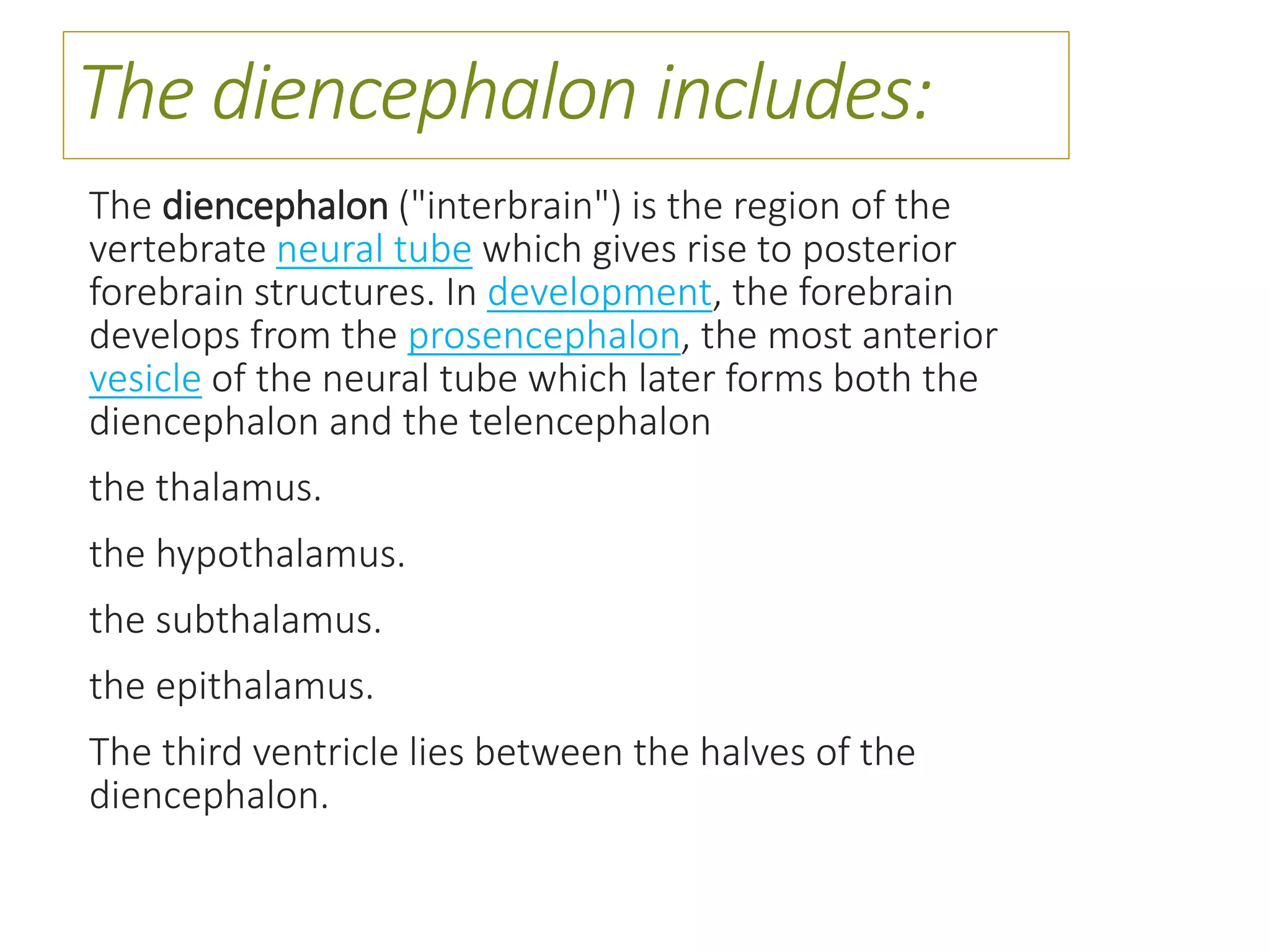
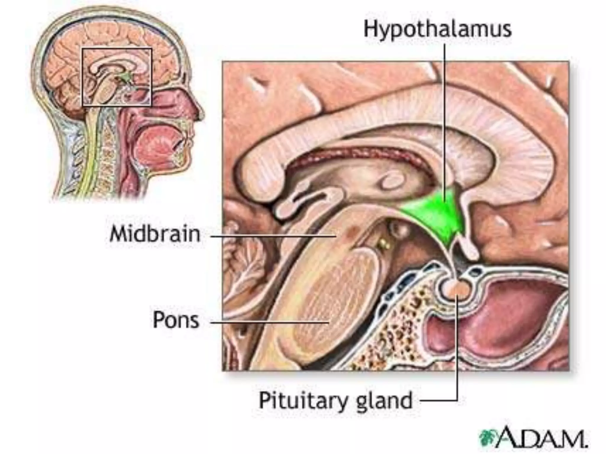
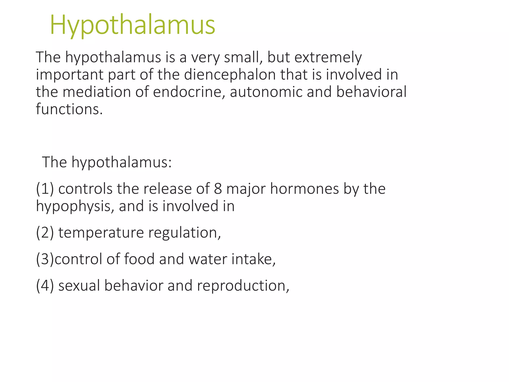
The diencephalon includes structures like the thalamus, hypothalamus, epithalamus, and subthalamus. The thalamus relays sensory and motor signals to the cerebral cortex. It contains nuclei that relay specific sensations like vision, hearing, and somatosensation. The hypothalamus controls autonomic functions and regulates behaviors related to hunger, thirst, temperature, sleep, and reproduction. It also controls the pituitary gland. The epithalamus includes the pineal gland and habenular nucleus. The subthalamus contains the subthalamic nucleus and is involved in motor control.
The diencephalon is the deep part of the forebrain located above the midbrain. It consists of four key parts: the thalamus, hypothalamus, epithalamus, and subthalamus. The thalamus acts as a relay station for all sensory systems except smell. The hypothalamus regulates functions like thirst, hunger, autonomic functions, and temperature. The epithalamus contains the pineal gland which regulates circadian rhythms. The subthalamus connects to motor control areas and the reticular activating system.
The document describes the anatomy and functions of the medulla oblongata. It is the lowest part of the brainstem located in the posterior cranial fossa. It connects the spinal cord to the forebrain and contains nuclei of cranial nerves III-XII. Key structures in the medulla include the pyramids, olives, inferior cerebellar peduncles, and nuclei that control vital functions like respiration and cardiovascular regulation. The document discusses the medulla at different transverse section levels to describe its internal organization and pathways for motor and sensory signals.
Development of Nervous System (Special Embryology)Dr. Sherif Fahmy
The document summarizes the development of the nervous system from the neural tube through formation of the spinal cord and brain. It discusses how the neural tube forms and closes, followed by differentiation of the spinal cord mantle layer. It also covers development of the brain vesicles and flexures, formation of the cerebellum from the rhombic lip, and development of the diencephalon, cerebral hemispheres, and common congenital anomalies.
EXTERNAL FEATURES OF MIDBRAIN, ANATOMY OF INTERNAL FEATURES OF MIDBRAIN, CRUS CEREBRI, SUBSTANTIA NIGRA, CEREBRAL PEDUNCLE,INFERIOR COLLICULUS,LEMNISCI
The document provides an overview of the diencephalon, which is the region of the brain between the cerebral hemispheres and below the thalamus. It describes the gross topography and borders of the diencephalon. The diencephalon includes the epithalamus, thalamus, metathalamus, subthalamus, and hypothalamus. Details are given on the structures and functions of these regions, including the thalamic nuclei and their connections. Clinical notes discuss lesions of the thalamus and how they can cause sensory loss or pain.
The brain stem is located between the cerebrum and spinal cord, and consists of the midbrain, pons, and medulla oblongata. The midbrain connects the forebrain to the pons and cerebellum. It contains important centers for visual and auditory reflexes, and gives rise to the trochlear nerve. Key structures in the midbrain include the superior and inferior colliculi, oculomotor nucleus, red nucleus, and substantia nigra. The midbrain serves to relay motor and sensory signals between the spinal cord and forebrain.
The document discusses the pituitary gland, also known as the hypophysis. It is a small gland located at the base of the brain that regulates several important body functions. The pituitary gland has two lobes - the anterior lobe which secretes hormones that control other endocrine glands, and the posterior lobe which stores and releases hormones involved in water balance and milk production. The pituitary gland is well protected in the sella turcica bone and receives blood flow through the hypophyseal portal system which allows the hypothalamus to regulate pituitary hormone secretion.
The document discusses the different types of white fibres in the brain, including association fibres, commissural fibres, and projection fibres. It describes key association fibre bundles like the cingulum and fornix. It also explains commissural fibres like the corpus callosum and anterior commissure. Projection fibres including the corticospinal tract are discussed, with details about fibre arrangement in the internal capsule and how lesions can cause motor or sensory deficits. Ascending and descending fibres passing through different parts of the internal capsule are identified.
The document summarizes key aspects of the cerebral cortex. It discusses the functional anatomy of the cerebral cortex, including its layers of neurons. It describes the relations of the cortex to the thalamus, and specific functions of motor, sensory, and association areas. Association areas integrate signals from multiple regions. Important association areas discussed include the parieto-occipitotemporal area, prefrontal area, and limbic area. The document also covers concepts such as the dominant hemisphere, functions in communication including language input and output, and thoughts, consciousness, and memory.
The pons is the bridge-shaped structure that connects the midbrain to the medulla oblongata. It contains fibers that connect the cerebellum and midbrain. The pons has a ventral surface with sulcus basilaris and lateral continuations with the middle cerebellar peduncle. It attaches to the 6th, 7th and 8th cranial nerves ventrally. Dorsally, it forms the upper part of the floor of the 4th ventricle. Internally, it contains longitudinal and transverse fibers, pontine nuclei in the basilar part, and ascending and descending tracts in the tegmental part such as the medial lemniscus and trigeminal lemn
MIDBRAIN basic anatomy and applied aspects.Pulak Agrawal
The document provides an overview of the anatomy and structures of the midbrain. It notes that the midbrain connects the pons and cerebellum to the forebrain, is about 0.8 inches long, and is traversed by the cerebral aqueduct filled with CSF. Key structures discussed include the crus cerebri, oculomotor nerve, superior and inferior colliculi, brachium, and trochlear nerve. The midbrain is divided into cerebral peduncles, tectum, and tegmentum. Transverse sections show structures like the substantia nigra, red nucleus, and tracts. Blood supply comes from the posterior cerebral, superior cerebellar, and basilar arteries.
The brain contains four interconnected fluid-filled cavities called ventricles that produce and circulate cerebrospinal fluid (CSF). The ventricles include the left and right lateral ventricles, the third ventricle, and the fourth ventricle. CSF is produced by choroid plexus tissue within the ventricles and flows between the ventricles through connecting openings before circulating around the brain and spinal cord to provide cushioning. Blockages within the ventricles or connections between them can cause excess fluid buildup and issues like hydrocephalus.
The document discusses the thalamus and hypothalamus. It provides details on the anatomy, internal structure, nuclei, connections and blood supply of the thalamus. It describes the relations, boundaries, nuclei and connections of the hypothalamus. The thalamus acts as a relay station and integrative center for sensory information to the cortex. The hypothalamus regulates the autonomic nervous system, endocrine system and limbic system through its nuclei and connections. Lesions of the thalamus can cause sensory loss, involuntary movements and effects on motor control through disruption of thalamic circuits.
The cerebellum plays an important role in motor control and coordination. It receives input from various sources, including the spinal cord, brainstem, and cerebral cortex. This input is processed within the cerebellar cortex and nuclei. The cerebellum then sends output to motor areas of the brainstem and cerebral cortex to coordinate movement, balance, and posture. It acts as a comparator, receiving feedback on actual movements and comparing them to intended movements, in order to calibrate motor output and prevent overshooting during voluntary motor acts such as walking and running. Damage to the cerebellum can cause ataxia or lack of coordination.
The document summarizes key anatomical structures related to the dura mater and cavernous sinus. It describes the layers of the meninges and identifies four dural folds - the falx cerebri, tentorium cerebelli, falx cerebelli, and diaphragma sella. It also details the location, relations, tributaries, and communications of the cavernous sinus.
The fourth ventricle is located ventral to the cerebellum and dorsal to the pons and medulla. It is bounded laterally by the gracile and cuneate tubercles and inferior cerebellar peduncles, and superiorly by the superior cerebellar peduncle. Its roof is formed by the superior cerebellar peduncle and medullary velum. Its floor contains landmarks like the median sulcus, facial colliculus, and hypoglossal triangle. Cerebrospinal fluid circulates from the fourth ventricle through the median aperture and exits into the subarachnoid space through the foramina of Luschka and Magendi.
The document summarizes the development, anatomy, and histology of the pons and midbrain. It describes that the pons develops from the metencephalon and receives cells from the myelencephalon. The midbrain develops from the mesencephalon. The document then provides detailed descriptions of the structures, tracts, nuclei, and blood supply of both the pons and midbrain through multiple sections and diagrams.
The cerebrum is the largest part of the forebrain and is divided into left and right cerebral hemispheres. Each hemisphere has four lobes - frontal, parietal, temporal, and occipital - which are involved in different cognitive functions like motor control, sensory processing, memory, and vision. The cerebral cortex is the outermost layer and consists of grey matter, while the deeper white matter contains axons connecting different areas. Key structures include the lateral ventricles and basal ganglia. Brodmann's areas map the histological regions of the cortex.
The cervical plexus is formed from the ventral rami of the upper four cervical nerves. It has superficial branches that supply skin of the head and neck, and deep branches that innervate muscles in the neck. The phrenic nerve arises from cervical nerves C3-C5 and innervates the diaphragm. It passes behind the neck vessels and scalenus anterior muscle into the thorax. The ansa cervicalis is formed by the union of superior and inferior roots, and it supplies infrahyoid neck muscles.
Lateral ventricle of Brain. By Dr.N.Mugunthan.M.Smgmcri1234
Lateral ventricle of brain. Lecture by Dr.N.Mugunthan.
Associate Professor,
Mahatma Gandhi Medical College & Research Institute,
Sri Balaji Vidyapeeth, Pondicherry.
The midbrain is located above the pons and below the thalamus. It consists of the tectum and tegmentum. The tectum includes the superior and inferior colliculi which are involved in visual and auditory processing. The tegmentum contains nuclei for cranial nerves 3 and 4. Major tracts passing through the midbrain include the cerebral peduncles, medial longitudinal fasciculus, and spinothalamic tract. The substantia nigra and red nucleus are motor control centers. The midbrain receives its blood supply from the posterior, anterior choroidal, and superior cerebellar arteries. Common midbrain lesions include Claude's syndrome and Holmes' tremor.
The three main arteries that develop in early human embryos are:
1) The dorsal aortae, which are the first major blood vessels to form and connect the heart to the developing vascular system.
2) The aortic arch arteries, which develop from the aortic sac to supply the pharyngeal arches as the embryo grows.
3) The umbilical arteries, which develop from the dorsal aortae and connect to the placenta to allow nutrient exchange with the mother.
As the embryo develops, these major arteries and the accompanying vascular networks are refined through vasculogenesis and angiogenesis guided by growth factors to establish the adult circulatory system.
The document discusses the development of the esophagus and stomach. It notes that the esophagus develops from the foregut caudal to the laryngo-tracheal groove until the stomach. The muscles of the esophagus are derived from surrounding mesoderm. The stomach initially forms as a fusiform dilatation of the foregut with narrow ends that are connected to the abdominal walls by mesenteries. The stomach undergoes rotation to its final position in the abdomen.
The diencephalon is the area surrounding the third ventricle of the brain. It contains several parts including the thalamus, hypothalamus, epithalamus, and subthalamus. The thalamus relays sensory information to the cortex and is involved in visual and auditory processing. The hypothalamus regulates autonomic functions and homeostasis through connections to the pituitary gland and autonomic nervous system. Lesions in the diencephalon can disrupt temperature regulation, appetite, water balance, and other homeostatic processes.
The thalamus is a large mass of gray matter located in the diencephalon that acts as a relay center for sensory and motor signals sent to the cerebral cortex. It has several nuclei that receive input from various sensory systems and subcortical structures and projects this information to different areas of the cortex via thalamic radiations. Damage to the thalamus can result in thalamic syndrome, where a person experiences hypersensitivity to pain throughout the body due to disruptions in sensory processing.
The document provides information on the gross anatomy and internal structure of the midbrain and cerebellum. It discusses:
- The midbrain connects the pons and cerebellum to the forebrain. It contains four colliculi, cranial nerve nuclei, motor and sensory tracts, and the cerebral aqueduct runs through it. Injuries can cause specific deficits depending on the structures involved.
- The cerebellum coordinates voluntary movement and balance. It has three lobes and is connected to the brainstem by three peduncles. It is divided into vermis, paravermis and hemispheres that serve different motor functions. Injuries can impact coordination and balance.
There are 12 pairs of cranial nerves in the brain with motor and/or sensory nuclei in the brain stem. Each cranial nerve has its own nucleus of origin or termination. These nuclei are arranged medio-laterally and include somatic, visceral, special, and general fibers. The medio-lateral arrangement includes sensory, special visceral efferent, general visceral efferent, general visceral afferent, special visceral afferent, general somatic afferent, and special somatic afferent. Cranial nerve nuclei are located in the midbrain, pons, and medulla.
The thalamus is a structure located in the middle of the brain between the cerebral cortex and midbrain. It is the largest component of the diencephalon. The thalamus acts as a relay station for sensory information (except smell) sending signals to the appropriate areas of the cerebral cortex. It is divided into nuclei that each have distinct functions and connections related to motor control, sensory processing and integration, arousal, memory and cognition. Damage to specific thalamic nuclei can disrupt functions like sensory perception, movement, consciousness and memory formation.
The thalamus is a structure located in the middle of the brain between the cerebral cortex and midbrain. It is the largest component of the diencephalon. The thalamus acts as a relay station for sensory information (except smell) sending signals to the appropriate areas of the cerebral cortex. It is divided into nuclei that each have distinct functions and connections related to motor control, sensory processing and integration, arousal, memory and cognition. Damage to different thalamic nuclei can disrupt various functions and result in sensory deficits, movement problems or changes to consciousness.
The document discusses the diencephalon region of the forebrain, focusing on the thalamus and hypothalamus. It describes the thalamus as the major part of the diencephalon, located on each side of the third ventricle. The thalamus is divided into anterior, medial, and lateral parts containing different thalamic nuclei. The hypothalamus lies ventral to the thalamus and is divided into preoptic, supraoptic, tuberal, and mamillary regions containing important nuclei that regulate functions like autonomic control, endocrine control, temperature, hunger and thirst, emotions, and circadian rhythms.
Thalamus which is the Relay center in our Body.
Anatomy & Physiology of Thalamus
Book references:- Snell's Anatomy and K. and prema Sembuligum
Medical
-Yash Bhandari (Physiotherapist)
The thalamus is an egg-shaped structure atop the brain stem that acts as a sensory relay station. It is composed of several discrete nuclei that are classified anatomically and physiologically. Anatomically, the thalamus is divided into anterior, medial, and lateral groups of nuclei. Physiologically, the nuclei are grouped into specific relay nuclei, association nuclei, nonspecific nuclei, and motor nuclei. The thalamus receives ascending sensory inputs and projects them to sensory cortical areas, functioning as a relay for somatic sensations and special senses. It also plays roles in arousal, memory, emotion, motor functions, and sleep-wake cycles.
The document summarizes key structures and functions of the forebrain and brainstem. It discusses the major components of the forebrain - the telencephalon including the cerebral hemispheres, limbic system, and basal ganglia. It also describes the diencephalon including the thalamus, hypothalamus, and epithalamus. The brainstem is formed of the medulla, pons, and midbrain. Key structures in the midbrain include the tectum, tegmentum, red nucleus, and substantia nigra. The document outlines functions of sensory processing, motor control, arousal, autonomic functions, and other roles of different brain regions.
The thalamus is a paired structure located in the brain that serves as a relay center and integrator for sensory and motor signals. It receives input from various areas of the body and brain and relays this information to the appropriate regions of the cerebral cortex. The thalamus is divided into several nuclei that serve functions like relaying sensory information, regulating states of consciousness, and participating in memory and emotion. Damage to certain thalamic nuclei can cause syndromes with loss of sensory abilities and motor impairments on the opposite side of the body.
The thalamus is located in the center of the brain and functions as a major relay station, integrating and routing sensory and motor information to different areas of the cerebral cortex and brainstem. It is composed of gray matter and is divided into medial and lateral halves by the internal medullary lamina. The thalamus receives input from various sensory pathways and projects this information to different regions of the cortex involved in sensory, motor, and cognitive functions. Damage to the thalamus can result in thalamic syndrome, coma, or various neurological disorders.
The thalamus is a large structure located in the diencephalon that serves as a relay center for sensory and motor signals to and from the cerebral cortex. It is divided into several nuclear groups based on location and connectivity. The anterior, medial dorsal, lateral, and intralaminar nuclei are association nuclei that connect to association areas of cortex. The ventral anterior, ventral lateral, and ventral posterior nuclei are modality-specific relay nuclei that connect primary sensory and motor areas. The thalamus plays key roles in sensory processing, motor control, cognition and memory through its extensive reciprocal connections throughout the brain.
The thalamus is a large structure in the diencephalon that serves as a relay center between various brain regions. It is subdivided into several nuclear groups including anterior, medial, lateral, intralaminar and reticular nuclei. The thalamus receives sensory information from ascending tracts and projects to different areas of the cerebral cortex, playing roles in motor, sensory, cognitive and limbic functions. Specific thalamic nuclei have reciprocal connections with cortical and subcortical regions to integrate various neural systems.
Thalamus-Anatomy,Physiology,Applied aspectsRanadhi Das
Thalamus is a very important relay station.
All general and special sensory impulses (except smell) & afferent impulses from RAS are integrated here.
Thalamus however is the center of pain and protopathic sensations.
It has other non sensory functions as well, like motor control, sleep, wakefulness.
It is the largest structure deriving from the embryonic diencephalon, the posterior part of the forebrain situated between the midbrain and the cerebrum.
The thalamus is part of a nuclear complex structured of 4 parts, the hypothalamus, epithalamus, prethalamus (formerly called ventral thalamus) and dorsal thalamus.
The thalamus is a paired symmetrical structure located in the center of the brain that relays sensory and motor signals between the brainstem and cerebral cortex. It is divided into several nuclei that have distinct connections and functions. The document provides detailed information on the anatomy, physiology, functional organization and clinical syndromes associated with lesions of different thalamic nuclei. Key points include a description of the gross anatomy and location of the thalamus, its blood supply, the nuclei and their connections, and syndromes associated with infarcts in the posterolateral and medial thalamic territories.
The thalamus is a midline paired structure in the brain that relays sensory and motor signals to the cerebral cortex. It contains several nuclei that subserve different functions. The ventral posterior nuclei relay sensory information from the body and face. The ventral anterior and ventral lateral nuclei are major motor relay nuclei. The medial geniculate and lateral geniculate bodies relay auditory and visual information respectively. Damage to specific nuclei causes localized neurological deficits, while more widespread damage can impact functions like arousal, memory and behavior.
The central nervous system consists of the brain and spinal cord. The brain is divided into the forebrain, midbrain, and hindbrain. The forebrain contains structures like the cerebral cortex that control functions like thinking and producing language. The midbrain regulates sensory processes and body movement. The hindbrain includes the cerebellum, pons, and medulla, which are involved in motor control, balance, and vital functions. The spinal cord connects the brain to the rest of the body and transmits motor and sensory signals.
The thalamus is a paired, oval structure located in the diencephalon that serves as a relay center for sensory and motor signals to and from the cerebral cortex. It is divided into several nuclei that process different sensory modalities. The thalamus receives input from various areas and projects to specific regions of the cortex. Damage to certain thalamic nuclei can disrupt sensory and motor functions and cause syndromes like thalamic pain. Surgical procedures targeting thalamic nuclei have been used to treat intractable pain.
The thalamus is a paired, oval structure located in the diencephalon that serves as a relay center for sensory and motor signals to and from the cerebral cortex. It is divided into several nuclei that process different sensory modalities. The thalamus receives input from various areas and projects to specific regions of the cortex. Damage to certain thalamic nuclei can disrupt motor control, sensory processing, and cause syndromes like thalamic pain. Surgical procedures targeting thalamic nuclei have been used to treat chronic pain conditions.
The thalamus is a paired structure located in the diencephalon that relays sensory and motor information to and from the cerebral cortex. It contains several nuclei that can be categorized as specific relay nuclei, association nuclei, and nonspecific nuclei. The thalamus plays a key role in sensory relay, feedback loops between cortex and subcortical structures, and regulating arousal and attention. Lesions of the thalamus can cause motor, sensory, cognitive and arousal disturbances depending on the location within the thalamus.
The limbic system is a set of brain structures located on top of the brainstem that are involved in emotions, motivations, and memory formation and storage. Key structures include the hippocampus, which forms and stores memories; the amygdala, which is involved in processing fear and emotions; and the septal nuclei, which is the pleasure center. Damage to limbic structures can result in disorders like amnesia, epilepsy, and schizophrenia.
Overview of the anatomy of the brain and its physiologyssuser1d880f
The document provides information about the anatomy and structures of the human brain. It discusses the main parts and lobes of the brain including the cerebrum, cerebellum, diencephalon, and brainstem. It describes the ventricles and basal ganglia. Key structures mentioned include the cerebral cortex, motor and sensory areas, Broca's area, thalamus, hypothalamus, pineal gland, lateral ventricles, caudate nucleus, lentiform nucleus, amygdala, and claustrum. The relationships between these structures and their functions are summarized.
The facial nerve, also known as cranial nerve VII, is one of the 12 cranial nerves originating from the brain. It's a mixed nerve, meaning it contains both sensory and motor fibres, and it plays a crucial role in controlling various facial muscles, as well as conveying sensory information from the taste buds on the anterior two-thirds of the tongue.
International Cancer Survivors Day is celebrated during June, placing the spotlight not only on cancer survivors, but also their caregivers.
CANSA has compiled a list of tips and guidelines of support:
https://cansa.org.za/who-cares-for-cancer-patients-caregivers/
The best massage spa Ajman is Chandrima Spa Ajman, which was founded in 2023 and is exclusively for men 24 hours a day. As of right now, our parent firm has been providing massage services to over 50,000+ clients in Ajman for the past 10 years. It has about 8+ branches. This demonstrates that Chandrima Spa Ajman is among the most reasonably priced spas in Ajman and the ideal place to unwind and rejuvenate. We provide a wide range of Spa massage treatments, including Indian, Pakistani, Kerala, Malayali, and body-to-body massages. Numerous massage techniques are available, including deep tissue, Swedish, Thai, Russian, and hot stone massages. Our massage therapists produce genuinely unique treatments that generate a revitalized sense of inner serenely by fusing modern techniques, the cleanest natural substances, and traditional holistic therapists.
Hypertension and it's role of physiotherapy in it.Vishal kr Thakur
This particular slides consist of- what is hypertension,what are it's causes and it's effect on body, risk factors, symptoms,complications, diagnosis and role of physiotherapy in it.
This slide is very helpful for physiotherapy students and also for other medical and healthcare students.
Here is summary of hypertension -
Hypertension, also known as high blood pressure, is a serious medical condition that occurs when blood pressure in the body's arteries is consistently too high. Blood pressure is the force of blood pushing against the walls of blood vessels as the heart pumps it. Hypertension can increase the risk of heart disease, brain disease, kidney disease, and premature death.
Letter to MREC - application to conduct studyAzreen Aj
Application to conduct study on research title 'Awareness and knowledge of oral cancer and precancer among dental outpatient in Klinik Pergigian Merlimau, Melaka'
At Apollo Hospital, Lucknow, U.P., we provide specialized care for children experiencing dehydration and other symptoms. We also offer NICU & PICU Ambulance Facility Services. Consult our expert today for the best pediatric emergency care.
For More Details:
Map: https://cutt.ly/BwCeflYo
Name: Apollo Hospital
Address: Singar Nagar, LDA Colony, Lucknow, Uttar Pradesh 226012
Phone: 08429021957
Opening Hours: 24X7
Chandrima Spa Ajman is one of the leading Massage Center in Ajman, which is open 24 hours exclusively for men. Being one of the most affordable Spa in Ajman, we offer Body to Body massage, Kerala Massage, Malayali Massage, Indian Massage, Pakistani Massage Russian massage, Thai massage, Swedish massage, Hot Stone Massage, Deep Tissue Massage, and many more. Indulge in the ultimate massage experience and book your appointment today. We are confident that you will leave our Massage spa feeling refreshed, rejuvenated, and ready to take on the world.
Visit : https://massagespaajman.com/
Call : 052 987 1315
Can coffee help me lose weight? Yes, 25,422 users in the USA use it for that ...nirahealhty
The South Beach Coffee Java Diet is a variation of the popular South Beach Diet, which was developed by cardiologist Dr. Arthur Agatston. The original South Beach Diet focuses on consuming lean proteins, healthy fats, and low-glycemic index carbohydrates. The South Beach Coffee Java Diet adds the element of coffee, specifically caffeine, to enhance weight loss and improve energy levels.
LGBTQ+ Adults: Unique Opportunities and Inclusive Approaches to CareVITASAuthor
This webinar helps clinicians understand the unique healthcare needs of the LGBTQ+ community, primarily in relation to end-of-life care. Topics include social and cultural background and challenges, healthcare disparities, advanced care planning, and strategies for reaching the community and improving quality of care.
TEST BANK For Accounting Information Systems, 3rd Edition by Vernon Richardso...rightmanforbloodline
TEST BANK For Accounting Information Systems, 3rd Edition by Vernon Richardson, Verified Chapters 1 - 18, Complete Newest Version
TEST BANK FOR Health Assessment in Nursing 7th Edition by Weber Chapters 1 - ...rightmanforbloodline
TEST BANK FOR Health Assessment in Nursing 7th Edition by Weber Chapters 1 - 34.
Can Allopathy and Homeopathy Be Used Together in India.pdfDharma Homoeopathy
This article explores the potential for combining allopathy and homeopathy in India, examining the benefits, challenges, and the emerging field of integrative medicine.
2. The diencephalon includes:
The diencephalon ("interbrain") is the region of the
vertebrate neural tube which gives rise to posterior
forebrain structures. In development, the forebrain
develops from the prosencephalon, the most anterior
vesicle of the neural tube which later forms both the
diencephalon and the telencephalon
the thalamus.
the hypothalamus.
the subthalamus.
the epithalamus.
The third ventricle lies between the halves of the
diencephalon.
3.
4. THALAMUS IN HORIZONTAL
SECTION
The anterior end with anterior nucleus & forms the posterior
boundary of the interventricular foramen
• Thalami form the lateral walls of the third ventricle.
• Posterior limb of the internal capsule separates thalamus
from the lentiform nucleus.
• Pulvinar is demonstrated in the artwork below
5.
6.
7. Thalamus
It is a large ovoid mass of grey matter that forms major
part of diencephalon.
It is a station for all main sensory systems.
Its anterior end forms the posterior boundary of
interventricular foramen.
Its posterior end forms pulvinar.
Its medial surface forms the lateral wall of the third
ventricle.& 2 surfaces are interconnected by an
interthalamic adhesion
Inferior surface rests on the subthalamus
8.
9. Its lateral surface separated from lentiform nucleus by internal
capsule.
It is sub-divided into anterior, medial and lateral parts, in each
we have a group of thalamic nuclei.
Function:-
Relaying sensation, spatial sense and motor signals to the
cerebral cortex, along with the regulation of consciousness,
sleep and alertness
10.
11. Organization oftheDorsalThalamus
The thalamus is the largest component of the
diencephalon. It is the primary site of relay for all of the
sensory pathways except olfaction on their way to the
cerebral cortex. Even olfactory signals reach the thalamus
via indirect connections with the cortical regions initially
receiving the olfactory signal.
Thalamic nuclei contain many inhibitory interneurons
(GABAergic and peptidergic) that can modulate the
transmission of signals through the thalamus. Additionally,
many neuromodulatory neurotransmitter systems (such as
serotonin and norepinephrine systems) have terminations
within thalamic nuclei.
12. There are three basic types of thalamic nuclei:
i) relay nuclei;
ii) association nuclei; and
iii) nonspecific nuclei.
Relay nuclei receive very well defined inputs and project this
signal to functionally distinct areas of the cerebral cortex.
These include :
The nuclei that relay primary sensations (the ventral
posterolateral - VPL,
ventral posteromedial - VPM,
medial geniculate and lateral geniculate nuclei) and also the
nuclei involved in feedback of cerebellar signals (ventral
lateral - VL) and in feedback of basal gangliar output (part of
the VL and the ventral anterior nucleus - VA).
13. The association nuclei are the second type of thalamic
nuclei and receive most of their input from the cerebral
cortex and project back to the cerebral cortex in the
association areas where they appear to regulate activity.
The third type of thalamic nuclei are the Nonspecific
nuclei, including many of the intralaminar and midline
thalamic nuclei that project quite broadly through the
cerebral cortex, may be involved in general functions
such as alerting.
14. Relay Thalamic Nuclei
The VPL and VPM nuclei are part of the somatosensory
system. The VPL relays medial lemniscal and spinothalamic
connections to the cerebral cortex. The VPM receives
trigeminothalamic input and relays to the inferior portion of
the postcentral gyrus.
The lateral and medial geniculate nuclei are specific nuclei
that relay vision and hearing, respectively. The lateral
geniculate receives retinotopic input via the optic tract from
the contralateral homonomous visual world. This projects in
a topographic manner to the primary visual cortex via the
optic radiations
15. The optic radiations from the upper visual world loop
through the temporal lobe white matter on the way to the
visual cortex (Meyer's loop), while optic radiations from
the lower visual world pass just deep to the parietal lobe.
The medial geniculate receives tonotopically organized
auditory afferents from the inferior colliculus via the
brachium of the inferior colliculus. This projects to the
primary auditory cortex on the superior temporal gyrus
(transverse gyrus of Heschel).
16. The VL receives input from the cerebellum, mainly from
the dentate nucleus. There is a small input from the
basal ganglia to the rostral part of the VL, as well. The VL
projects to the primary motor area, area 4, of the
precentral gyrus and also has a smaller projection to
premotor areas. The VL is thus involved in motor
feedback from the cerebellum and basal ganglia to the
cerebral cortex.
17.
18. The VA nucleus receives most of its input from the basal
ganglia especially the medial globus pallidus and
substantia nigra, parts reticulata. This projects to
premotor cortex including the supplementary motor
area of the frontal lobes and is involved in planning and
initiating movements. The centromedian nucleus (one of
the intralaminar nuclei) has reciprocal connections with
the globus pallidus and with the premotor cortex. It
appears to function as part of the basal gangliar
feedback system.
22. Association Thalamic Nuclei
These nuclei receive the largest input directly from the
cerebral cortex.
The pulvinar is the largest of these association nuclei,
occupying the posterior part of the dorsal tier of the
thalamus. This receives afferent projections from the
superior colliculus as well as from the association cortex. It
projects to secondary visual areas and to association areas in
the parietotemporal region. This contributes to visual
perception and eye movements, probably relating to
attention to these stimuli.
23.
24. ASSOCIATION THALAMICNUCLEI-
mediodorsal nucleus
The medial portion of the MD, along with the midline
nuclei, receives inputs from several brain areas including
the solitary nucleus, substantia nigra reticulata,
amygdala and ventral pallidum. It projects to limbic areas
of the cortex, including insular cortex, orbital frontal
cortex and subcallosal region. These cortical areas are
involved in autonomic regulation and emotions. Damage
to this area can also impair memory as may happen with
the amnestic syndrome due to alcoholism
25.
26. Nonspecific Thalamic Nuclei
The reticular thalamic nucleus receives afferents from the
brain stem reticular formation as well as from the cerebral
cortex and thalamus. This makes a strongly inhibitory input
to thalamic nuclei. This nucleus may be important in sleep
wake cycles and maybe an important regulator of signals
relaying through the thalamus.
Many of the intralaminar nuclei and midline nuclei have
diffuse projections to the cortex and have been termed
"nonspecific". These nuclei are probably mostly involved in
arousal and alertness.
27. Connections of thalamus
Afferent impulses from a large number of subcortical
centres converge on thalamus.
Extroceptive and proprioceptive impulses ascend to it
through the medial lemniscus, Spinothalamic tract and
trigeminothalamic tract
Visual and auditory impulses reach the medial and lateral
geniculate bodies respectively.
Sensation of tastes are conveyed to it through
solitariothalamic fibers.
Although the thalamus does not receive direct olfactory
impulses, they reach it through the amygdaloid complex
28.
29. Lesions of the thalamus
SENSORY LOSS
These lesions usually result from thrombosis or
hemorrhage in branches of MCA which is supplying the
thalamus.
Damage to ventral posteromedial nucleus and the
posterolateral nucleus will result in the loss of all forms of
sensation,including light touch, tactile localisation and
discrimination and muscle joint sense from the opposite
side of the body
30.
31. Thalamic pain
It occurs when the patient is recovering from a thalamic
infarct.
Spontaneous pain i.e. excessive thalamic overreaction,
occurs on the opposite side of the body.
The painful sensation may be aroused by light touch or by
cold and may fail to respond to powerful analgesic drugs
The intralaminar nuclei of thalamus takes part in
the relay of pain to cerebral cortex. Cauterization
of these nuclei has shown to relieve severe and
intractable pain associated with terminal cancer
32. Abnormal involuntary
movements
Choreoathetosis with ataxia may follow vascular lesions
of the thalamus
The ataxia may arise as a result of loss of appriciation of
muscle and joint movement caused due to the thalamic
lesion.
33. Thalamic hand
The contralateral hand is held in an abnormal posture in some
patients with thalamic lesions.
The wrist is pronated and flexed, the metacarpophalengeal
joints are flexed and interphalangeal joints are extended
The fingers can be moved actively but slowly
The condition is due to altered muscle tone in the different
muscle groups.
34.
35.
36.
37. Metathalamus
It provides the relay for special sensation of vision and
hearing :-
Lateral geniculate body for relay of visual sensation :-Part
of lateral geniculate nucleus that receives major input
from the retina & has reciprocal connections with the
primary visual cortex & the thalamic reticular nucleus
Medial geniculate body for relay of auditory sensationsit
occupies the ventro-medial quadrant, extending into the
fibers of the brachium of the inferior colliculus and
adjoining the ventral thalamic nucleus antero-medially.
38.
39. Subthalamus
Lies inferior to thalamus.
It is situated between the thalamus and the tegmentum
of the midbrain; craniomedially it is related to the
hypothalamus.
Nerve cells found in it are cranial ends of the red nucleus
and the substantia nigra
Its structure is extremely complex.
It has subthalamic nucleus which has connection with
corpus striatum.
Subthalamus also contains many important tracts.
40. Clinical anatomy
Discrete lesions of the subthalamus nucleus result in
hemiballismus characterised by:-
Involuntary choreiform movements on the opposite side
of the body.
The condition is abolished by ablation/removal of the
globus pallidus,or of its efferent tracts, the anterior
ventral nucleus of the thalamus, area 4 of the cerebral
cortex, or of corticospinal tract.
From these facts it appears that subthalamic nucleus has
inhibitory control on the globus pallidus & on cerebral
cortex
41. Epithalamus
Consists of habenular nucleus and the pineal gland.
Pineal body or epiphysis is a small organ,projecting
backwards and downward b/w superior colliculi
It consists of a body & stalk which divides into superior
lamina that contains habenular commissure & inferior
contains the posterior commisssure
i.e. is related to gonadal functions, through secretion of
a hormone – melatonin[skin colour].
42. Habenular nucleus is the center for integration of olfactory ,
visceral and somatic pathways
Habenular complex- habenular nucleus & its connection to the
interpedunclar nucleus and tegmentum of the midbrain by
means of well defined tracts
It forms the part of limbic system
43. hypothalamus
It extends from the region of optic chiasma to the mammillary
bodies
lies below the hypothalamic sulcus, on the lateral wall of third
ventricle.
There is hardly any activity in the body that is not influenced by
the hypothalamus.
44.
45. Hypothalamus
The hypothalamus is a very small, but extremely
important part of the diencephalon that is involved in
the mediation of endocrine, autonomic and behavioral
functions.
The hypothalamus:
(1) controls the release of 8 major hormones by the
hypophysis, and is involved in
(2) temperature regulation,
(3)control of food and water intake,
(4) sexual behavior and reproduction,
46. (5) control of daily cycles in physiological state and
behavior,
(6) mediation of emotional responses.
The hypothalamus is on either side of the third ventricle,
with the hypothalamic sulcus delineating its dorsal border.
The ventral aspect of the hypothalamus is exposed on the
base of the brain. It extends from the rostral limit of the
optic chiasm to the caudal limit of the mammillary bodies.
47.
48. hypothalamus
It extends from the rostral limit of the optic chiasm to the
caudal limit of the mammillary bodies.
Three rostral to caudal regions are distinguished in the
hypothalamus that correspond to three prominent features
on its ventral surface:
1) The supraoptic or anterior region at the level of the optic
chiasm,
2) the tuberal or middle region at the level of the tuber
cinereum (also known as the median eminence—the bulge
from which the infundibulum extends to the hypophysis),
and
3)the mammillary or posterior region at the level of the
mammillary bodies.
49. Parts of hypothalamus
Optic part supraoptic nucleus above optic chiasma and
paraventricular nucleus above supraoptic nucleus
Tuberal region (at the level of the tuber cinereum) is
commonly divided into medial and
lateral parts by a plane passing through the fornix
Mammillary Region
Posterior nucleus, and lateral nucleus
The mammillary part of the hypothalamus consists of
the posterior hypothalamic nucleus and the prominent
mammillary nuclei. The posterior nucleus is a large, ill-
defined group of cells that may play a role in
thermoregulation
50.
51. Hypothalamus has important
regulatory functions
Temperature
Emotional regulation
Hunger and thirst
Sexual behaviour
Neurosecretion
Endocrine control
52. Functions
Hypothalamus is concerned with many visceral activities,
involving a coordinated and balancing of sympathetic
and parasympathetic nervous system:-
Temperature control, with a heat loss area in the
preoptic nucleus and heat conservation area in the
posterior hypothalamic area.
Neural control of the neurohypophysis, with secretion of
antidiuretic hormone[ADH] by the supraoptic nucleus. It
helps in regulation of water balance
53. Contii..
Hormonal control of the adenohypophysis in form of
secretions of ACTH, TSH…
Control of eating, in form of feeding centre in the lateral
hypothalamic area and satiety centre in the ventromedial
nucleus
Regulates certain body functions that vary at
Different times of the day (e.g., body temperature,
hormone secretion, hunger)
or those that vary over a period of many days (e.g.,
menstrual cycle).
The projection from the retina to the suprachiasmatic
nucleus is thought to supply the clock with day-night
information needed for synchronizing diurnal (daily)
rhythms (also known as circadian rhythms)
55. Lesions to hypothalamus
Damage to the anterior hypothalamus blocks the production
of ADH, resulting in diabetes insipidus,
which is characterized by:-
rapid water loss from the kidneys.
CRH is released by the paraventricular and
taken up by the portal system where it has its action on the
anterior lobe of the pituitary.
Obesity. Frolich’s syndrome, Laurence-Moon-Biedl syndrome
Disrupt the state of the sleep-waking cycle: Somnolence
[persistent sleep]
56. When body temperature increases, neurons in the
anterior part of the hypothalamus turn on mechanisms
for heat dissipation that include sweating and dilation of
blood vessels in the skin. When body temperature
decreases, neurons in the posterior part of the
hypothalamus are responsible for heat production
through shivering, vasoconstriction in the skin,and
blockage of perspiration.
Lesions in the anterior part can result in hyperthermia
(increase in body temperature) and lesions in the caudal
part can result in hypothermia when the environmental
temperature is low.
57. Diencephalic autonomic epilepsy
is characterised by :
Flushing, sweating, salivation,lacrimation, tachycardia,
retardation of respiratory rate, unconsciosness.
Sexual disturbance- impotence,precocity.
Acute ulcerations in the upper part of the gastrointestinal
tract
58. THALAMIC STROKE
A thalamic stroke or hemorrhage is a potentially life-
threatening type of intracerebral hemorrhage. Since quick
medical treatment is the best way to prevent permanent
damage to the brain, it is important to understand the
symptoms of thalamic strokes.
Dejerine–Roussy syndrome or thalamic pain syndrome is a
condition developed after a thalamic stroke, a stroke
causing damage to the thalamus.Ischemic
strokes and Hemorrhagic strokes can cause lesioning in the
thalamus.
59. The lesions, usually present in one hemisphere of the brain,
most often cause an initial lack of sensation and tingling in the
opposite side of the body. Weeks to months later, numbness
can develop into severe and chronic pain that is not
proportional to an environmental stimulus,
called dysaesthesia or allodynia.[1] As initial stroke symptoms,
numbness and tingling, dissipate, an imbalance in sensation
causes these later syndromes, characterizing Dejerine–Roussy
syndrome. Although some treatments exist, they are often
expensive, chemically based, invasive, and only treat patients
for some time before they need more treatment, called
"refractory treatment.
60. Identification
A thalamic stroke occurs when a blood vessel inside a structure
in the lower part of your brain known as the thalamus suddenly
ruptures. This causes bleeding that seeps into other parts of your
brain, damaging cells.
Causes
Possible causes of thalamic stroke include aneurysms, high blood
pressure, protein deposits within your brain or a sudden brain
injury.
Time Frame
The symptoms of a thalamic stroke are most common during
times of activity. Typically, symptoms come on all at once and
rather suddenly.
61. Types of Symptoms
Common symptoms of thalamic stroke include loss of
coordination or balance, numbness, tingling, facial
paralysis, double vision, drooping eyelids, headaches,
nausea, vomiting and difficulty speaking, swallowing,
reading and writing. Some patients become very sleepy or
completely lose consciousness.
Treatment
Sometimes, surgery is necessary to remove the ruptured
blood vessel that caused the hemorrhage. After a
thalamic stroke, it is common to need physical,
occupational or speech therapy.