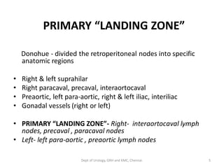
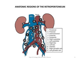
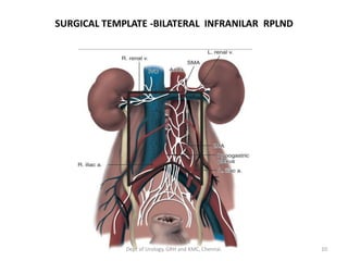
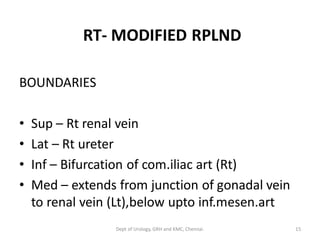
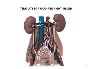
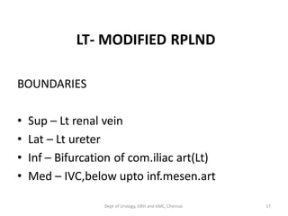
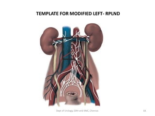
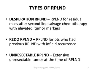
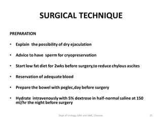
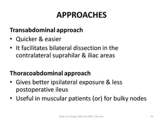
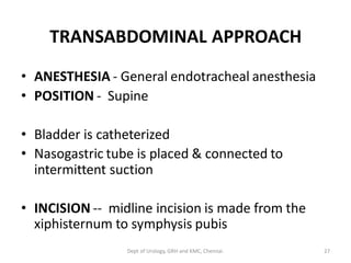
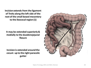
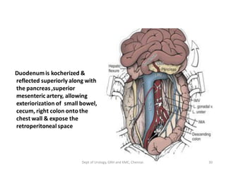
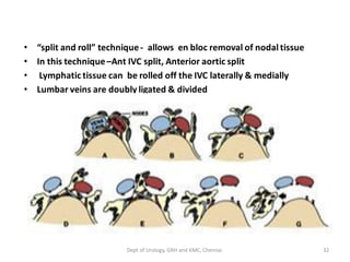
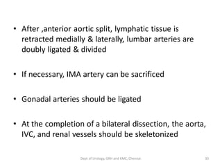
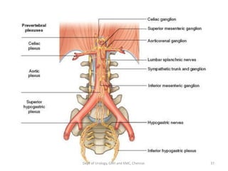
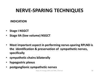
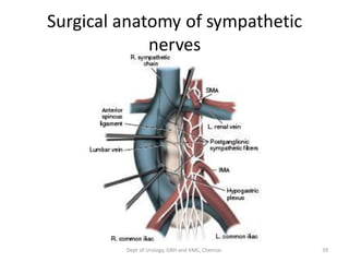
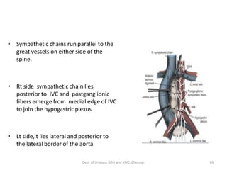
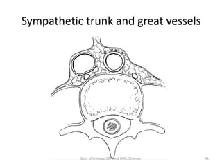
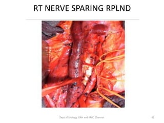
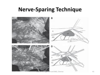
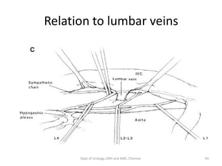
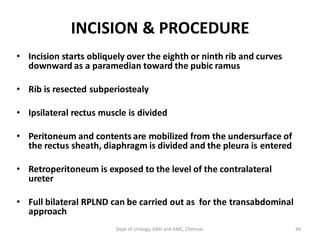
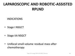
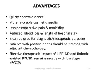
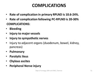
RPLND AND IT’S
COMPLICATIONS
Dept of Urology
Govt Royapettah Hospital and Kilpauk Medical College
Chennai
1
Moderators:
Professors:
• Prof. Dr. G. Sivasankar, M.S., M.Ch.,
• Prof. Dr. A. Senthilvel, M.S., M.Ch.,
Asst Professors:
• Dr. J. Sivabalan, M.S., M.Ch.,
• Dr. R. Bhargavi, M.S., M.Ch.,
• Dr. S. Raju, M.S., M.Ch.,
• Dr. K. Muthurathinam, M.S., M.Ch.,
• Dr. D. Tamilselvan, M.S., M.Ch.,
• Dr. K. Senthilkumar, M.S., M.Ch.
Dept of Urology, GRH and KMC, Chennai. 2
HISTORY
• 1910 - Jamieson - first to describe the
primary lymphatic drainage of the testis.
• 1914- Hinman – who underwent RPLND on
the basis of above mapping.
• 1977 – Donohue- First described the extended
bilateral suprahilar RPLND.
• 1980- Jewett- published template for
preserving para aortic sympathetic nerves.
3
Dept of Urology, GRH and KMC, Chennai.
Lymphatics of testis – clinical
implications
• Pattern of lymph drainage is from Right To Left
• Contralateral spread is common with right side
• For the left side contralateral spread occurs in
bulky disease
• Caudal deposit reflects retrograde spread in
large volume disease
– Distal iliac and inguinal nodes
• Aberrant testicular lymphatic drainage is rare but
possible
4
Dept of Urology, GRH and KMC, Chennai.
PRIMARY “LANDING ZONE”
Donohue - divided the retroperitoneal nodes into specific
anatomic regions
• Right & left suprahilar
• Right paracaval, precaval, interaortocaval
• Preaortic, left para-aortic, right & left iliac, interiliac
• Gonadal vessels (right or left)
• PRIMARY “LANDING ZONE”- Right- interaortocaval lymph
nodes, precaval , paracaval nodes
• Left- left para-aortic , preaortic lymph nodes
5
Dept of Urology, GRH and KMC, Chennai.
ANATOMIC REGIONS OF THE RETROPERITONEUM
6
Dept of Urology, GRH and KMC, Chennai.
RATIONALE FOR TREATMENT OF RETROPERITONEAL LYMPH NODES
It is based on several factors
• Nodal spread is usually the first & often the only site of metastatic
disease ,nodes treated by RPLND increases survival rates
• 15% - 40% of patients are clinically understaged - 20% - 30%
incidence of pathologic stage II disease in clinical stage I - cause of
25% relapse during surveillance protocols in clinical stage I
• 20% incidence of teratoma /or viable carcinoma in patients with
radiographically normal CT
• Untreated nodal metastases are usually fatal
• Most common site of late recurrence of both teratoma & viable
GCT is the retroperitoneum, Late recurrences are usually
chemorefractory, decreases the survival rates
7
Dept of Urology, GRH and KMC, Chennai.
EVOLUTION OF SURGICAL TEMPLATES AND
TECHNIQUES
• BILATERAL SUPRAHILAR DISSECTIONS
• Removal of all the nodal tissue between both ureters down
to the bifurcation of the common iliac arteries,superiorly
upto crus of diaphragm
It was associated with increased
• Pancreatic injury,lymphatic
• Renovascular complications
• Not routinly done
• Now, suprahilar dissections are indicated for residual hilar
or suprahilar masses after cytoreductive chemotherapy for
advanced stage NSGCT
8
Dept of Urology, GRH and KMC, Chennai.
MODIFICATION
• RATIONALE - Suprahilar metastases are rare in low-stage
NSGCT, suprahilar dissection not necessary & high
morbidity
• To reduce surgical morbidity, suprahilar dissection is
modified to
BILATERAL INFRAHILAR RPLND
BOUNDARIES
• Sup – Renal hilum
• Lat – Ureter
• Inf – Bifurcation of common iliac vessels
• This bilateral infrahilar RPLND also have long-term
morbidity of loss of antegrade ejaculation due to damage
of sympathetic nerve fibers
9
Dept of Urology, GRH and KMC, Chennai.
SURGICAL TEMPLATE -BILATERAL INFRANILAR RPLND
10
Dept of Urology, GRH and KMC, Chennai.
ANTEGRADE EJACULATION
It requires the coordination of three events
(1) Closure of the bladder neck
(2) Seminal emission
(3) Ejaculation
The sympathetic fibers from T12 - L3 form the
hypogastric plexus near the origin of inferior
mesenteric artery just above the aortic
bifurcation
From the hypogastric plexus, the sympathetic
fibers travel via the pelvic plexus to innervate
the seminal vesicles, vas deferens, prostate,
bladder neck
11
Dept of Urology, GRH and KMC, Chennai.
12
Dept of Urology, GRH and KMC, Chennai.
FURTHER MODIFICATIONS OF SURGICAL TEMPLATES
• Further modifications were developed to
reduce the incidence of ejaculatory
dysfunction by preserve the sympathetic
nerves
13
Dept of Urology, GRH and KMC, Chennai.
GOAL OF MODIFIED TEMPLATES
(1) Resect all interaortocaval & ipsilateral lymph nodes
between the level of the renal vessels & bifurcation of the
common iliac artery
(2) To minimize contralateral dissection, particularly below the
level of IMA
• “Nerve-sparing” techniques can be utilized either in the
primary or post-chemotherapy RPLND
• Margins of resection should never be compromised in an
attempt to maintain ejaculatory function
14
Dept of Urology, GRH and KMC, Chennai.
RT- MODIFIED RPLND
BOUNDARIES
• Sup – Rt renal vein
• Lat – Rt ureter
• Inf – Bifurcation of com.iliac art (Rt)
• Med – extends from junction of gonadal vein
to renal vein (Lt),below upto inf.mesen.art
15
Dept of Urology, GRH and KMC, Chennai.
TEMPLATE FOR MODIFIED RIGHT -RPLND
16
Dept of Urology, GRH and KMC, Chennai.
LT- MODIFIED RPLND
BOUNDARIES
• Sup – Lt renal vein
• Lat – Lt ureter
• Inf – Bifurcation of com.iliac art(Lt)
• Med – IVC,below upto inf.mesen.art
17
Dept of Urology, GRH and KMC, Chennai.
TEMPLATE FOR MODIFIED LEFT- RPLND
18
Dept of Urology, GRH and KMC, Chennai.
INDICATION
• Clinical stage I (CS I) NSGCT- risk factors-PT2-
T4,LV invasion,>40% emb carcinoma
• CS IIA NSGCT (single lymph node < 2 cm)
• CS IIB NSGCT (single or multiple lymph nodes
2 to 5 cm).
• Postchemotherapy residual mass with
normal tumor markers
19
Dept of Urology, GRH and KMC, Chennai.
INDICATION
MODIFIED TEMPLATE RPLND
• Clinical stage-I
BILATERAL RPLND
• Clinical stage- IIA,IIB
• Postchemotherapy residual mass with normal
tumor markers
20
Dept of Urology, GRH and KMC, Chennai.
INDICATION FOR POSTCHEMOTHERAPY RPLND
If the residual mass is > 3cm
• 50% viable GCT
• 40% teratoma
• 10% fibrosis
• Residual mass with normal tumor marker
after primary chemotherapy
• Residual mass irrespective of serum marker
after salvage chemo
• Recurrent resectable mass after salvage
chemo
21
Dept of Urology, GRH and KMC, Chennai.
TYPES OF RPLND
• STANDARD RPLND – Primary RPLND. either unilat
modified template/ Bilat RPLND in stage I,IIA,IIB
• POSTCHEMOTHERAPY RPLND – postchemo
residual mass with normal tumor markers after
induction chemotherapy
• SALVAGE RPLND – RPLND for residual mass after
second line salvage chemotherapy with normal
tumor markers
22
Dept of Urology, GRH and KMC, Chennai.
TYPES OF RPLND
• DESPERATION RPLND – RPLND for residual
mass after second line salvage chemotherapy
with elevated tumor markers
• REDO RPLND – RPLND for pts who had
previous RPLND with infield recurrence
• UNRESECTABLE RPLND – Extensive
unresectable tumor at the time of RPLND
23
Dept of Urology, GRH and KMC, Chennai.
ADVANTAGES OF RPLND
• Provides accurate pathological staging
• Complete excision of nodes, relapse rate is
very low
• Gives information regarding adjuvant
chemotherapy
• Remove chemoresistant teratoma
• Remove occult metastasis (15-30% in stage I)
24
Dept of Urology, GRH and KMC, Chennai.
SURGICAL TECHNIQUE
PREPARATION
• Explain the possibility of dry ejaculation
• Advice to have sperm for cryopreservation
• Start low fat diet for 2wks before surgery,to reduce chylous ascites
• Reservation of adequateblood
• Prepare the bowel with peglec,day before surgery
• Hydrate intravenouslywith 5% dextrose in half-normal saline at 150
ml/hr the night before surgery
25
Dept of Urology, GRH and KMC, Chennai.
APPROACHES
Transabdominal approach
• Quicker & easier
• It facilitates bilateral dissection in the
contralateral suprahilar & iliac areas
Thoracoabdominal approach
• Gives better ipsilateral exposure & less
postoperative ileus
• Useful in muscular patients (or) for bulky nodes
26
Dept of Urology, GRH and KMC, Chennai.
TRANSABDOMINAL APPROACH
• ANESTHESIA - General endotracheal anesthesia
• POSITION - Supine
• Bladder is catheterized
• Nasogastric tube is placed & connected to
intermittent suction
• INCISION -- midline incision is made from the
xiphisternum to symphysis pubis
27
Dept of Urology, GRH and KMC, Chennai.
PROCEDURE
• Peritoneum is opened
• Falciform ligament is divided between
ligatures
• Inspect abdomen, retroperitoneum, to assess
resectability , presence of metastatic disease
• Greater omentum & transverse colon are
displaced superiorly onto the chest
• Small bowel is reflected to the right, incision
is made in the posterior peritoneum
28
Dept of Urology, GRH and KMC, Chennai.
Incision extends from the ligament
of Treitz along the left side of the
root of the small bowel mesentery
to the ileocecal region (1)
It may be extended superiorly &
medially to the duodenojejunal
flexure
Incision is extended around the
cecum up to the right paracolic
gutter
29
Dept of Urology, GRH and KMC, Chennai.
Duodenumis kocherized &
reflected superiorly along with
the pancreas,superior
mesenteric artery, allowing
exteriorization of small bowel,
cecum, right colon onto the
chest wall & expose the
retroperitoneal space
30
Dept of Urology, GRH and KMC, Chennai.
LYMPHADENECTOMY
• Lat -- Ureters
• Sup -- Upper edge of the origin of renal arteries
• Inf -- Bifurcation of ipsilateral common iliac arteries
• Post -- Psoas muscle fascia
• If possible, lumbar vessels are preserved to reduce
postoperative back pain
31
Dept of Urology, GRH and KMC, Chennai.
• “split and roll” technique- allows en bloc removal of nodal tissue
• In this technique–Ant IVC split, Anterior aortic split
• Lymphatic tissue can be rolled off the IVC laterally & medially
• Lumbar veins are doubly ligated & divided
32
Dept of Urology, GRH and KMC, Chennai.
• After ,anterior aortic split, lymphatic tissue is
retracted medially & laterally, lumbar arteries are
doubly ligated & divided
• If necessary, IMA artery can be sacrificed
• Gonadal arteries should be ligated
• At the completion of a bilateral dissection, the aorta,
IVC, and renal vessels should be skeletonized
33
Dept of Urology, GRH and KMC, Chennai.
Nerve-Sparing Techniques
• Highest rates of preserved ejaculation
• The sympathetic chains, the postganglionic sympathetic
fibers, and the hypogastric plexus are identified,
meticulously dissected, and preserved
• Technique can be utilised during the primary procedure
• Can be combined with standard or modifeid templates
• Can be utilised in post chemo setting
“Margins of dissection should never be compromised for
nerve preservation”
34
Dept of Urology, GRH and KMC, Chennai.
Nerve-Sparing Techniques - requisites
1) In depth understanding of retroperitoneal
anatomy
2) Ability to recognise variations in anatomy
3) Excellent exposure of the retroperitoneum
4) Meticulous application of “split and roll “
technique
35
Dept of Urology, GRH and KMC, Chennai.
Prospective Nerve-Sparing Technique
• The emphasis is in identification and
preservation of relevant sympathetic nerves
– (1) The sympathetic chains bilaterally
– (2) The postganglionic sympathetic nerves arising
from the sympathetic chains
– (3) The hypogastric plexus (anastomosing network
of nerve fibers anterior to the lower aorta)
36
Dept of Urology, GRH and KMC, Chennai.
37
Dept of Urology, GRH and KMC, Chennai.
NERVE-SPARING TECHNIQUES
INDICATION
• Stage I NSGCT
• Stage IIA (low volume) NSGCT
• Most important aspect in performing nerve-sparing RPLND is
the identification & preservation of sympathetic nerves,
specifically
• sympathetic chains bilaterally
• hypogastric plexus
• postganglionic sympathetic nerves
38
Dept of Urology, GRH and KMC, Chennai.
Surgical anatomy of sympathetic
nerves
39
Dept of Urology, GRH and KMC, Chennai.
• Sympathetic chains run parallel to the
great vessels on either side of the
spine.
• Rt side sympathetic chain lies
posterior to IVC and postganglionic
fibers emerge from medial edge of IVC
to join the hypogastric plexus
• Lt side,it lies lateral and posterior to
the lateral border of the aorta
40
Dept of Urology, GRH and KMC, Chennai.
Sympathetic trunk and great vessels
41
Dept of Urology, GRH and KMC, Chennai.
RT NERVE SPARING RPLND
42
Dept of Urology, GRH and KMC, Chennai.
Nerve-Sparing Technique
43
Dept of Urology, GRH and KMC, Chennai.
Relation to lumbar veins
44
Dept of Urology, GRH and KMC, Chennai.
• Anterior “split” maneuver over the IVC does not
damage these fibers
• But, dissection along the aorta before isolating
and preserving these nerves results in
disruption of these fibres
• Proper nerve-sparing techniques result in greater
than 95% rates of antegrade ejaculation
45
Dept of Urology, GRH and KMC, Chennai.
THORACOABDOMINAL APPROACH
• It is extraperitoneal approach
ADVANTAGES
• It allow easy visualization & dissection of the
suprahilar lymphatic tissues
• Less risk of postoperative small bowel
obstruction
• Simultaneous thoracic procedures can be
performed
46
Dept of Urology, GRH and KMC, Chennai.
POSITION
• Patient is placed in an oblique
position with the upper part
at a 45-degree angle to the
table & lower part flat on the
table
• Ipsilat arm is placed in a sling
with the arm bent at the
elbow
• Contralat arm is secured to an
arm board
47
Dept of Urology, GRH and KMC, Chennai.
INCISION & PROCEDURE
• Incision starts obliquely over the eighth or ninth rib and curves
downward as a paramedian toward the pubic ramus
• Rib is resected subperiostealy
• Ipsilateral rectus muscle is divided
• Peritoneum and contents are mobilized from the undersurface of
the rectus sheath, diaphragm is divided and the pleura is entered
• Retroperitoneum is exposed to the level of the contralateral
ureter
• Full bilateral RPLND can be carried out as for the transabdominal
approach
48
Dept of Urology, GRH and KMC, Chennai.
LAPAROSCOPIC AND ROBOTIC-ASSISTED
RPLND
INDICATIONS
• Stage I NSGCT
• Stage IIA NSGCT
• Unifocal small-volume residual mass after
chemotherapy
49
Dept of Urology, GRH and KMC, Chennai.
ADVANTAGES
• Quicker convalescence
• More favorable cosmetic results
• Less postoperative pain & morbidity.
• Reduced blood loss & length of hospital stay
• It can be used for diagnostic/therapeutic purposes
• Patients with positive nodes should be treated with
adjuvant chemotherapy.
• Effective therapeutic impact of L-RPLND and Robotic-
assisted RPLND remains mostly with low stage
NSGCTs .
50
Dept of Urology, GRH and KMC, Chennai.
COMPLICATIONS
• Rate of complication in primary RPLND is 10.6-24%.
• Rate of complication following PC-RPLND is 20-30%
COMPLICATIONS:
• Bleeding
• Injury to major vessels
• Injury to sympathetic nerves
• Injury to adjacent organs (duodenum, bowel, kidney,
pancreas)
• Pulmonary
• Paralytic ileus
• Chylous ascites
• Peripheral Nerve injury
51
Dept of Urology, GRH and KMC, Chennai.
RPLND AND FERTILITY
• Preserving fertility in men undergoing RPLND is more
complex than simply sparing their postganglionic
sympathetic nerves.
• Subfertility in a significant proportion of patients
presenting with newly diagnosed testicular cancer is well
documented.
• When including all stages of disease, approximately 40%
to 60% of patients presenting with testicular GCT have
been reported to demonstrate abnormal parameters on
semen analysis.
• Baseline subfertility needs to be taken into account when
evaluating paternity rates after RPLND.
52
Dept of Urology, GRH and KMC, Chennai.
• Before the development of unilateral modified
RPLND templates and nerve-sparing techniques,
most patients undergoing bilateral RPLND were
rendered anejaculatory .
• Techniques were altered in two ways: (1) changing
the boundaries of dissection and (2) prospectively
identifying postganglionic sympathetic fibers and
the superior hypogastric plexus .
• Recent studies, reported preservation of antegrade
ejaculation in 97% of men undergoing modified
unilateral template dissection without ipsilateral
nerve sparing technique. 53
Dept of Urology, GRH and KMC, Chennai.
• Nerve sparing RPLND results in preservation
of antegrade ejaculation in 90-100 % of
patients.
• Postoperative paternity can be expected in
approximately 75% of men undergoing
primary nerve-sparing RPLND.
• Fertility after PC-RPLND has not been
established because chemotherapy-induced
disruption of spermatogenesis can persist for
several years after completion of therapy.
54
Dept of Urology, GRH and KMC, Chennai.
Pulmonary Complications
• Major pulmonary complications are extremely rare after primary
RPLND but have been reported to occur in approximately 3% to
5% of patients after PC-RPLND .
• Because most patients who undergo PC-RPLND have received
bleomycin containing induction chemotherapy, acute
respiratory distress syndrome and prolonged postoperative
ventilation account for most of these major complications.
• The incidence of bleomycin-related perioperative pulmonary
complications can be minimized by avoiding aggressive
intraoperative and postoperative intravenous fluid
resuscitation and keeping FiO2 as low as is safely possible .
55
Dept of Urology, GRH and KMC, Chennai.
Paralytic Ileus
• The reported rates of postoperative paralytic ileus
range widely in the primary RPLND (0% to 18%) and
PC-RPLND (2.2% to 21%) .
• In relatively low-volume PC-RPLND, an orogastric tube
is used and removed at the conclusion of the
procedure.
• In Retroperitoneal higher volume disease, the
probability of significant ileus is greater, and a
nasogastric tube should be used.
56
Dept of Urology, GRH and KMC, Chennai.
Lymphocele
• The incidence of subclinical lymphocele after RPLND is unknown.
• Symptomatic retroperitoneal lymphoceles are extremely rare with
reported rates ranging from 0 % to 1.7 %.
• Symptoms can be related to ureteral compression, displacement of
abdominal viscera (if very large), or secondary infection.
• Meticulous attention to ligation of large-caliber lymphatics during
resection likely decreases the risk of developing a symptomatic
lymphocele.
• Treatment of symptomatic and/or infected lymphoceles includes
percutaneous drainage with systemic antibiotics reserved for
infected lymphoceles.
57
Dept of Urology, GRH and KMC, Chennai.
Chylous Ascites
• Chylous ascites has been reported to occur in 0.2% to 2.1% of pts
undergoing primary RPLND and 2% to 7% of patients undergoing
PC-RPLND .
• Suprahilar resections are thought to carry a higher risk for chylous
ascites because of disruption of the cisterna chyli and its
contributing lymphatics.
• Patients with symptomatic chylous ascites should first be managed
with simple paracentesis with consideration of low-fat/medium-
chain triglyceride diet and intramuscular octreotide.
• If Persistent high-volume chylous drainage (>100 mL/24 hr) despite
these modifications if occur, placement of a peritoneovenous
(LeVeen) shunt, or surgical exploration with attempted ligation of
the lymphatic leak to be done.
58
Dept of Urology, GRH and KMC, Chennai.
Venous Thromboembolism
• The rate of pulmonary embolism after primary RPLND has been
reported to be less than 1% ,After PC-RPLND, the rates range from
0.1% to 3.1%.
• All patients undergoing RPLND should have sequential
compression devices placed before induction, which should be
maintained throughout the hospital course along with early
ambulatory practice.
• Prophylactic subcutaneous low-dose unfractionated heparin or
low-molecular-weight heparin has demonstrated efficacy in
decreasing VTE rates in patients with a personal history of VTE,
obesity, known hypercoagulable condition, or older age.
59
Dept of Urology, GRH and KMC, Chennai.
Neurologic Complication
• Peripheral nerve injury were secondary to patient
positioning and potentially retractor placement (femoral
neurapraxia).
• Careful attention to appropriate patient positioning by
the surgical and anesthesia teams is important in
minimizing peripheral nerve damage
• Patients with bulky mediastinal and retroperitoneal
disease are at an increased risk of developing paraplegia.
• The likelihood of neurologic complications increases
with the scale of para-aortic resection.
60
Dept of Urology, GRH and KMC, Chennai.
COMPLICATIONS OF RPLND
61
Dept of Urology, GRH and KMC, Chennai.
SUMMARY
• RPLND is the choice for low stage NSGCT
• Modified template RPLND is choice for clinical stage I
• In Modified templates, surgical margins should never
be compromised in an effort to preserve ejaculation
• In stage IIA,B NSGCT bilateral INFRA HILAR RPLND
remains the standard
62
Dept of Urology, GRH and KMC, Chennai.
• If positive nodes are noted at the time of RPLND, bilateral
dissection is warrented, particularly for right-sided
tumors (contralateral crossover)
• L- RPLND is a difficult procedure even for the well-trained
laparoscopic surgeon (because dissection is near major
vessels,retrocaval & retroaortic nodes dissection is
difficult)
• Therapeutic impact of L-RPLND remains effective for low
stage NSGCTs.
• Major complications are rare after primary RPLND and PC-
RPLND. A significant proportion of major complications at
PC-RPLND are pulmonary and are related to prior
bleomycin and thoracic disease burden.
63
Dept of Urology, GRH and KMC, Chennai.
64
Dept of Urology, GRH and KMC, Chennai.

Testis carcinoma- management- rplnd

  • 1.
    RPLND AND IT’S COMPLICATIONS Deptof Urology Govt Royapettah Hospital and Kilpauk Medical College Chennai 1
  • 2.
    Moderators: Professors: • Prof. Dr.G. Sivasankar, M.S., M.Ch., • Prof. Dr. A. Senthilvel, M.S., M.Ch., Asst Professors: • Dr. J. Sivabalan, M.S., M.Ch., • Dr. R. Bhargavi, M.S., M.Ch., • Dr. S. Raju, M.S., M.Ch., • Dr. K. Muthurathinam, M.S., M.Ch., • Dr. D. Tamilselvan, M.S., M.Ch., • Dr. K. Senthilkumar, M.S., M.Ch. Dept of Urology, GRH and KMC, Chennai. 2
  • 3.
    HISTORY • 1910 -Jamieson - first to describe the primary lymphatic drainage of the testis. • 1914- Hinman – who underwent RPLND on the basis of above mapping. • 1977 – Donohue- First described the extended bilateral suprahilar RPLND. • 1980- Jewett- published template for preserving para aortic sympathetic nerves. 3 Dept of Urology, GRH and KMC, Chennai.
  • 4.
    Lymphatics of testis– clinical implications • Pattern of lymph drainage is from Right To Left • Contralateral spread is common with right side • For the left side contralateral spread occurs in bulky disease • Caudal deposit reflects retrograde spread in large volume disease – Distal iliac and inguinal nodes • Aberrant testicular lymphatic drainage is rare but possible 4 Dept of Urology, GRH and KMC, Chennai.
  • 5.
    PRIMARY “LANDING ZONE” Donohue- divided the retroperitoneal nodes into specific anatomic regions • Right & left suprahilar • Right paracaval, precaval, interaortocaval • Preaortic, left para-aortic, right & left iliac, interiliac • Gonadal vessels (right or left) • PRIMARY “LANDING ZONE”- Right- interaortocaval lymph nodes, precaval , paracaval nodes • Left- left para-aortic , preaortic lymph nodes 5 Dept of Urology, GRH and KMC, Chennai.
  • 6.
    ANATOMIC REGIONS OFTHE RETROPERITONEUM 6 Dept of Urology, GRH and KMC, Chennai.
  • 7.
    RATIONALE FOR TREATMENTOF RETROPERITONEAL LYMPH NODES It is based on several factors • Nodal spread is usually the first & often the only site of metastatic disease ,nodes treated by RPLND increases survival rates • 15% - 40% of patients are clinically understaged - 20% - 30% incidence of pathologic stage II disease in clinical stage I - cause of 25% relapse during surveillance protocols in clinical stage I • 20% incidence of teratoma /or viable carcinoma in patients with radiographically normal CT • Untreated nodal metastases are usually fatal • Most common site of late recurrence of both teratoma & viable GCT is the retroperitoneum, Late recurrences are usually chemorefractory, decreases the survival rates 7 Dept of Urology, GRH and KMC, Chennai.
  • 8.
    EVOLUTION OF SURGICALTEMPLATES AND TECHNIQUES • BILATERAL SUPRAHILAR DISSECTIONS • Removal of all the nodal tissue between both ureters down to the bifurcation of the common iliac arteries,superiorly upto crus of diaphragm It was associated with increased • Pancreatic injury,lymphatic • Renovascular complications • Not routinly done • Now, suprahilar dissections are indicated for residual hilar or suprahilar masses after cytoreductive chemotherapy for advanced stage NSGCT 8 Dept of Urology, GRH and KMC, Chennai.
  • 9.
    MODIFICATION • RATIONALE -Suprahilar metastases are rare in low-stage NSGCT, suprahilar dissection not necessary & high morbidity • To reduce surgical morbidity, suprahilar dissection is modified to BILATERAL INFRAHILAR RPLND BOUNDARIES • Sup – Renal hilum • Lat – Ureter • Inf – Bifurcation of common iliac vessels • This bilateral infrahilar RPLND also have long-term morbidity of loss of antegrade ejaculation due to damage of sympathetic nerve fibers 9 Dept of Urology, GRH and KMC, Chennai.
  • 10.
    SURGICAL TEMPLATE -BILATERALINFRANILAR RPLND 10 Dept of Urology, GRH and KMC, Chennai.
  • 11.
    ANTEGRADE EJACULATION It requiresthe coordination of three events (1) Closure of the bladder neck (2) Seminal emission (3) Ejaculation The sympathetic fibers from T12 - L3 form the hypogastric plexus near the origin of inferior mesenteric artery just above the aortic bifurcation From the hypogastric plexus, the sympathetic fibers travel via the pelvic plexus to innervate the seminal vesicles, vas deferens, prostate, bladder neck 11 Dept of Urology, GRH and KMC, Chennai.
  • 12.
    12 Dept of Urology,GRH and KMC, Chennai.
  • 13.
    FURTHER MODIFICATIONS OFSURGICAL TEMPLATES • Further modifications were developed to reduce the incidence of ejaculatory dysfunction by preserve the sympathetic nerves 13 Dept of Urology, GRH and KMC, Chennai.
  • 14.
    GOAL OF MODIFIEDTEMPLATES (1) Resect all interaortocaval & ipsilateral lymph nodes between the level of the renal vessels & bifurcation of the common iliac artery (2) To minimize contralateral dissection, particularly below the level of IMA • “Nerve-sparing” techniques can be utilized either in the primary or post-chemotherapy RPLND • Margins of resection should never be compromised in an attempt to maintain ejaculatory function 14 Dept of Urology, GRH and KMC, Chennai.
  • 15.
    RT- MODIFIED RPLND BOUNDARIES •Sup – Rt renal vein • Lat – Rt ureter • Inf – Bifurcation of com.iliac art (Rt) • Med – extends from junction of gonadal vein to renal vein (Lt),below upto inf.mesen.art 15 Dept of Urology, GRH and KMC, Chennai.
  • 16.
    TEMPLATE FOR MODIFIEDRIGHT -RPLND 16 Dept of Urology, GRH and KMC, Chennai.
  • 17.
    LT- MODIFIED RPLND BOUNDARIES •Sup – Lt renal vein • Lat – Lt ureter • Inf – Bifurcation of com.iliac art(Lt) • Med – IVC,below upto inf.mesen.art 17 Dept of Urology, GRH and KMC, Chennai.
  • 18.
    TEMPLATE FOR MODIFIEDLEFT- RPLND 18 Dept of Urology, GRH and KMC, Chennai.
  • 19.
    INDICATION • Clinical stageI (CS I) NSGCT- risk factors-PT2- T4,LV invasion,>40% emb carcinoma • CS IIA NSGCT (single lymph node < 2 cm) • CS IIB NSGCT (single or multiple lymph nodes 2 to 5 cm). • Postchemotherapy residual mass with normal tumor markers 19 Dept of Urology, GRH and KMC, Chennai.
  • 20.
    INDICATION MODIFIED TEMPLATE RPLND •Clinical stage-I BILATERAL RPLND • Clinical stage- IIA,IIB • Postchemotherapy residual mass with normal tumor markers 20 Dept of Urology, GRH and KMC, Chennai.
  • 21.
    INDICATION FOR POSTCHEMOTHERAPYRPLND If the residual mass is > 3cm • 50% viable GCT • 40% teratoma • 10% fibrosis • Residual mass with normal tumor marker after primary chemotherapy • Residual mass irrespective of serum marker after salvage chemo • Recurrent resectable mass after salvage chemo 21 Dept of Urology, GRH and KMC, Chennai.
  • 22.
    TYPES OF RPLND •STANDARD RPLND – Primary RPLND. either unilat modified template/ Bilat RPLND in stage I,IIA,IIB • POSTCHEMOTHERAPY RPLND – postchemo residual mass with normal tumor markers after induction chemotherapy • SALVAGE RPLND – RPLND for residual mass after second line salvage chemotherapy with normal tumor markers 22 Dept of Urology, GRH and KMC, Chennai.
  • 23.
    TYPES OF RPLND •DESPERATION RPLND – RPLND for residual mass after second line salvage chemotherapy with elevated tumor markers • REDO RPLND – RPLND for pts who had previous RPLND with infield recurrence • UNRESECTABLE RPLND – Extensive unresectable tumor at the time of RPLND 23 Dept of Urology, GRH and KMC, Chennai.
  • 24.
    ADVANTAGES OF RPLND •Provides accurate pathological staging • Complete excision of nodes, relapse rate is very low • Gives information regarding adjuvant chemotherapy • Remove chemoresistant teratoma • Remove occult metastasis (15-30% in stage I) 24 Dept of Urology, GRH and KMC, Chennai.
  • 25.
    SURGICAL TECHNIQUE PREPARATION • Explainthe possibility of dry ejaculation • Advice to have sperm for cryopreservation • Start low fat diet for 2wks before surgery,to reduce chylous ascites • Reservation of adequateblood • Prepare the bowel with peglec,day before surgery • Hydrate intravenouslywith 5% dextrose in half-normal saline at 150 ml/hr the night before surgery 25 Dept of Urology, GRH and KMC, Chennai.
  • 26.
    APPROACHES Transabdominal approach • Quicker& easier • It facilitates bilateral dissection in the contralateral suprahilar & iliac areas Thoracoabdominal approach • Gives better ipsilateral exposure & less postoperative ileus • Useful in muscular patients (or) for bulky nodes 26 Dept of Urology, GRH and KMC, Chennai.
  • 27.
    TRANSABDOMINAL APPROACH • ANESTHESIA- General endotracheal anesthesia • POSITION - Supine • Bladder is catheterized • Nasogastric tube is placed & connected to intermittent suction • INCISION -- midline incision is made from the xiphisternum to symphysis pubis 27 Dept of Urology, GRH and KMC, Chennai.
  • 28.
    PROCEDURE • Peritoneum isopened • Falciform ligament is divided between ligatures • Inspect abdomen, retroperitoneum, to assess resectability , presence of metastatic disease • Greater omentum & transverse colon are displaced superiorly onto the chest • Small bowel is reflected to the right, incision is made in the posterior peritoneum 28 Dept of Urology, GRH and KMC, Chennai.
  • 29.
    Incision extends fromthe ligament of Treitz along the left side of the root of the small bowel mesentery to the ileocecal region (1) It may be extended superiorly & medially to the duodenojejunal flexure Incision is extended around the cecum up to the right paracolic gutter 29 Dept of Urology, GRH and KMC, Chennai.
  • 30.
    Duodenumis kocherized & reflectedsuperiorly along with the pancreas,superior mesenteric artery, allowing exteriorization of small bowel, cecum, right colon onto the chest wall & expose the retroperitoneal space 30 Dept of Urology, GRH and KMC, Chennai.
  • 31.
    LYMPHADENECTOMY • Lat --Ureters • Sup -- Upper edge of the origin of renal arteries • Inf -- Bifurcation of ipsilateral common iliac arteries • Post -- Psoas muscle fascia • If possible, lumbar vessels are preserved to reduce postoperative back pain 31 Dept of Urology, GRH and KMC, Chennai.
  • 32.
    • “split androll” technique- allows en bloc removal of nodal tissue • In this technique–Ant IVC split, Anterior aortic split • Lymphatic tissue can be rolled off the IVC laterally & medially • Lumbar veins are doubly ligated & divided 32 Dept of Urology, GRH and KMC, Chennai.
  • 33.
    • After ,anterioraortic split, lymphatic tissue is retracted medially & laterally, lumbar arteries are doubly ligated & divided • If necessary, IMA artery can be sacrificed • Gonadal arteries should be ligated • At the completion of a bilateral dissection, the aorta, IVC, and renal vessels should be skeletonized 33 Dept of Urology, GRH and KMC, Chennai.
  • 34.
    Nerve-Sparing Techniques • Highestrates of preserved ejaculation • The sympathetic chains, the postganglionic sympathetic fibers, and the hypogastric plexus are identified, meticulously dissected, and preserved • Technique can be utilised during the primary procedure • Can be combined with standard or modifeid templates • Can be utilised in post chemo setting “Margins of dissection should never be compromised for nerve preservation” 34 Dept of Urology, GRH and KMC, Chennai.
  • 35.
    Nerve-Sparing Techniques -requisites 1) In depth understanding of retroperitoneal anatomy 2) Ability to recognise variations in anatomy 3) Excellent exposure of the retroperitoneum 4) Meticulous application of “split and roll “ technique 35 Dept of Urology, GRH and KMC, Chennai.
  • 36.
    Prospective Nerve-Sparing Technique •The emphasis is in identification and preservation of relevant sympathetic nerves – (1) The sympathetic chains bilaterally – (2) The postganglionic sympathetic nerves arising from the sympathetic chains – (3) The hypogastric plexus (anastomosing network of nerve fibers anterior to the lower aorta) 36 Dept of Urology, GRH and KMC, Chennai.
  • 37.
    37 Dept of Urology,GRH and KMC, Chennai.
  • 38.
    NERVE-SPARING TECHNIQUES INDICATION • StageI NSGCT • Stage IIA (low volume) NSGCT • Most important aspect in performing nerve-sparing RPLND is the identification & preservation of sympathetic nerves, specifically • sympathetic chains bilaterally • hypogastric plexus • postganglionic sympathetic nerves 38 Dept of Urology, GRH and KMC, Chennai.
  • 39.
    Surgical anatomy ofsympathetic nerves 39 Dept of Urology, GRH and KMC, Chennai.
  • 40.
    • Sympathetic chainsrun parallel to the great vessels on either side of the spine. • Rt side sympathetic chain lies posterior to IVC and postganglionic fibers emerge from medial edge of IVC to join the hypogastric plexus • Lt side,it lies lateral and posterior to the lateral border of the aorta 40 Dept of Urology, GRH and KMC, Chennai.
  • 41.
    Sympathetic trunk andgreat vessels 41 Dept of Urology, GRH and KMC, Chennai.
  • 42.
    RT NERVE SPARINGRPLND 42 Dept of Urology, GRH and KMC, Chennai.
  • 43.
    Nerve-Sparing Technique 43 Dept ofUrology, GRH and KMC, Chennai.
  • 44.
    Relation to lumbarveins 44 Dept of Urology, GRH and KMC, Chennai.
  • 45.
    • Anterior “split”maneuver over the IVC does not damage these fibers • But, dissection along the aorta before isolating and preserving these nerves results in disruption of these fibres • Proper nerve-sparing techniques result in greater than 95% rates of antegrade ejaculation 45 Dept of Urology, GRH and KMC, Chennai.
  • 46.
    THORACOABDOMINAL APPROACH • Itis extraperitoneal approach ADVANTAGES • It allow easy visualization & dissection of the suprahilar lymphatic tissues • Less risk of postoperative small bowel obstruction • Simultaneous thoracic procedures can be performed 46 Dept of Urology, GRH and KMC, Chennai.
  • 47.
    POSITION • Patient isplaced in an oblique position with the upper part at a 45-degree angle to the table & lower part flat on the table • Ipsilat arm is placed in a sling with the arm bent at the elbow • Contralat arm is secured to an arm board 47 Dept of Urology, GRH and KMC, Chennai.
  • 48.
    INCISION & PROCEDURE •Incision starts obliquely over the eighth or ninth rib and curves downward as a paramedian toward the pubic ramus • Rib is resected subperiostealy • Ipsilateral rectus muscle is divided • Peritoneum and contents are mobilized from the undersurface of the rectus sheath, diaphragm is divided and the pleura is entered • Retroperitoneum is exposed to the level of the contralateral ureter • Full bilateral RPLND can be carried out as for the transabdominal approach 48 Dept of Urology, GRH and KMC, Chennai.
  • 49.
    LAPAROSCOPIC AND ROBOTIC-ASSISTED RPLND INDICATIONS •Stage I NSGCT • Stage IIA NSGCT • Unifocal small-volume residual mass after chemotherapy 49 Dept of Urology, GRH and KMC, Chennai.
  • 50.
    ADVANTAGES • Quicker convalescence •More favorable cosmetic results • Less postoperative pain & morbidity. • Reduced blood loss & length of hospital stay • It can be used for diagnostic/therapeutic purposes • Patients with positive nodes should be treated with adjuvant chemotherapy. • Effective therapeutic impact of L-RPLND and Robotic- assisted RPLND remains mostly with low stage NSGCTs . 50 Dept of Urology, GRH and KMC, Chennai.
  • 51.
    COMPLICATIONS • Rate ofcomplication in primary RPLND is 10.6-24%. • Rate of complication following PC-RPLND is 20-30% COMPLICATIONS: • Bleeding • Injury to major vessels • Injury to sympathetic nerves • Injury to adjacent organs (duodenum, bowel, kidney, pancreas) • Pulmonary • Paralytic ileus • Chylous ascites • Peripheral Nerve injury 51 Dept of Urology, GRH and KMC, Chennai.
  • 52.
    RPLND AND FERTILITY •Preserving fertility in men undergoing RPLND is more complex than simply sparing their postganglionic sympathetic nerves. • Subfertility in a significant proportion of patients presenting with newly diagnosed testicular cancer is well documented. • When including all stages of disease, approximately 40% to 60% of patients presenting with testicular GCT have been reported to demonstrate abnormal parameters on semen analysis. • Baseline subfertility needs to be taken into account when evaluating paternity rates after RPLND. 52 Dept of Urology, GRH and KMC, Chennai.
  • 53.
    • Before thedevelopment of unilateral modified RPLND templates and nerve-sparing techniques, most patients undergoing bilateral RPLND were rendered anejaculatory . • Techniques were altered in two ways: (1) changing the boundaries of dissection and (2) prospectively identifying postganglionic sympathetic fibers and the superior hypogastric plexus . • Recent studies, reported preservation of antegrade ejaculation in 97% of men undergoing modified unilateral template dissection without ipsilateral nerve sparing technique. 53 Dept of Urology, GRH and KMC, Chennai.
  • 54.
    • Nerve sparingRPLND results in preservation of antegrade ejaculation in 90-100 % of patients. • Postoperative paternity can be expected in approximately 75% of men undergoing primary nerve-sparing RPLND. • Fertility after PC-RPLND has not been established because chemotherapy-induced disruption of spermatogenesis can persist for several years after completion of therapy. 54 Dept of Urology, GRH and KMC, Chennai.
  • 55.
    Pulmonary Complications • Majorpulmonary complications are extremely rare after primary RPLND but have been reported to occur in approximately 3% to 5% of patients after PC-RPLND . • Because most patients who undergo PC-RPLND have received bleomycin containing induction chemotherapy, acute respiratory distress syndrome and prolonged postoperative ventilation account for most of these major complications. • The incidence of bleomycin-related perioperative pulmonary complications can be minimized by avoiding aggressive intraoperative and postoperative intravenous fluid resuscitation and keeping FiO2 as low as is safely possible . 55 Dept of Urology, GRH and KMC, Chennai.
  • 56.
    Paralytic Ileus • Thereported rates of postoperative paralytic ileus range widely in the primary RPLND (0% to 18%) and PC-RPLND (2.2% to 21%) . • In relatively low-volume PC-RPLND, an orogastric tube is used and removed at the conclusion of the procedure. • In Retroperitoneal higher volume disease, the probability of significant ileus is greater, and a nasogastric tube should be used. 56 Dept of Urology, GRH and KMC, Chennai.
  • 57.
    Lymphocele • The incidenceof subclinical lymphocele after RPLND is unknown. • Symptomatic retroperitoneal lymphoceles are extremely rare with reported rates ranging from 0 % to 1.7 %. • Symptoms can be related to ureteral compression, displacement of abdominal viscera (if very large), or secondary infection. • Meticulous attention to ligation of large-caliber lymphatics during resection likely decreases the risk of developing a symptomatic lymphocele. • Treatment of symptomatic and/or infected lymphoceles includes percutaneous drainage with systemic antibiotics reserved for infected lymphoceles. 57 Dept of Urology, GRH and KMC, Chennai.
  • 58.
    Chylous Ascites • Chylousascites has been reported to occur in 0.2% to 2.1% of pts undergoing primary RPLND and 2% to 7% of patients undergoing PC-RPLND . • Suprahilar resections are thought to carry a higher risk for chylous ascites because of disruption of the cisterna chyli and its contributing lymphatics. • Patients with symptomatic chylous ascites should first be managed with simple paracentesis with consideration of low-fat/medium- chain triglyceride diet and intramuscular octreotide. • If Persistent high-volume chylous drainage (>100 mL/24 hr) despite these modifications if occur, placement of a peritoneovenous (LeVeen) shunt, or surgical exploration with attempted ligation of the lymphatic leak to be done. 58 Dept of Urology, GRH and KMC, Chennai.
  • 59.
    Venous Thromboembolism • Therate of pulmonary embolism after primary RPLND has been reported to be less than 1% ,After PC-RPLND, the rates range from 0.1% to 3.1%. • All patients undergoing RPLND should have sequential compression devices placed before induction, which should be maintained throughout the hospital course along with early ambulatory practice. • Prophylactic subcutaneous low-dose unfractionated heparin or low-molecular-weight heparin has demonstrated efficacy in decreasing VTE rates in patients with a personal history of VTE, obesity, known hypercoagulable condition, or older age. 59 Dept of Urology, GRH and KMC, Chennai.
  • 60.
    Neurologic Complication • Peripheralnerve injury were secondary to patient positioning and potentially retractor placement (femoral neurapraxia). • Careful attention to appropriate patient positioning by the surgical and anesthesia teams is important in minimizing peripheral nerve damage • Patients with bulky mediastinal and retroperitoneal disease are at an increased risk of developing paraplegia. • The likelihood of neurologic complications increases with the scale of para-aortic resection. 60 Dept of Urology, GRH and KMC, Chennai.
  • 61.
    COMPLICATIONS OF RPLND 61 Deptof Urology, GRH and KMC, Chennai.
  • 62.
    SUMMARY • RPLND isthe choice for low stage NSGCT • Modified template RPLND is choice for clinical stage I • In Modified templates, surgical margins should never be compromised in an effort to preserve ejaculation • In stage IIA,B NSGCT bilateral INFRA HILAR RPLND remains the standard 62 Dept of Urology, GRH and KMC, Chennai.
  • 63.
    • If positivenodes are noted at the time of RPLND, bilateral dissection is warrented, particularly for right-sided tumors (contralateral crossover) • L- RPLND is a difficult procedure even for the well-trained laparoscopic surgeon (because dissection is near major vessels,retrocaval & retroaortic nodes dissection is difficult) • Therapeutic impact of L-RPLND remains effective for low stage NSGCTs. • Major complications are rare after primary RPLND and PC- RPLND. A significant proportion of major complications at PC-RPLND are pulmonary and are related to prior bleomycin and thoracic disease burden. 63 Dept of Urology, GRH and KMC, Chennai.
  • 64.
    64 Dept of Urology,GRH and KMC, Chennai.