Downloaded 101 times


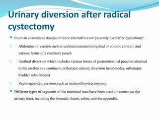


![Ureterocolonic diversion
The oldest and most common form was primarily a refluxive and later an antirefluxive
connection of ureters into the intact rectosigmoideum (uretero[recto]sigmoidostomy) .
most of the indicationsfor this procedure have become obsolete due to a high incidence
of upper urinary tract infectionsand the long-term risk of developing colon cancer .
Bowel frequency and urge incontinence were additional side-effectsof this type of
urinary diversion.
however, it may be possibleto circumvent the above-mentionedproblems by interposing
a segment of ileum between ureters and rectum or sigmoid in order to augment capacity
and to avoid a direct interactionbetween urothelium, colonic mucosa, together with
faeces and urine .](https://image.slidesharecdn.com/bladdersx-171212163040/85/Bladder-cancer-surgery-59-320.jpg)


This document outlines indications and techniques for radical cystectomy in the treatment of bladder cancer. It indicates radical cystectomy involves removal of the bladder and adjacent organs. Lymphadenectomy is also performed to remove pelvic lymph nodes. The extent of lymphadenectomy is controversial but removal of more than 15 nodes may provide prognostic benefits. Post-cystectomy urinary diversion options include abdominal conduits, orthotopic neobladders using bowel segments, and rectosigmoid diversions. Patient selection factors and oncologic outcomes are discussed.


![Muscle invasive bladder Cancer [Dr.Edmond Wong]](https://cdn.slidesharecdn.com/ss_thumbnails/muscleinvasivebladdertumoredmond-140716213247-phpapp01-thumbnail.jpg?width=640&height=640&fit=bounds)


![Chapter 39 role of radiotherapy in benign diseases.pptx [read only]](https://cdn.slidesharecdn.com/ss_thumbnails/chapter39roleofradiotherapyinbenigndiseases-191105205437-thumbnail.jpg?width=640&height=640&fit=bounds)


