SlideShare a Scribd company logo
ELBOW INSTABILITY AND
TERRIBLE TRAID INJURIES
DR.M.S.DHINESH
DEPARTMENT OF ORTHOPAEDICS
MAPIMS
ELBOW STABILISERS
• STATIC STABILISERS
• PRIMARY CONSTRAINTS
• SECONDARY CONSTRAINTS
• DYNAMIC STABILISERS
STATIC CONSTRAINTS OF ELBOW
LATERAL COLLATERAL LIGAMENT
COMPLEX
MEDIAL COLLATERAL LIGAMENT
COMPLEX
DYNAMIC STABILISERS OF ELBOW
TRAUMATIC ELBOW INSTABILITY
• Mechanism of injury
• Primary mechanism is posterolateral
• Other mechanisms postulated - controversial
• Valgus-external rotation
• Valgus-hyperextension
• Sequence of injury progression also debated
• Medial dislocations
Posterolateral Mechanism - Injury progression
O’DRISCOLL’S RING OF INSTABILITY
O'Driscoll et al. described a valgus, axial, and posterolateral force that
results in the typical posterolateral dislocation of the elbow joint
LATERAL PIVOT SHIFT TEST
O’DRISCOLL’S RING OF INSTABILITY
TRAUMATIC ELBOW INSTABILITY
• Simple Dislocations
• Complex Dislocations – Fracture-
dislocations
• Coronoid fractures
• Radial Head Fractures
• Monteggia Fractures
• Trans-olecranon dislocations
• Terrible Triad
• Chronic Dislocations
• Not associated with fracture
• Posterior and Posterolateral (M/C)– all directions and divergent
dislocations can occur
• Beware patterns other than posterior/posterolateral – can be more unstable
after reduction
• Mechanism of Injury
• Partially flexed elbow
• Axial load, supination, and valgus
• Varus mechanisms also described
• Medial ligamentous injuries in most cases
SIMPLE DISLOCATION
REDUCTION MANNEOVUR
REHABILITATION PROTOCOL
56/M, POSTEROLATERAL ELBOW D/L
56/M,POST REDUCTION
UNSTABLE AFTER REDUCTION
• Uncommon in simple dislocations
• May require soft tissue reconstruction
• Do what needs to be done to hold a concentric
reduction
• Splint in more flexion
• External fixator – static vs hinged
• Elbow cross pinning
• Internal fixator
ALGORITHM
FOR
SIMPLE
ELBOW D/L
• Associated with a fracture
• Sub-Types
• Coronoid Fracture
• Monteggia Fracture
• Radial Head Fracture
• Epicondyle fractures - adolescents
• Trans-Olecranon fracture-Dislocations
• Terrible Triad Elbow
COMPLEX ELBOW DISLOCATION
VALGUS INSTABILITY
RADIAL HEAD FRACTURE
Mechanism of Injury:
Valgus force + axial load
RADIAL HEAD FRACTURE
• Comprehensive classification of radial head fracture with
description of associated injuries (van Riet and Morrey)
Valgus stability-MCL and Radial head
Importance: Valgus force
Primary stabilisers – anterior bundle of MCL
Secondary stabilisers- Radial Head
So,whenever excise the radial head in case of MCL rupture- no
resist to valgus force- valgus instability
Radial head
communited
fracture
Radial head
excised
Intact
intraosseous
membrane
Radial head
excised
Ruptured
intraosseous
membrane
Radius migrated
proximally-
causing damage
to elbow joint,
Ulna move
towards distally-
causing damage
to ulnocarpal jt.
So, whenever Radial
head fracture
Always look for
DRUJ
Disruption/intraoss
eous membrane
damage
ESSEX LOPRESSETI #
UNSTABLE RADIAL HEAD #
No MCL + Radial head excision = valgus instability =
Early osteoarthritis
UNSTABLE RADIAL HEAD FRACTURE
DECISION MAKING
• Fragment number
• Displacement
• Articular surface
• Age and bone quality
• Dislocation
• Ass. Ligamentous injury
• Ass. Elbow fractures
DECISION MAKING
STABLE FIXATION AND EARLY MOBILISATION
RADIAL HEAD- SMALL NON ARTICULAR
PORTION
RADIAL HEAD AND NECK- SAFE ZONE
• 240° of circumference
articulates with ulna at
lesser sigmoid notch
• ~90-100 ° arc of safe
hardware placement
Caput et al recommended using the radial styloid and listers
tubercle as guides
FRAGMENT EXCISION
RADIAL HEAD EXCISION
ALGORITHM FOR RADIAL HEAD FRACTURE
COMMINUTED RADIAL HEAD FRACTURE
ROLE OF THE RADIAL HEAD ARTHROPLASTY
▪ Excision will lead to instability
▪ Functional spacer
▪ Creates stability by increasing radial length & restoring valgus restraint
RADIAL HEAD FRACTURE-
OVERSTUFFING
Radial Head Should be in line with
proximal edge of the lesser sigmaoid
notch to avoid overstuffing
RADIAL HEAD FRACTURE-
OVERSTUFFING
RADIAL HEAD FRACTURE-
OVERSTUFFING
▪ Direct visualization
▪ Most accurate way to
determine appropriate head
size
▪ Radial head should be just
at or proximal to radial
notch of the ulna
▪ Intra-op Fluoro
• Needs to be assessed in
flexion and extension
• Less reliable • > 6mm
overstuffing must be present
to consistently be seen on
fluoro
RADIAL HEAD FRACTURE-OVERSTUFFING
POST-OP PROTOCOL
▪ For all stabilized fxs and dislocations regardless of fixation
• Initially
• Immobilization for 10-14 days
• Secondarily
• Early ACTIVE range of motion
• Allows dynamic stabilizers to help hold reduction of joint
▪ Will reduce pseudosubluxations
• Limits elbow stiffness
• Some limit active shoulder abduction if LUCL was repaired
APPROACHES
• KAPLAN APPROACH
• KOCHER APPROACH
• EDC Split
• Modified Boyd
• Posterior approach
• Elevate LUCL from lateral epicondyle
• Can be used for combined olecranon/radial head
fxs
RARELY USED APPROACH
KOCHER APPROACH
Plane Between ECU and anconeus
Most often utilized for radial head
• Interval
• Anconeus – Radial Nerve
• ECU – PIN
• 5cm incision from lateral epicondyle distally
• Angled posteriorly 30-45 degrees
• Often deep soft tissues will be disrupted by injury
• Damage to LUCL
• Stay on anterior half of radial
head
• Damage to PIN
• Pronate the arm to move nerve
distally
• Carefully dissect distal to annular
ligament
KOCHER PITFALLS
• Distal extension becomes dorsal
Thompson approach
• More often used for radial neck/proximal
radial shaft fxs
• Interval
• ECRB – Radial nerve or PIN (variable) •
EDC – PIN
10cm incision from lateral epicondyle to
Lister’s Tubercle
KAPLAN APPROACH
CORONOID PROCESS FRACTURE
CLASSIFICATION: CORONOID
FRACTURES
▪ Regan & Morrey
• Type 1: tip
• Type 2 : < 50%y be ste
• Type 3 > 50%
▪ Highly unstable
CLASSIFICATION: CORONOID
FRACTURES
▪O’Driscoll Classification
▪Type I: Tip
▪Type II: anteromedial facet
▪Type III: base
PROXIMAL ULNA - ANTERIOR
CORONOID
•Anterior capsule
•Brachialis
•Anterior bundle of MCL
•Anteromedial facet of coronoid
▪ Fx propagation into this region
may cause functional MCL
incompetancy
CORONOID FRACTURE-
INDICATION
1.An isolated fracture (Type 3)
2.As a part of fracture dislocation
3.As a part of communited fractures of
proximal ulna
CORONOID FRACTURE
Complex instability of the
elbow results from posterior
directed forces
• TR(Triceps)
• BR(Brachialis)
• BC(Biceps)
OLECRANON FRACTURE
Mechanism of Injury
• Acute Tension overload: Tension applied by
the triceps with flexion of the elbow
• Direct Trauma
• Chronic overload: eg. stress fractures seen
commonly with osteopaenic or pediatric
patients
CLASSIFICATION
Many Classifications:
– Colton
– Morrey
– Schatzker
– AO/ASIF
– OTA
Criteria
– Displacement
– Direction of fracture
– Degree of comminution
– Percent involvement
– Associated injuries
OLECRANON
FRACTURE
TREATMENT -AIM
Restoration of elbow motion and prevention
of stiffness
– Goal is to begin early ROM
• Restoration and preservation of the elbow
extensor mechanism.
• Restoration of the articular surface.
• Prevention of complications.
TREATMENT METHOD
• Non operative method
• Operative method
• Excision of olecranon and triceps repair
• Open reduction with internal fiaxtion
• TBW with pins or intramedullary
screws
• Plate
PUDA
Olecranon Process
– 96% of proximal ulna
exhibit a ~ 4 degrees dorsal
angulation (PUDA)
VARUS ANGULATION
Proximally the ulna
demonstrates ~ 12 degrees
varus angulation
– The articular surface
extends beyond the “joint
space” visualized on the
lateral radiograph
SURGICAL ANATOMY
• Coronoid process: preserve
height
– Coronoid Height ~ 2 x Olecranon
height
– Tip of Coronoid to tip of
Olecranon subtends angle of ~30
degrees from long axis of ulnar
shaft
Articular cartilage
– Sigmoid notch of ulna: bare
spot centrally between tip and
coronoid
– Pearl: Beware of narrowing
sigmoid fossa when treating
comminuted olecranon fx’s.
TBW
For most simple, transverse,
non-comminuted fractures
• Use 18- or 20-gauge steel
wire or small braided cable.
– Be sure wires cross over
dorsal cortex.
– 2 smaller (22 gauge) wires
may be less prominent
• May use with either parallel
K-wires or an
intramedullary screw
For simple and transverse fracture fracture
If fracture goes beyond the coronoid,TBW
principle not work
INTRAMEDULLARY SCREWS
Need to add tension band
wire
• Long/large screw required
– 6.5mm cancellous
– 85-110 mm long
• Risk of shortening…
osteopaenic bone, oblique
fracture and comminution
ANATOMY OF PROXIMAL ULNA
HOW TO DECIDE
TBW/PLATES?
PLATE FIXATION
LOCKED ANATOMICAL PLATE
Disadvantages
• More difficult to contour
anatomically
• Locking configurations do
not prevent violation of
proximal articulations
• More expensive
• Not necessarily less
prominent
Advantages
• Simplify plate fixation
May accommodate
- Slight varus proximal angulation
- Slight rocker bottom proximal
subcutaneous border
- requires extensive triceps split
- may improve proximal fixation
“TERRIBLE TRIAD” FRACTURE-DISLOCATIONS OF THE ELBOW
WHAT IS A TERRIBLE TRIAD?
1. Elbow dislocation
2. Coronoid fracture
3. Radial head fracture
TERRIBLE TRIAD INJURIES: MECHANISM OF
INJURY
▪ Fall on an outstretched hand
▪ Axial load
▪ Relative elbow extension
▪ Valgus
▪ Forearm rotation
▪ Supination
The ultimate
“Posterolateral rotatory instability”
TERRIBLE TRIAD FRACTURE-
DISLOCATION
▪ What is so terrible about it?
▪ Extremely unstable
▪ Loss of joint congruency
▪ Instability
▪ Fracture fragments are usually quite small
▪ Difficult to repair
▪ Patients don’t routinely do “well”
▪ Unaware of the magnitude of the injury
for the elbow
▪ Residual instability
▪ Stiffness
TERRIBLE TRIAD INJURIES
PATIENT AND INJURY ASSESSMENT
• Patient evaluation
▪ Associated injuries
▪ Mechanism of injury
▪ Soft tissue status
▪ Radiographs (possible traction views)
▪ Post-reduction CT w/ 3D recons
• Operative timing
▪ As urgently as possible but during the
daytime
▪ Pre-op planning for appropriate equipment
INJURY PATTERNS
•Posterior dislocation & radial head fracture
INJURY PATTERNS
▪Posterior dislocation, radial head & coronoid fractures
▪ “Terrible Triad”
INJURY PATTERNS
▪Posterior dislocation & radial
head fracture
▪Posterior dislocation, radial head
& coronoid fractures
▪ “Terrible Triad”
▪Transolecranon fracture-
dislocations
▪ Anterior
▪ Posterior
47 yo trip and fall down stairs
TERRIBLE TRIAD –TREATMENT PROTOCOL
(MCKEE, PUGH, SCHEMITSCH,ET AL JBJS(A) ’04)
▪ 36 consecutive patients treated:
1. Fix or suture coronoid
2. Repair / replace radial head
3. Repair LCL
4. If still unstable, repair MCL
5. If still unstable, hinged ex-fix
SURGICAL PLANNING: APPROACHES
▪What’s injured?
▪ Radial head only
▪ Radial head
▪ type 1 coronoid
▪ Radial head
▪ type 2 or 3 coronoid
▪ Proximal ulna / olecranon
●Medial Approach Needed if:
▪ plate coronoid fracture
▪ transpose ulnar nerve
▪ repair or reconstruct MCL
Radial head replacement &
common proximal ulna fracture
exposes coronoid tip
INTERNAL FIXATION
▪3 steps:
▪ Repair radial head
▪ Secure radial head to the radial neck
▪ Avoid impingement of plates during
forearm rotation.
▪Small K wires used provisionally.
▪“mini-fragment” screws (1.5 to
2.7 mm), countersink heads
▪Secure radial head to neck with
2.0 or 2.7 L-shaped plates or mini
blade plates
TERRIBLE TRIAD: MEDIAL INSTABILITY ?
▪ Repair MCL
▪ Reconstruct through bone tunnels
▪ Suture Anchors
▪ Palmaris autograft or allograft tendon
▪ Repair muscle origins
FC
U
Ulnar
Nerve
Ulnohumeral
joint reduced
TERRIBLE TRIAD: PERSISTENT
INSTABILITY ?
▪ Hinges
Uniplanar Lateral Frame Multiplanar Compass Hinge
SURGICAL PLANNING
▪ Positioning: supine vs lateral
▪ Supine:
▪ Better access and visualization of anterior joint & coronoid
▪ Lateral
▪ facilitates ulnar length, lessens needs for assistants
▪ Surgical approach:
▪ Midline Posterior
▪ Kocher (posterolateral) vs Kaplan
(anterolateral)
▪ Anteromedial
▪ Posteromedial
▪ Percutaneous coronoid fixation
INCISION MIDLINE POSTERIOR
LATERAL: KAPLAN APPROACH
• Anterior column
exposure
▪ Supracondylar ridge
▪ Anterior to mid-axis of
radiocapitellar joint
▪ Utilize LCL tear
▪ Incise anterior capsule
▪ Exposes anterior
coronoid
▪ Replacement or
fixation
LATERAL APPROACH: DEEP DISSECTION
• Access to anterior ulno-
humeral joint
▪ Elevate the extensors
▪ Stay superior to the LCL
▪ Able to visualize the PIN
• Arthrotomy
▪ Release of the lateral
capsule and annular
ligament
ANTEROMEDIAL APPROACH TO CORONOID
•Medial supracondylar ridge
•Pronator teres - brachialis interval
•Incise anterior 1/2 flexor-pronator
mass
•Anterior capsule
ANTEROMEDIAL APPROACH TO CORONOID
•Medial supracondylar ridge
•Pronator teres - brachialis interval
•Incise anterior 1/2 flexor-pronator
mass
•Anterior capsule
ANTEROMEDIAL APPROACH TO CORONOID
•Medial supracondylar ridge
•Pronator teres - brachialis interval
•Incise anterior 1/2 flexor-pronator mass
•Anterior capsule
POSTEROMEDIAL APPROACH TO
CORONOID
Exposure of:
• Coronoid
• Sublime tubercle
• MCL
• Proximal ulna
▪MCL reconstruction or repair
▪ORIF AM facet of coronoid
▪Buttress plating of coronoid
POSTEROMEDIAL APPROACH TO CORONOID
▪Necessitates ulnar nerve exposure and transposition
▪Palpate sublime tubercle
▪Incise FCU ulnar attachment distal to sublime tubercle and proceed proximally ->
anterior bundle of MCL.
TERRIBLE TRIAD INJURIES:
REHABILITATION
▪ Rehab
▪ Stiffness vs. Instability
▪ Cautious
▪ Posterior splint
▪ 14 days post-op
▪ Cuff and collar
▪ Guided rehab is essential
▪ Flexion first!
▪ Active and passive
▪ Active and passive forearm rotation at
90°
▪ Begin extension at 3 weeks, active only
▪ Start supine—active against gravity
TERRIBLE TRIAD INJURIES: SUMMARY
▪ Not so Terrible
▪ Isolated injury & cooperative patient
▪ Stable repairs & motion
▪ Coronoid fixation
▪ Radial head arthroplasty vs. ORIF
▪ LCL repair
▪ Terrible
▪ Poor stability after repairs complete
▪ Multi-trauma
▪ ICU stay
▪ Head injuries
▪ Non-weight bearing on lower extremities
▪ Uncooperative patient
THANK YOU.

More Related Content

What's hot

Elbow instability
Elbow instabilityElbow instability
Tuberculosis of the hip
Tuberculosis of the hipTuberculosis of the hip
Tuberculosis of the hip
kesarkar88
 
Supracondylar humerus fractures in children
Supracondylar humerus fractures in childrenSupracondylar humerus fractures in children
Supracondylar humerus fractures in children
docortho Patel
 
Fracture talus
Fracture talusFracture talus
Fracture talus
orthoprince
 
Proximal humerus fractures
Proximal humerus fracturesProximal humerus fractures
Proximal humerus fractures
MONTHER ALKHAWLANY
 
TL Spine Injury 2
TL Spine Injury 2TL Spine Injury 2
TL Spine Injury 2
Mahmood Hassan
 
DHS vs PFNA for Intertrochanteric fractures - Dr Chintan N Patel
DHS vs PFNA for Intertrochanteric fractures - Dr Chintan N PatelDHS vs PFNA for Intertrochanteric fractures - Dr Chintan N Patel
DHS vs PFNA for Intertrochanteric fractures - Dr Chintan N Patel
DrChintan Patel
 
Management of knee dislocation
Management of knee dislocationManagement of knee dislocation
Management of knee dislocation
Soliudeen Arojuraye
 
Management of neglected monteggia fracture
Management of neglected monteggia fractureManagement of neglected monteggia fracture
Management of neglected monteggia fracture
Raul Bhardwaj
 
Ankle arthrodesis
Ankle arthrodesisAnkle arthrodesis
Ankle arthrodesis
Dr venkatesh v
 
Distal radius fracture slssh feb 2022
Distal radius fracture slssh feb 2022Distal radius fracture slssh feb 2022
Distal radius fracture slssh feb 2022
Vaikunthan Rajaratnam
 
Principles of deformity correction
Principles of deformity correctionPrinciples of deformity correction
Principles of deformity correction
Abdulla Kamal
 
Intertrochanteric fractures and its management with DHS or PFN or Arthroplast...
Intertrochanteric fractures and its management with DHS or PFN or Arthroplast...Intertrochanteric fractures and its management with DHS or PFN or Arthroplast...
Intertrochanteric fractures and its management with DHS or PFN or Arthroplast...
ASRAM Medical College, Eluru, Andhra Pradesh
 
Humerus Shaft fractures -PAWAN
Humerus Shaft fractures -PAWANHumerus Shaft fractures -PAWAN
Humerus Shaft fractures -PAWAN
Pawan Yadav
 
Radial head fracture
Radial head  fractureRadial head  fracture
Radial head fracture
Bahaa Kornah
 
Humeral shaft
Humeral shaftHumeral shaft
Humeral shaft
Orthosurg2016
 
Posterior approach to humerus
Posterior approach to humerusPosterior approach to humerus
Posterior approach to humerus
BipulBorthakur
 
SLAC & SNAC WRIST
SLAC & SNAC WRISTSLAC & SNAC WRIST
SLAC & SNAC WRIST
Benthungo Tungoe
 
Complex elbow injury 2013
Complex elbow injury 2013Complex elbow injury 2013
Complex elbow injury 2013
Sumroeng Neti
 
Acetabular fractures
Acetabular fracturesAcetabular fractures
Acetabular fractures
Arshad Shaikh
 

What's hot (20)

Elbow instability
Elbow instabilityElbow instability
Elbow instability
 
Tuberculosis of the hip
Tuberculosis of the hipTuberculosis of the hip
Tuberculosis of the hip
 
Supracondylar humerus fractures in children
Supracondylar humerus fractures in childrenSupracondylar humerus fractures in children
Supracondylar humerus fractures in children
 
Fracture talus
Fracture talusFracture talus
Fracture talus
 
Proximal humerus fractures
Proximal humerus fracturesProximal humerus fractures
Proximal humerus fractures
 
TL Spine Injury 2
TL Spine Injury 2TL Spine Injury 2
TL Spine Injury 2
 
DHS vs PFNA for Intertrochanteric fractures - Dr Chintan N Patel
DHS vs PFNA for Intertrochanteric fractures - Dr Chintan N PatelDHS vs PFNA for Intertrochanteric fractures - Dr Chintan N Patel
DHS vs PFNA for Intertrochanteric fractures - Dr Chintan N Patel
 
Management of knee dislocation
Management of knee dislocationManagement of knee dislocation
Management of knee dislocation
 
Management of neglected monteggia fracture
Management of neglected monteggia fractureManagement of neglected monteggia fracture
Management of neglected monteggia fracture
 
Ankle arthrodesis
Ankle arthrodesisAnkle arthrodesis
Ankle arthrodesis
 
Distal radius fracture slssh feb 2022
Distal radius fracture slssh feb 2022Distal radius fracture slssh feb 2022
Distal radius fracture slssh feb 2022
 
Principles of deformity correction
Principles of deformity correctionPrinciples of deformity correction
Principles of deformity correction
 
Intertrochanteric fractures and its management with DHS or PFN or Arthroplast...
Intertrochanteric fractures and its management with DHS or PFN or Arthroplast...Intertrochanteric fractures and its management with DHS or PFN or Arthroplast...
Intertrochanteric fractures and its management with DHS or PFN or Arthroplast...
 
Humerus Shaft fractures -PAWAN
Humerus Shaft fractures -PAWANHumerus Shaft fractures -PAWAN
Humerus Shaft fractures -PAWAN
 
Radial head fracture
Radial head  fractureRadial head  fracture
Radial head fracture
 
Humeral shaft
Humeral shaftHumeral shaft
Humeral shaft
 
Posterior approach to humerus
Posterior approach to humerusPosterior approach to humerus
Posterior approach to humerus
 
SLAC & SNAC WRIST
SLAC & SNAC WRISTSLAC & SNAC WRIST
SLAC & SNAC WRIST
 
Complex elbow injury 2013
Complex elbow injury 2013Complex elbow injury 2013
Complex elbow injury 2013
 
Acetabular fractures
Acetabular fracturesAcetabular fractures
Acetabular fractures
 

Similar to ELBOW INJURY AND TERRIBLE TRAID.pptx

Olecronon and radial head fractures (1).pptx
Olecronon and radial head fractures (1).pptxOlecronon and radial head fractures (1).pptx
Olecronon and radial head fractures (1).pptx
HarshitPaliwal13
 
Management of Elbow Fracture Dislocation.pptx
Management of Elbow Fracture Dislocation.pptxManagement of Elbow Fracture Dislocation.pptx
Management of Elbow Fracture Dislocation.pptx
Bedrumohammed2
 
terribletriad-SMA.pptx
terribletriad-SMA.pptxterribletriad-SMA.pptx
terribletriad-SMA.pptx
SebastianMihardja1
 
Fracture calcaneum and talus by dr ashutosh
Fracture calcaneum and talus by dr ashutoshFracture calcaneum and talus by dr ashutosh
Fracture calcaneum and talus by dr ashutosh
Ashutosh Kumar
 
Upperlimb fractures bpt
Upperlimb fractures bptUpperlimb fractures bpt
Upperlimb fractures bpt
varuntandra
 
radial head fracture_and OLECRANONfracture.pptx
radial head fracture_and OLECRANONfracture.pptxradial head fracture_and OLECRANONfracture.pptx
radial head fracture_and OLECRANONfracture.pptx
manasil1
 
Clavicle fracture
Clavicle fractureClavicle fracture
Clavicle fracture
Yeswanth Mohan
 
Distal radius fractures
Distal radius fracturesDistal radius fractures
Distal radius fractures
Dr Padma C Anand
 
Talus body fracture management
Talus body fracture managementTalus body fracture management
Talus body fracture management
Arjun Kouloth
 
2TROCHANTERIC FRACTURES VIGNESH.pptx
2TROCHANTERIC FRACTURES VIGNESH.pptx2TROCHANTERIC FRACTURES VIGNESH.pptx
2TROCHANTERIC FRACTURES VIGNESH.pptx
VigneshwarArumugam1
 
Calcaneal fractures
Calcaneal fracturesCalcaneal fractures
Calcaneal fractures
Ahmad Jafar
 
Hand fractures ppt by Dr Ahsan
Hand fractures ppt by Dr AhsanHand fractures ppt by Dr Ahsan
Hand fractures ppt by Dr Ahsan
SunnyArmaan
 
Elbow dislocations and terrible triad
Elbow dislocations and terrible triadElbow dislocations and terrible triad
Elbow dislocations and terrible triad
Mohammad Mahdi Shater
 
Elbow instability
Elbow instabilityElbow instability
Elbow instability
Ayush Arora
 
Named fractures of forearm ,wrist &and hand
Named fractures of forearm ,wrist &and handNamed fractures of forearm ,wrist &and hand
Named fractures of forearm ,wrist &and hand
Jim Jacob Roy
 
Condyles of Femur Fractures.pptx. .
Condyles of Femur Fractures.pptx.         .Condyles of Femur Fractures.pptx.         .
Condyles of Femur Fractures.pptx. .
AkshayBadore2
 
Closed ankle injuries
Closed ankle injuriesClosed ankle injuries
Closed ankle injuries
Asi-oqua Bassey
 
Talus neck fracture of Ankle in leg injuries.pptx
Talus  neck fracture of Ankle in leg injuries.pptxTalus  neck fracture of Ankle in leg injuries.pptx
Talus neck fracture of Ankle in leg injuries.pptx
Arya RaveendranNair
 
319 thoracolumbar trauma
319 thoracolumbar trauma319 thoracolumbar trauma
319 thoracolumbar trauma
Neurosurgery Vajira
 
Elbow Injuries.pptx
Elbow Injuries.pptxElbow Injuries.pptx
Elbow Injuries.pptx
esicOrtho1
 

Similar to ELBOW INJURY AND TERRIBLE TRAID.pptx (20)

Olecronon and radial head fractures (1).pptx
Olecronon and radial head fractures (1).pptxOlecronon and radial head fractures (1).pptx
Olecronon and radial head fractures (1).pptx
 
Management of Elbow Fracture Dislocation.pptx
Management of Elbow Fracture Dislocation.pptxManagement of Elbow Fracture Dislocation.pptx
Management of Elbow Fracture Dislocation.pptx
 
terribletriad-SMA.pptx
terribletriad-SMA.pptxterribletriad-SMA.pptx
terribletriad-SMA.pptx
 
Fracture calcaneum and talus by dr ashutosh
Fracture calcaneum and talus by dr ashutoshFracture calcaneum and talus by dr ashutosh
Fracture calcaneum and talus by dr ashutosh
 
Upperlimb fractures bpt
Upperlimb fractures bptUpperlimb fractures bpt
Upperlimb fractures bpt
 
radial head fracture_and OLECRANONfracture.pptx
radial head fracture_and OLECRANONfracture.pptxradial head fracture_and OLECRANONfracture.pptx
radial head fracture_and OLECRANONfracture.pptx
 
Clavicle fracture
Clavicle fractureClavicle fracture
Clavicle fracture
 
Distal radius fractures
Distal radius fracturesDistal radius fractures
Distal radius fractures
 
Talus body fracture management
Talus body fracture managementTalus body fracture management
Talus body fracture management
 
2TROCHANTERIC FRACTURES VIGNESH.pptx
2TROCHANTERIC FRACTURES VIGNESH.pptx2TROCHANTERIC FRACTURES VIGNESH.pptx
2TROCHANTERIC FRACTURES VIGNESH.pptx
 
Calcaneal fractures
Calcaneal fracturesCalcaneal fractures
Calcaneal fractures
 
Hand fractures ppt by Dr Ahsan
Hand fractures ppt by Dr AhsanHand fractures ppt by Dr Ahsan
Hand fractures ppt by Dr Ahsan
 
Elbow dislocations and terrible triad
Elbow dislocations and terrible triadElbow dislocations and terrible triad
Elbow dislocations and terrible triad
 
Elbow instability
Elbow instabilityElbow instability
Elbow instability
 
Named fractures of forearm ,wrist &and hand
Named fractures of forearm ,wrist &and handNamed fractures of forearm ,wrist &and hand
Named fractures of forearm ,wrist &and hand
 
Condyles of Femur Fractures.pptx. .
Condyles of Femur Fractures.pptx.         .Condyles of Femur Fractures.pptx.         .
Condyles of Femur Fractures.pptx. .
 
Closed ankle injuries
Closed ankle injuriesClosed ankle injuries
Closed ankle injuries
 
Talus neck fracture of Ankle in leg injuries.pptx
Talus  neck fracture of Ankle in leg injuries.pptxTalus  neck fracture of Ankle in leg injuries.pptx
Talus neck fracture of Ankle in leg injuries.pptx
 
319 thoracolumbar trauma
319 thoracolumbar trauma319 thoracolumbar trauma
319 thoracolumbar trauma
 
Elbow Injuries.pptx
Elbow Injuries.pptxElbow Injuries.pptx
Elbow Injuries.pptx
 

Recently uploaded

Integrating Ayurveda into Parkinson’s Management: A Holistic Approach
Integrating Ayurveda into Parkinson’s Management: A Holistic ApproachIntegrating Ayurveda into Parkinson’s Management: A Holistic Approach
Integrating Ayurveda into Parkinson’s Management: A Holistic Approach
Ayurveda ForAll
 
Adhd Medication Shortage Uk - trinexpharmacy.com
Adhd Medication Shortage Uk - trinexpharmacy.comAdhd Medication Shortage Uk - trinexpharmacy.com
Adhd Medication Shortage Uk - trinexpharmacy.com
reignlana06
 
OCT Training Course for clinical practice Part 1
OCT Training Course for clinical practice Part 1OCT Training Course for clinical practice Part 1
OCT Training Course for clinical practice Part 1
KafrELShiekh University
 
CHEMOTHERAPY_RDP_CHAPTER 4_ANTI VIRAL DRUGS.pdf
CHEMOTHERAPY_RDP_CHAPTER 4_ANTI VIRAL DRUGS.pdfCHEMOTHERAPY_RDP_CHAPTER 4_ANTI VIRAL DRUGS.pdf
CHEMOTHERAPY_RDP_CHAPTER 4_ANTI VIRAL DRUGS.pdf
rishi2789
 
Hemodialysis: Chapter 4, Dialysate Circuit - Dr.Gawad
Hemodialysis: Chapter 4, Dialysate Circuit - Dr.GawadHemodialysis: Chapter 4, Dialysate Circuit - Dr.Gawad
Hemodialysis: Chapter 4, Dialysate Circuit - Dr.Gawad
NephroTube - Dr.Gawad
 
Netter's Atlas of Human Anatomy 7.ed.pdf
Netter's Atlas of Human Anatomy 7.ed.pdfNetter's Atlas of Human Anatomy 7.ed.pdf
Netter's Atlas of Human Anatomy 7.ed.pdf
BrissaOrtiz3
 
Tests for analysis of different pharmaceutical.pptx
Tests for analysis of different pharmaceutical.pptxTests for analysis of different pharmaceutical.pptx
Tests for analysis of different pharmaceutical.pptx
taiba qazi
 
8 Surprising Reasons To Meditate 40 Minutes A Day That Can Change Your Life.pptx
8 Surprising Reasons To Meditate 40 Minutes A Day That Can Change Your Life.pptx8 Surprising Reasons To Meditate 40 Minutes A Day That Can Change Your Life.pptx
8 Surprising Reasons To Meditate 40 Minutes A Day That Can Change Your Life.pptx
Holistified Wellness
 
Part II - Body Grief: Losing parts of ourselves and our identity before, duri...
Part II - Body Grief: Losing parts of ourselves and our identity before, duri...Part II - Body Grief: Losing parts of ourselves and our identity before, duri...
Part II - Body Grief: Losing parts of ourselves and our identity before, duri...
bkling
 
Osteoporosis - Definition , Evaluation and Management .pdf
Osteoporosis - Definition , Evaluation and Management .pdfOsteoporosis - Definition , Evaluation and Management .pdf
Osteoporosis - Definition , Evaluation and Management .pdf
Jim Jacob Roy
 
Muscles of Mastication by Dr. Rabia Inam Gandapore.pptx
Muscles of Mastication by Dr. Rabia Inam Gandapore.pptxMuscles of Mastication by Dr. Rabia Inam Gandapore.pptx
Muscles of Mastication by Dr. Rabia Inam Gandapore.pptx
Dr. Rabia Inam Gandapore
 
CHEMOTHERAPY_RDP_CHAPTER 3_ANTIFUNGAL AGENT.pdf
CHEMOTHERAPY_RDP_CHAPTER 3_ANTIFUNGAL AGENT.pdfCHEMOTHERAPY_RDP_CHAPTER 3_ANTIFUNGAL AGENT.pdf
CHEMOTHERAPY_RDP_CHAPTER 3_ANTIFUNGAL AGENT.pdf
rishi2789
 
K CỔ TỬ CUNG.pdf tự ghi chép, chữ hơi xấu
K CỔ TỬ CUNG.pdf tự ghi chép, chữ hơi xấuK CỔ TỬ CUNG.pdf tự ghi chép, chữ hơi xấu
K CỔ TỬ CUNG.pdf tự ghi chép, chữ hơi xấu
HongBiThi1
 
Ear and its clinical correlations By Dr. Rabia Inam Gandapore.pptx
Ear and its clinical correlations By Dr. Rabia Inam Gandapore.pptxEar and its clinical correlations By Dr. Rabia Inam Gandapore.pptx
Ear and its clinical correlations By Dr. Rabia Inam Gandapore.pptx
Dr. Rabia Inam Gandapore
 
Top 10 Best Ayurvedic Kidney Stone Syrups in India
Top 10 Best Ayurvedic Kidney Stone Syrups in IndiaTop 10 Best Ayurvedic Kidney Stone Syrups in India
Top 10 Best Ayurvedic Kidney Stone Syrups in India
Swastik Ayurveda
 
Cell Therapy Expansion and Challenges in Autoimmune Disease
Cell Therapy Expansion and Challenges in Autoimmune DiseaseCell Therapy Expansion and Challenges in Autoimmune Disease
Cell Therapy Expansion and Challenges in Autoimmune Disease
Health Advances
 
Vestibulocochlear Nerve by Dr. Rabia Inam Gandapore.pptx
Vestibulocochlear Nerve by Dr. Rabia Inam Gandapore.pptxVestibulocochlear Nerve by Dr. Rabia Inam Gandapore.pptx
Vestibulocochlear Nerve by Dr. Rabia Inam Gandapore.pptx
Dr. Rabia Inam Gandapore
 
The Best Ayurvedic Antacid Tablets in India
The Best Ayurvedic Antacid Tablets in IndiaThe Best Ayurvedic Antacid Tablets in India
The Best Ayurvedic Antacid Tablets in India
Swastik Ayurveda
 
share - Lions, tigers, AI and health misinformation, oh my!.pptx
share - Lions, tigers, AI and health misinformation, oh my!.pptxshare - Lions, tigers, AI and health misinformation, oh my!.pptx
share - Lions, tigers, AI and health misinformation, oh my!.pptx
Tina Purnat
 
Cardiac Assessment for B.sc Nursing Student.pdf
Cardiac Assessment for B.sc Nursing Student.pdfCardiac Assessment for B.sc Nursing Student.pdf
Cardiac Assessment for B.sc Nursing Student.pdf
shivalingatalekar1
 

Recently uploaded (20)

Integrating Ayurveda into Parkinson’s Management: A Holistic Approach
Integrating Ayurveda into Parkinson’s Management: A Holistic ApproachIntegrating Ayurveda into Parkinson’s Management: A Holistic Approach
Integrating Ayurveda into Parkinson’s Management: A Holistic Approach
 
Adhd Medication Shortage Uk - trinexpharmacy.com
Adhd Medication Shortage Uk - trinexpharmacy.comAdhd Medication Shortage Uk - trinexpharmacy.com
Adhd Medication Shortage Uk - trinexpharmacy.com
 
OCT Training Course for clinical practice Part 1
OCT Training Course for clinical practice Part 1OCT Training Course for clinical practice Part 1
OCT Training Course for clinical practice Part 1
 
CHEMOTHERAPY_RDP_CHAPTER 4_ANTI VIRAL DRUGS.pdf
CHEMOTHERAPY_RDP_CHAPTER 4_ANTI VIRAL DRUGS.pdfCHEMOTHERAPY_RDP_CHAPTER 4_ANTI VIRAL DRUGS.pdf
CHEMOTHERAPY_RDP_CHAPTER 4_ANTI VIRAL DRUGS.pdf
 
Hemodialysis: Chapter 4, Dialysate Circuit - Dr.Gawad
Hemodialysis: Chapter 4, Dialysate Circuit - Dr.GawadHemodialysis: Chapter 4, Dialysate Circuit - Dr.Gawad
Hemodialysis: Chapter 4, Dialysate Circuit - Dr.Gawad
 
Netter's Atlas of Human Anatomy 7.ed.pdf
Netter's Atlas of Human Anatomy 7.ed.pdfNetter's Atlas of Human Anatomy 7.ed.pdf
Netter's Atlas of Human Anatomy 7.ed.pdf
 
Tests for analysis of different pharmaceutical.pptx
Tests for analysis of different pharmaceutical.pptxTests for analysis of different pharmaceutical.pptx
Tests for analysis of different pharmaceutical.pptx
 
8 Surprising Reasons To Meditate 40 Minutes A Day That Can Change Your Life.pptx
8 Surprising Reasons To Meditate 40 Minutes A Day That Can Change Your Life.pptx8 Surprising Reasons To Meditate 40 Minutes A Day That Can Change Your Life.pptx
8 Surprising Reasons To Meditate 40 Minutes A Day That Can Change Your Life.pptx
 
Part II - Body Grief: Losing parts of ourselves and our identity before, duri...
Part II - Body Grief: Losing parts of ourselves and our identity before, duri...Part II - Body Grief: Losing parts of ourselves and our identity before, duri...
Part II - Body Grief: Losing parts of ourselves and our identity before, duri...
 
Osteoporosis - Definition , Evaluation and Management .pdf
Osteoporosis - Definition , Evaluation and Management .pdfOsteoporosis - Definition , Evaluation and Management .pdf
Osteoporosis - Definition , Evaluation and Management .pdf
 
Muscles of Mastication by Dr. Rabia Inam Gandapore.pptx
Muscles of Mastication by Dr. Rabia Inam Gandapore.pptxMuscles of Mastication by Dr. Rabia Inam Gandapore.pptx
Muscles of Mastication by Dr. Rabia Inam Gandapore.pptx
 
CHEMOTHERAPY_RDP_CHAPTER 3_ANTIFUNGAL AGENT.pdf
CHEMOTHERAPY_RDP_CHAPTER 3_ANTIFUNGAL AGENT.pdfCHEMOTHERAPY_RDP_CHAPTER 3_ANTIFUNGAL AGENT.pdf
CHEMOTHERAPY_RDP_CHAPTER 3_ANTIFUNGAL AGENT.pdf
 
K CỔ TỬ CUNG.pdf tự ghi chép, chữ hơi xấu
K CỔ TỬ CUNG.pdf tự ghi chép, chữ hơi xấuK CỔ TỬ CUNG.pdf tự ghi chép, chữ hơi xấu
K CỔ TỬ CUNG.pdf tự ghi chép, chữ hơi xấu
 
Ear and its clinical correlations By Dr. Rabia Inam Gandapore.pptx
Ear and its clinical correlations By Dr. Rabia Inam Gandapore.pptxEar and its clinical correlations By Dr. Rabia Inam Gandapore.pptx
Ear and its clinical correlations By Dr. Rabia Inam Gandapore.pptx
 
Top 10 Best Ayurvedic Kidney Stone Syrups in India
Top 10 Best Ayurvedic Kidney Stone Syrups in IndiaTop 10 Best Ayurvedic Kidney Stone Syrups in India
Top 10 Best Ayurvedic Kidney Stone Syrups in India
 
Cell Therapy Expansion and Challenges in Autoimmune Disease
Cell Therapy Expansion and Challenges in Autoimmune DiseaseCell Therapy Expansion and Challenges in Autoimmune Disease
Cell Therapy Expansion and Challenges in Autoimmune Disease
 
Vestibulocochlear Nerve by Dr. Rabia Inam Gandapore.pptx
Vestibulocochlear Nerve by Dr. Rabia Inam Gandapore.pptxVestibulocochlear Nerve by Dr. Rabia Inam Gandapore.pptx
Vestibulocochlear Nerve by Dr. Rabia Inam Gandapore.pptx
 
The Best Ayurvedic Antacid Tablets in India
The Best Ayurvedic Antacid Tablets in IndiaThe Best Ayurvedic Antacid Tablets in India
The Best Ayurvedic Antacid Tablets in India
 
share - Lions, tigers, AI and health misinformation, oh my!.pptx
share - Lions, tigers, AI and health misinformation, oh my!.pptxshare - Lions, tigers, AI and health misinformation, oh my!.pptx
share - Lions, tigers, AI and health misinformation, oh my!.pptx
 
Cardiac Assessment for B.sc Nursing Student.pdf
Cardiac Assessment for B.sc Nursing Student.pdfCardiac Assessment for B.sc Nursing Student.pdf
Cardiac Assessment for B.sc Nursing Student.pdf
 

ELBOW INJURY AND TERRIBLE TRAID.pptx

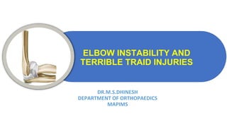
  • 1. ELBOW INSTABILITY AND TERRIBLE TRAID INJURIES DR.M.S.DHINESH DEPARTMENT OF ORTHOPAEDICS MAPIMS
  • 2. ELBOW STABILISERS • STATIC STABILISERS • PRIMARY CONSTRAINTS • SECONDARY CONSTRAINTS • DYNAMIC STABILISERS
  • 4.
  • 8.
  • 9.
  • 10. TRAUMATIC ELBOW INSTABILITY • Mechanism of injury • Primary mechanism is posterolateral • Other mechanisms postulated - controversial • Valgus-external rotation • Valgus-hyperextension • Sequence of injury progression also debated • Medial dislocations
  • 11. Posterolateral Mechanism - Injury progression
  • 12. O’DRISCOLL’S RING OF INSTABILITY O'Driscoll et al. described a valgus, axial, and posterolateral force that results in the typical posterolateral dislocation of the elbow joint
  • 15. TRAUMATIC ELBOW INSTABILITY • Simple Dislocations • Complex Dislocations – Fracture- dislocations • Coronoid fractures • Radial Head Fractures • Monteggia Fractures • Trans-olecranon dislocations • Terrible Triad • Chronic Dislocations
  • 16. • Not associated with fracture • Posterior and Posterolateral (M/C)– all directions and divergent dislocations can occur • Beware patterns other than posterior/posterolateral – can be more unstable after reduction • Mechanism of Injury • Partially flexed elbow • Axial load, supination, and valgus • Varus mechanisms also described • Medial ligamentous injuries in most cases SIMPLE DISLOCATION
  • 21. UNSTABLE AFTER REDUCTION • Uncommon in simple dislocations • May require soft tissue reconstruction • Do what needs to be done to hold a concentric reduction • Splint in more flexion • External fixator – static vs hinged • Elbow cross pinning • Internal fixator
  • 23.
  • 24. • Associated with a fracture • Sub-Types • Coronoid Fracture • Monteggia Fracture • Radial Head Fracture • Epicondyle fractures - adolescents • Trans-Olecranon fracture-Dislocations • Terrible Triad Elbow COMPLEX ELBOW DISLOCATION
  • 26. RADIAL HEAD FRACTURE Mechanism of Injury: Valgus force + axial load RADIAL HEAD FRACTURE
  • 28. • Comprehensive classification of radial head fracture with description of associated injuries (van Riet and Morrey)
  • 29.
  • 30.
  • 31. Valgus stability-MCL and Radial head Importance: Valgus force Primary stabilisers – anterior bundle of MCL Secondary stabilisers- Radial Head So,whenever excise the radial head in case of MCL rupture- no resist to valgus force- valgus instability
  • 32. Radial head communited fracture Radial head excised Intact intraosseous membrane Radial head excised Ruptured intraosseous membrane Radius migrated proximally- causing damage to elbow joint, Ulna move towards distally- causing damage to ulnocarpal jt. So, whenever Radial head fracture Always look for DRUJ Disruption/intraoss eous membrane damage ESSEX LOPRESSETI #
  • 33. UNSTABLE RADIAL HEAD # No MCL + Radial head excision = valgus instability = Early osteoarthritis UNSTABLE RADIAL HEAD FRACTURE
  • 34. DECISION MAKING • Fragment number • Displacement • Articular surface • Age and bone quality • Dislocation • Ass. Ligamentous injury • Ass. Elbow fractures DECISION MAKING
  • 35. STABLE FIXATION AND EARLY MOBILISATION
  • 36. RADIAL HEAD- SMALL NON ARTICULAR PORTION
  • 37. RADIAL HEAD AND NECK- SAFE ZONE • 240° of circumference articulates with ulna at lesser sigmoid notch • ~90-100 ° arc of safe hardware placement Caput et al recommended using the radial styloid and listers tubercle as guides
  • 38.
  • 40.
  • 42. ALGORITHM FOR RADIAL HEAD FRACTURE
  • 43. COMMINUTED RADIAL HEAD FRACTURE ROLE OF THE RADIAL HEAD ARTHROPLASTY ▪ Excision will lead to instability ▪ Functional spacer ▪ Creates stability by increasing radial length & restoring valgus restraint
  • 44.
  • 45. RADIAL HEAD FRACTURE- OVERSTUFFING Radial Head Should be in line with proximal edge of the lesser sigmaoid notch to avoid overstuffing
  • 48. ▪ Direct visualization ▪ Most accurate way to determine appropriate head size ▪ Radial head should be just at or proximal to radial notch of the ulna ▪ Intra-op Fluoro • Needs to be assessed in flexion and extension • Less reliable • > 6mm overstuffing must be present to consistently be seen on fluoro RADIAL HEAD FRACTURE-OVERSTUFFING
  • 49. POST-OP PROTOCOL ▪ For all stabilized fxs and dislocations regardless of fixation • Initially • Immobilization for 10-14 days • Secondarily • Early ACTIVE range of motion • Allows dynamic stabilizers to help hold reduction of joint ▪ Will reduce pseudosubluxations • Limits elbow stiffness • Some limit active shoulder abduction if LUCL was repaired
  • 50. APPROACHES • KAPLAN APPROACH • KOCHER APPROACH • EDC Split • Modified Boyd • Posterior approach • Elevate LUCL from lateral epicondyle • Can be used for combined olecranon/radial head fxs RARELY USED APPROACH
  • 51. KOCHER APPROACH Plane Between ECU and anconeus Most often utilized for radial head • Interval • Anconeus – Radial Nerve • ECU – PIN • 5cm incision from lateral epicondyle distally • Angled posteriorly 30-45 degrees • Often deep soft tissues will be disrupted by injury
  • 52. • Damage to LUCL • Stay on anterior half of radial head • Damage to PIN • Pronate the arm to move nerve distally • Carefully dissect distal to annular ligament KOCHER PITFALLS
  • 53. • Distal extension becomes dorsal Thompson approach • More often used for radial neck/proximal radial shaft fxs • Interval • ECRB – Radial nerve or PIN (variable) • EDC – PIN 10cm incision from lateral epicondyle to Lister’s Tubercle KAPLAN APPROACH
  • 55.
  • 56.
  • 57.
  • 58. CLASSIFICATION: CORONOID FRACTURES ▪ Regan & Morrey • Type 1: tip • Type 2 : < 50%y be ste • Type 3 > 50% ▪ Highly unstable
  • 59. CLASSIFICATION: CORONOID FRACTURES ▪O’Driscoll Classification ▪Type I: Tip ▪Type II: anteromedial facet ▪Type III: base
  • 60. PROXIMAL ULNA - ANTERIOR CORONOID •Anterior capsule •Brachialis •Anterior bundle of MCL •Anteromedial facet of coronoid ▪ Fx propagation into this region may cause functional MCL incompetancy
  • 61. CORONOID FRACTURE- INDICATION 1.An isolated fracture (Type 3) 2.As a part of fracture dislocation 3.As a part of communited fractures of proximal ulna
  • 62. CORONOID FRACTURE Complex instability of the elbow results from posterior directed forces • TR(Triceps) • BR(Brachialis) • BC(Biceps)
  • 63.
  • 64.
  • 65. OLECRANON FRACTURE Mechanism of Injury • Acute Tension overload: Tension applied by the triceps with flexion of the elbow • Direct Trauma • Chronic overload: eg. stress fractures seen commonly with osteopaenic or pediatric patients
  • 66.
  • 67. CLASSIFICATION Many Classifications: – Colton – Morrey – Schatzker – AO/ASIF – OTA Criteria – Displacement – Direction of fracture – Degree of comminution – Percent involvement – Associated injuries
  • 69. TREATMENT -AIM Restoration of elbow motion and prevention of stiffness – Goal is to begin early ROM • Restoration and preservation of the elbow extensor mechanism. • Restoration of the articular surface. • Prevention of complications.
  • 70. TREATMENT METHOD • Non operative method • Operative method • Excision of olecranon and triceps repair • Open reduction with internal fiaxtion • TBW with pins or intramedullary screws • Plate
  • 71.
  • 72. PUDA Olecranon Process – 96% of proximal ulna exhibit a ~ 4 degrees dorsal angulation (PUDA)
  • 73. VARUS ANGULATION Proximally the ulna demonstrates ~ 12 degrees varus angulation – The articular surface extends beyond the “joint space” visualized on the lateral radiograph
  • 74. SURGICAL ANATOMY • Coronoid process: preserve height – Coronoid Height ~ 2 x Olecranon height – Tip of Coronoid to tip of Olecranon subtends angle of ~30 degrees from long axis of ulnar shaft Articular cartilage – Sigmoid notch of ulna: bare spot centrally between tip and coronoid – Pearl: Beware of narrowing sigmoid fossa when treating comminuted olecranon fx’s.
  • 75. TBW For most simple, transverse, non-comminuted fractures • Use 18- or 20-gauge steel wire or small braided cable. – Be sure wires cross over dorsal cortex. – 2 smaller (22 gauge) wires may be less prominent • May use with either parallel K-wires or an intramedullary screw
  • 76.
  • 77.
  • 78.
  • 79. For simple and transverse fracture fracture If fracture goes beyond the coronoid,TBW principle not work
  • 80. INTRAMEDULLARY SCREWS Need to add tension band wire • Long/large screw required – 6.5mm cancellous – 85-110 mm long • Risk of shortening… osteopaenic bone, oblique fracture and comminution
  • 83.
  • 85. LOCKED ANATOMICAL PLATE Disadvantages • More difficult to contour anatomically • Locking configurations do not prevent violation of proximal articulations • More expensive • Not necessarily less prominent Advantages • Simplify plate fixation May accommodate - Slight varus proximal angulation - Slight rocker bottom proximal subcutaneous border - requires extensive triceps split - may improve proximal fixation
  • 87. WHAT IS A TERRIBLE TRIAD? 1. Elbow dislocation 2. Coronoid fracture 3. Radial head fracture
  • 88. TERRIBLE TRIAD INJURIES: MECHANISM OF INJURY ▪ Fall on an outstretched hand ▪ Axial load ▪ Relative elbow extension ▪ Valgus ▪ Forearm rotation ▪ Supination The ultimate “Posterolateral rotatory instability”
  • 89. TERRIBLE TRIAD FRACTURE- DISLOCATION ▪ What is so terrible about it? ▪ Extremely unstable ▪ Loss of joint congruency ▪ Instability ▪ Fracture fragments are usually quite small ▪ Difficult to repair ▪ Patients don’t routinely do “well” ▪ Unaware of the magnitude of the injury for the elbow ▪ Residual instability ▪ Stiffness
  • 90. TERRIBLE TRIAD INJURIES PATIENT AND INJURY ASSESSMENT • Patient evaluation ▪ Associated injuries ▪ Mechanism of injury ▪ Soft tissue status ▪ Radiographs (possible traction views) ▪ Post-reduction CT w/ 3D recons • Operative timing ▪ As urgently as possible but during the daytime ▪ Pre-op planning for appropriate equipment
  • 92. INJURY PATTERNS ▪Posterior dislocation, radial head & coronoid fractures ▪ “Terrible Triad”
  • 93. INJURY PATTERNS ▪Posterior dislocation & radial head fracture ▪Posterior dislocation, radial head & coronoid fractures ▪ “Terrible Triad” ▪Transolecranon fracture- dislocations ▪ Anterior ▪ Posterior
  • 94. 47 yo trip and fall down stairs
  • 95. TERRIBLE TRIAD –TREATMENT PROTOCOL (MCKEE, PUGH, SCHEMITSCH,ET AL JBJS(A) ’04) ▪ 36 consecutive patients treated: 1. Fix or suture coronoid 2. Repair / replace radial head 3. Repair LCL 4. If still unstable, repair MCL 5. If still unstable, hinged ex-fix
  • 96. SURGICAL PLANNING: APPROACHES ▪What’s injured? ▪ Radial head only ▪ Radial head ▪ type 1 coronoid ▪ Radial head ▪ type 2 or 3 coronoid ▪ Proximal ulna / olecranon ●Medial Approach Needed if: ▪ plate coronoid fracture ▪ transpose ulnar nerve ▪ repair or reconstruct MCL Radial head replacement & common proximal ulna fracture exposes coronoid tip
  • 97. INTERNAL FIXATION ▪3 steps: ▪ Repair radial head ▪ Secure radial head to the radial neck ▪ Avoid impingement of plates during forearm rotation. ▪Small K wires used provisionally. ▪“mini-fragment” screws (1.5 to 2.7 mm), countersink heads ▪Secure radial head to neck with 2.0 or 2.7 L-shaped plates or mini blade plates
  • 98. TERRIBLE TRIAD: MEDIAL INSTABILITY ? ▪ Repair MCL ▪ Reconstruct through bone tunnels ▪ Suture Anchors ▪ Palmaris autograft or allograft tendon ▪ Repair muscle origins FC U Ulnar Nerve Ulnohumeral joint reduced
  • 99. TERRIBLE TRIAD: PERSISTENT INSTABILITY ? ▪ Hinges Uniplanar Lateral Frame Multiplanar Compass Hinge
  • 100. SURGICAL PLANNING ▪ Positioning: supine vs lateral ▪ Supine: ▪ Better access and visualization of anterior joint & coronoid ▪ Lateral ▪ facilitates ulnar length, lessens needs for assistants ▪ Surgical approach: ▪ Midline Posterior ▪ Kocher (posterolateral) vs Kaplan (anterolateral) ▪ Anteromedial ▪ Posteromedial ▪ Percutaneous coronoid fixation
  • 102. LATERAL: KAPLAN APPROACH • Anterior column exposure ▪ Supracondylar ridge ▪ Anterior to mid-axis of radiocapitellar joint ▪ Utilize LCL tear ▪ Incise anterior capsule ▪ Exposes anterior coronoid ▪ Replacement or fixation
  • 103. LATERAL APPROACH: DEEP DISSECTION • Access to anterior ulno- humeral joint ▪ Elevate the extensors ▪ Stay superior to the LCL ▪ Able to visualize the PIN • Arthrotomy ▪ Release of the lateral capsule and annular ligament
  • 104. ANTEROMEDIAL APPROACH TO CORONOID •Medial supracondylar ridge •Pronator teres - brachialis interval •Incise anterior 1/2 flexor-pronator mass •Anterior capsule
  • 105. ANTEROMEDIAL APPROACH TO CORONOID •Medial supracondylar ridge •Pronator teres - brachialis interval •Incise anterior 1/2 flexor-pronator mass •Anterior capsule
  • 106. ANTEROMEDIAL APPROACH TO CORONOID •Medial supracondylar ridge •Pronator teres - brachialis interval •Incise anterior 1/2 flexor-pronator mass •Anterior capsule
  • 107. POSTEROMEDIAL APPROACH TO CORONOID Exposure of: • Coronoid • Sublime tubercle • MCL • Proximal ulna ▪MCL reconstruction or repair ▪ORIF AM facet of coronoid ▪Buttress plating of coronoid
  • 108. POSTEROMEDIAL APPROACH TO CORONOID ▪Necessitates ulnar nerve exposure and transposition ▪Palpate sublime tubercle ▪Incise FCU ulnar attachment distal to sublime tubercle and proceed proximally -> anterior bundle of MCL.
  • 109. TERRIBLE TRIAD INJURIES: REHABILITATION ▪ Rehab ▪ Stiffness vs. Instability ▪ Cautious ▪ Posterior splint ▪ 14 days post-op ▪ Cuff and collar ▪ Guided rehab is essential ▪ Flexion first! ▪ Active and passive ▪ Active and passive forearm rotation at 90° ▪ Begin extension at 3 weeks, active only ▪ Start supine—active against gravity
  • 110. TERRIBLE TRIAD INJURIES: SUMMARY ▪ Not so Terrible ▪ Isolated injury & cooperative patient ▪ Stable repairs & motion ▪ Coronoid fixation ▪ Radial head arthroplasty vs. ORIF ▪ LCL repair ▪ Terrible ▪ Poor stability after repairs complete ▪ Multi-trauma ▪ ICU stay ▪ Head injuries ▪ Non-weight bearing on lower extremities ▪ Uncooperative patient