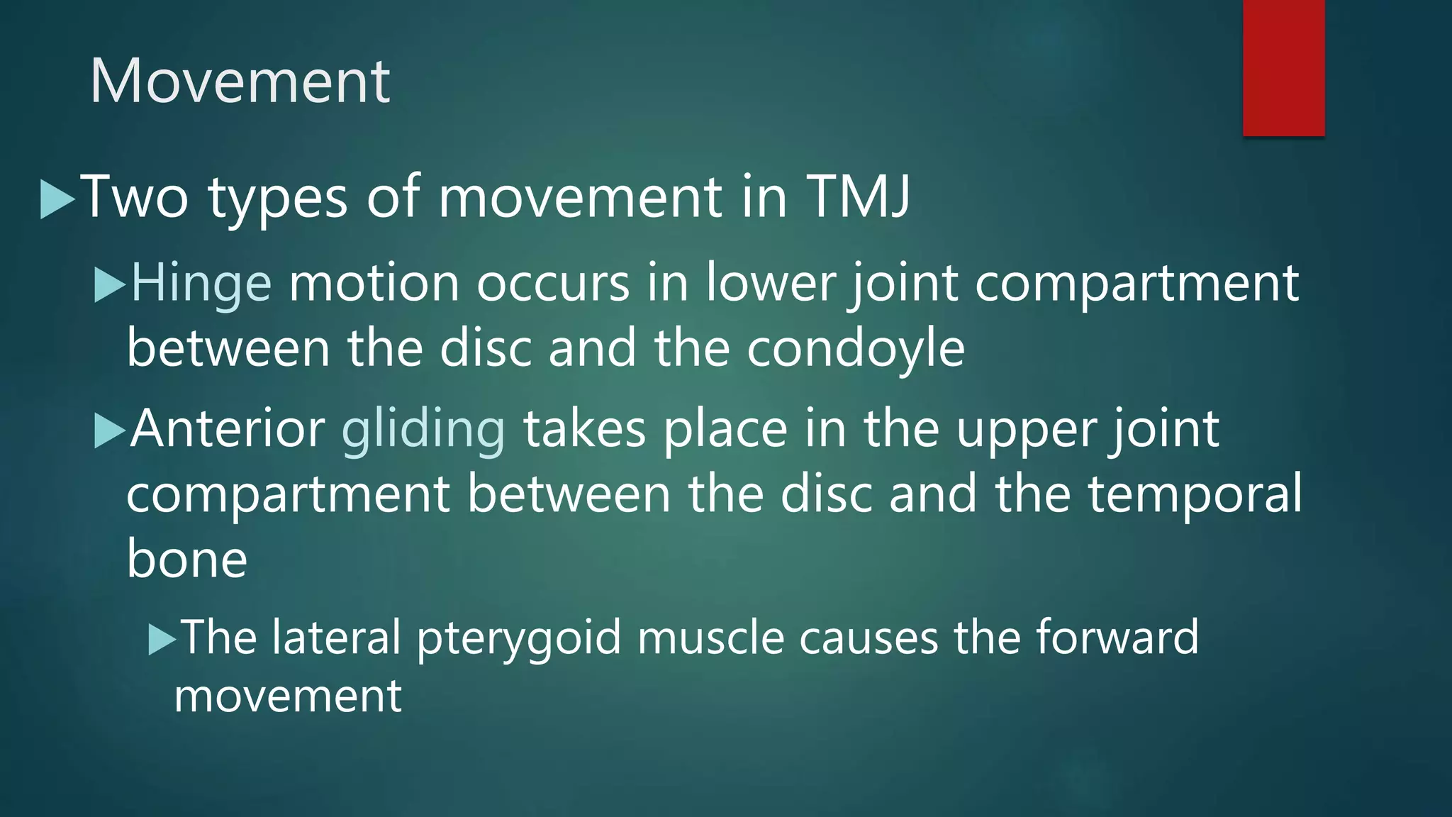
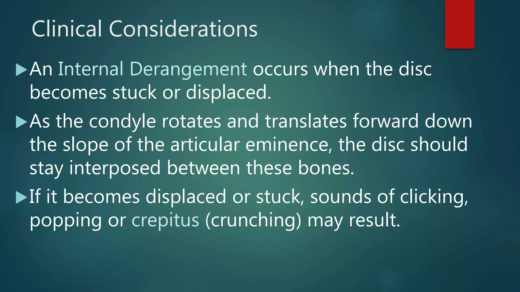
Temporomandibular Joint
(TMJ)
CHAPTER 22
Anatomy
Joint is the joining together of two bones
Temporomandibular joint (TMJ) is articulating
joint between temporal bone (skull) and mandible
(lower jaw)
Bilateral movement of right and left sides
interrelated and function as single unit
The articular disc separates the joint into upper and
lower compartments
Anatomy
The condoyle of the mandible articulates with the
mandibular (glenoid) fossa
Location is the posterior slope of the articular
eminence and anterior portion of the mandibular
fossa
Does not fit into the center of the mandibular fossa
but rests near the articular (eminence) tubercle
Articular disc (meniscus) rests between them
Anatomy
Anatomy
The articular disc is a pad of dense fibrous connective
tissue that is thickest at the posterior ends, thinnest in
the middle, and thicker again at the anterior end.
It separates the TMJ into upper and lower joint
compartments.
Laterally and medially, the disc is attached to the condyle
itself, so that whenever the condyle glides forward and
backward the disc moves with it.
Anatomy
The condyle and articular eminence are covered
by dense connective tissue
Contains no blood vessels or nerves
Nourished and lubricated by synovial fluid
Joint is covered by a thick fibrous capsule
The articular disc and capsule are connected by a
retrodiscal pad
A pad of loose connective tissue that allows for
anterior movement of the joint
Anatomy
Nerve and Blood Supply
Innervation supplied by two nerves
Auriculotemporal and Masseteric
Branches of mandibular nerve (V3)
Blood supply provided by branches of
superficial temporal and maxillary arteries
Movement
Two types of movement in TMJ
Hinge motion occurs in lower joint compartment
between the disc and the condoyle
Anterior gliding takes place in the upper joint
compartment between the disc and the temporal
bone
The lateral pterygoid muscle causes the forward
movement
Movement
Closing of the mouth is
accomplished by the lateral
pterygoid muscle, which
controls posterior movement
of disc
Clinical Considerations
An Internal Derangement occurs when the disc
becomes stuck or displaced.
As the condyle rotates and translates forward down
the slope of the articular eminence, the disc should
stay interposed between these bones.
If it becomes displaced or stuck, sounds of clicking,
popping or crepitus (crunching) may result.
Clinical Concerns
Catching or locking of the jaw, subluxation
The condoyle is displaced anteriorly and cannot be
returned voluntarily to normal position
Temporomandibular disorder (TMD), characterized
by muscle soreness
Bruxism (teeth grinding) from stress or tension are
usual causes
Clinical Concerns
Inflammation - arthritis may result from
wear or tear within the joint
Can also be a result of systemic disease
like rheumatoid arthritis
Cortisone is used to alleviate the pain of
arthritis
Clinical Concerns
TMD
https://youtu.be/mB468Jh9aAY
https://youtu.be/Cj3GVRlrgEQ
Suggested Homework
Answer the questions on pages 294-
295 in your textbook.
Complete pages 76-81 in your coloring
book

Temporomandibular Joint (TMJ)

  • 1.
  • 2.
    Anatomy Joint is thejoining together of two bones Temporomandibular joint (TMJ) is articulating joint between temporal bone (skull) and mandible (lower jaw) Bilateral movement of right and left sides interrelated and function as single unit The articular disc separates the joint into upper and lower compartments
  • 3.
    Anatomy The condoyle ofthe mandible articulates with the mandibular (glenoid) fossa Location is the posterior slope of the articular eminence and anterior portion of the mandibular fossa Does not fit into the center of the mandibular fossa but rests near the articular (eminence) tubercle Articular disc (meniscus) rests between them
  • 4.
  • 5.
    Anatomy The articular discis a pad of dense fibrous connective tissue that is thickest at the posterior ends, thinnest in the middle, and thicker again at the anterior end. It separates the TMJ into upper and lower joint compartments. Laterally and medially, the disc is attached to the condyle itself, so that whenever the condyle glides forward and backward the disc moves with it.
  • 6.
    Anatomy The condyle andarticular eminence are covered by dense connective tissue Contains no blood vessels or nerves Nourished and lubricated by synovial fluid Joint is covered by a thick fibrous capsule The articular disc and capsule are connected by a retrodiscal pad A pad of loose connective tissue that allows for anterior movement of the joint
  • 7.
  • 8.
    Nerve and BloodSupply Innervation supplied by two nerves Auriculotemporal and Masseteric Branches of mandibular nerve (V3) Blood supply provided by branches of superficial temporal and maxillary arteries
  • 9.
    Movement Two types ofmovement in TMJ Hinge motion occurs in lower joint compartment between the disc and the condoyle Anterior gliding takes place in the upper joint compartment between the disc and the temporal bone The lateral pterygoid muscle causes the forward movement
  • 10.
    Movement Closing of themouth is accomplished by the lateral pterygoid muscle, which controls posterior movement of disc
  • 11.
    Clinical Considerations An InternalDerangement occurs when the disc becomes stuck or displaced. As the condyle rotates and translates forward down the slope of the articular eminence, the disc should stay interposed between these bones. If it becomes displaced or stuck, sounds of clicking, popping or crepitus (crunching) may result.
  • 12.
    Clinical Concerns Catching orlocking of the jaw, subluxation The condoyle is displaced anteriorly and cannot be returned voluntarily to normal position Temporomandibular disorder (TMD), characterized by muscle soreness Bruxism (teeth grinding) from stress or tension are usual causes
  • 13.
    Clinical Concerns Inflammation -arthritis may result from wear or tear within the joint Can also be a result of systemic disease like rheumatoid arthritis Cortisone is used to alleviate the pain of arthritis
  • 14.
  • 15.
  • 16.
    Suggested Homework Answer thequestions on pages 294- 295 in your textbook. Complete pages 76-81 in your coloring book