Downloaded 214 times


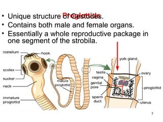
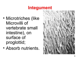
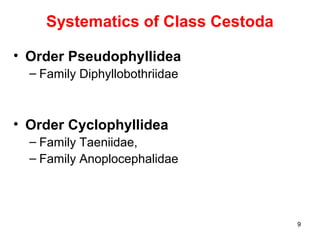


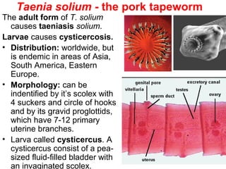


1. Tapeworms are parasitic flatworms that consist of a head called a scolex and a long segmented body called a strobila. They attach to the intestinal wall of their host and absorb nutrients. 2. Common tapeworms discussed include Diphyllobothrium latum, Taenia solium, Taenia saginata, Hymenolepis nana, Echinococcus granulosus, and Echinococcus multilocularis. Their life cycles involve an intermediate host and definitive host, usually transmitting between via ingestion of eggs from feces. 3. Symptoms from tapeworm infections can range from asymptomatic to abdominal pain, diarrhea, and nutritional deficiencies. Diagnosis


![[Micro] cestodes](https://cdn.slidesharecdn.com/ss_thumbnails/nvpl1fbyq2ofjfsbmped-signature-2127a2ca5368c7fdfd023e8d90dde3fc0b9fe7d91346a4189562c9f63dc0d19d-poli-150819190753-lva1-app6892-thumbnail.jpg?width=640&height=640&fit=bounds)


