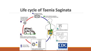
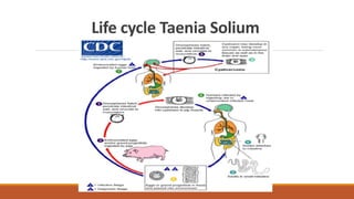
- Taenia saginata and Taenia solium are two common tapeworms that infect humans. T. saginata, the beef tapeworm, passes between humans and cattle, while T. solium, the pork tapeworm, passes between humans and pigs.
- Both have a life cycle involving an adult tapeworm in the human small intestine and a larval cyst stage in cattle (for T. saginata) or pigs (for T. solium). Humans can become infected by eating undercooked beef or pork containing the cysts.
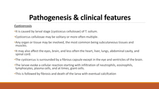
- Intestinal infection is usually asymptomatic but cysticercosis, occurring when the larval cysts establish in tissues like brain or muscle,