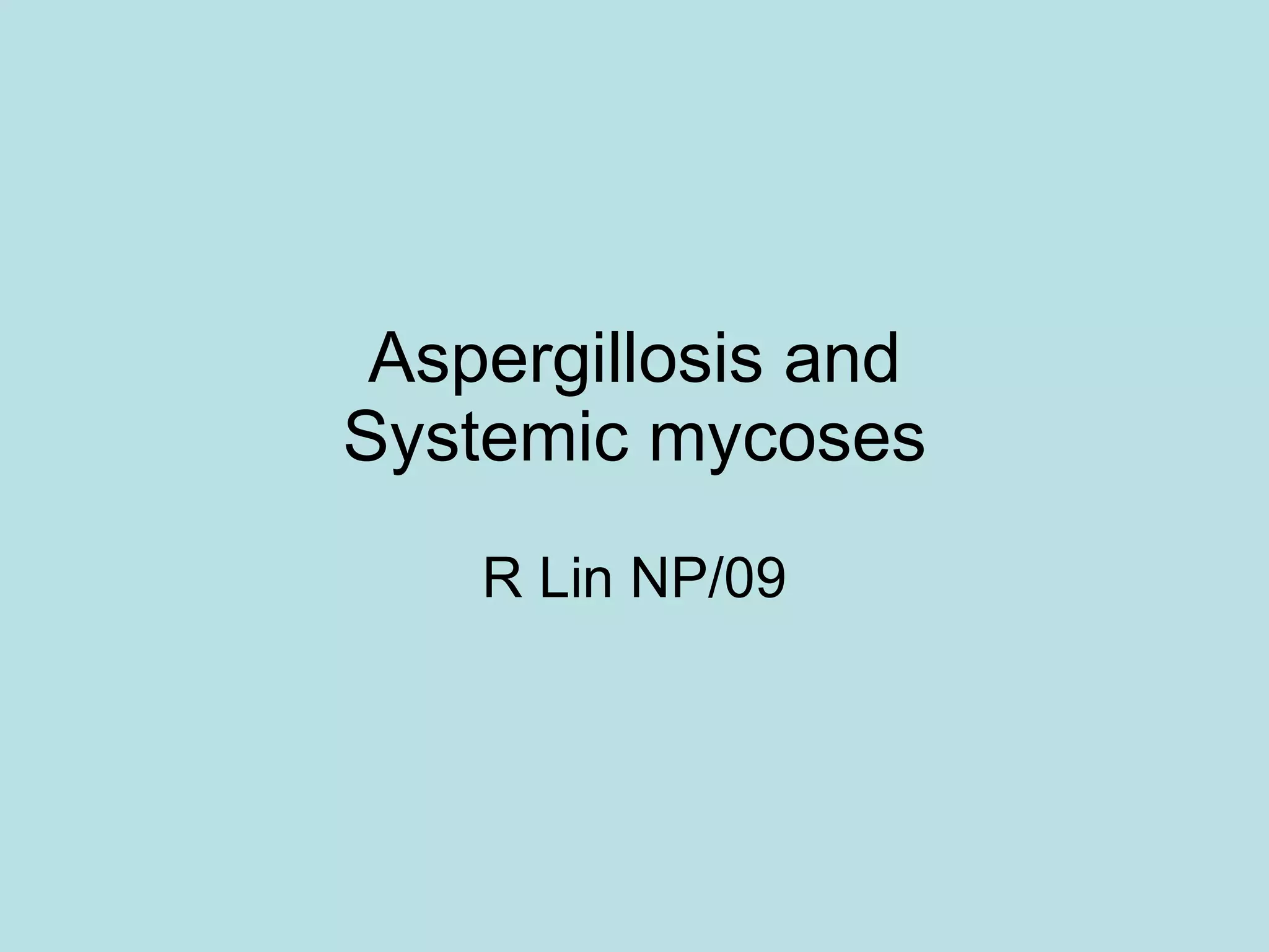
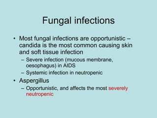
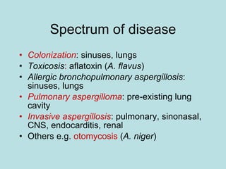
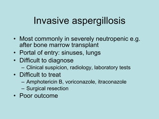
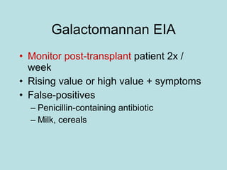
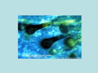
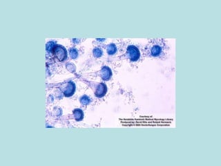
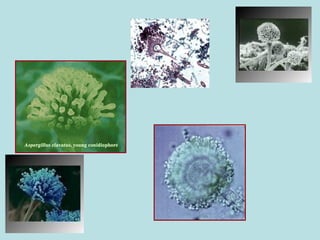
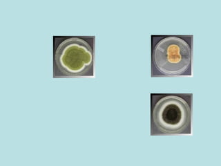
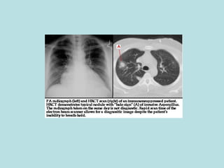
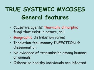
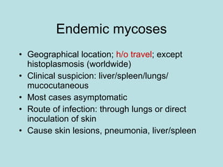
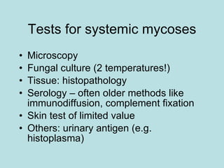
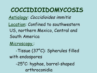
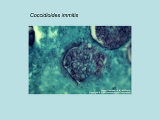
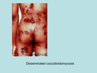
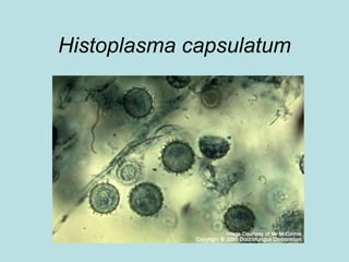
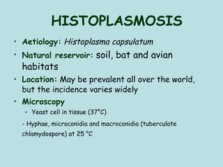
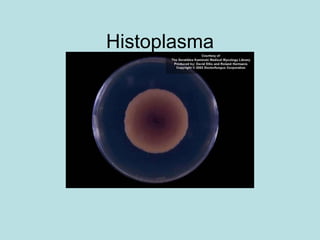
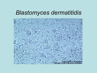
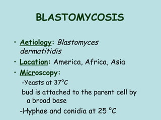
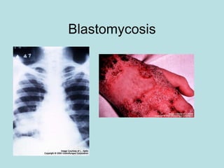
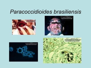
Aspergillosis and Systemic mycoses R Lin NP/09
Fungal infections Most fungal infections are opportunistic – candida is the most common causing skin and soft tissue infection Severe infection (mucous membrane, oesophagus) in AIDS Systemic infection in neutropenic Aspergillus Opportunistic, and affects the most  severely neutropenic
Aspergillosis Found in the air all around us Harmless to normal people Opportunistic disease in the immunocompromised patient Severe neutropenia Debilitating disease Disruption of normal flora Antibiotics, steroids Abnormal lung e.g. bronchiectasis, cancer
Spectrum of disease Colonization : sinuses, lungs Toxicosis :  aflatoxin ( A. flavus ) Allergic bronchopulmonary aspergillosis : sinuses, lungs Pulmonary aspergilloma : pre-existing lung cavity Invasive aspergillosis : pulmonary, sinonasal, CNS, endocarditis, renal Others e.g.  otomycosis  ( A. niger )
Invasive aspergillosis Most commonly in severely neutropenic e.g. after bone marrow transplant Portal of entry: sinuses, lungs Difficult to diagnose Clinical suspicion, radiology, laboratory tests Difficult to treat Amphotericin B, voriconazole, itraconazole Surgical resection Poor outcome
Laboratory investigation Microscopy: KOH prep, sputum, bronchoalveolar lavage Histology : lung Culture : sputum (contaminant?), BAL, tissue Galactomannan EIA PCR: 18sRNA gene, not always useful
Galactomannan EIA Monitor post-transplant  patient 2x / week Rising value or high value + symptoms False-positives Penicillin-containing antibiotic Milk, cereals


TRUE SYSTEMIC (ENDEMIC) MYCOSES Coccidioidomycosis Histoplasmosis Blastomycosis Paracoccidioidomycosis
TRUE SYSTEMIC MYCOSES General features Causative agents:  thermally dimorphic  fungi that exist in nature, soil Geographic  distribution varies Inhalation   pulmonary INFECTION    dissemination  No evidence of transmission among humans or animals Otherwise healthy individuals are infected
Endemic mycoses Geographical location;  h/o travel ; except histoplasmosis (worldwide) Clinical suspicion: liver/spleen/lungs/ mucocutaneous Most cases asymptomatic Route of infection: through lungs or direct inoculation of skin Cause skin lesions, pneumonia, liver/spleen
Tests for systemic mycoses Microscopy Fungal culture (2 temperatures!) Tissue: histopathology Serology – often older methods like immunodiffusion, complement fixation Skin test of limited value Others: urinary antigen (e.g. histoplasma)
COCCIDIOIDOMYCOSIS Aetiology :   Coccidioides immitis Location :  Confined to southwestern US, northern Mexico, Central and South America Microscopy. :  - Tissue (37°C): Spherules filled with endospores  -25°C: hyphae, barrel-shaped  arthroconidia
Case history A 71-year-old male subject regularly spends several winter months in Arizona to play golf in the sun. Last March he experienced a gradual onset of fever and a headache, followed by a non-productive cough, myalgia and profound fatigue. His local physician diagnosed bronchopneumonia on chest radiograph, and prescribed azithromycin. The antibiotic provided no benefit, and ultimately the patient received two more courses of different empiric antibiotics. He returned home with continued cough and fatigue, even though the fever had abated somewhat. Two months following the initial onset of symptoms, a bronchoscopy was performed, and cultures grew Coccidioides species.
Other presentations Months to years following a symptomatic or asymptomatic infection, the affected lung may show complete resolution or an area of calcified or uncalcified pulmonary nodule similar radiographically to cancer. Microscopic examination of excised tissue identifies the organism. Occasionally the nodule liquefies to form a thin-walled cavity, which may close spontaneously or remain and become a nidus for suprainfection or spontaneous pneumothorax. Extrapulmonary dissemination can be identified in nearly all tissues, although skin and soft tissue, bones and meninges are the most common sites of dissemination. Chronic fibrocavitary pneumonia is seen infrequently, with chronic cough and dyspnoea, night sweats and weight loss, and lung fibrosis with thick-walled cavities.  From BMJ Best Practice
Coccidioides immitis
Disseminated coccidioidomycosis
Histoplasma capsulatum
HISTOPLASMOSIS Aetiology:   Histoplasma capsulatum Natural reservoir:   soil, bat and avian habitats Location:  May be prevalent all over the world, but the incidence varies widely  Microscopy   Yeast cell in tissue (37°C) - Hyphae, microconidia and macroconidia (tuberculate chlamydospore) at 25 °C
Histoplasma capsulatum
Histoplasma


Blastomyces dermatitidis
BLASTOMYCOSIS Aetiology :   Blastomyces dermatitidis  Location :   America, Africa, Asia Micr oscopy:   -Yeasts at 37°C bud is attached to the parent cell by a broad base   -Hyphae and conidia at 25 °C
Blastomycosis
Paracoccidioides brasiliensis
PARACOCCIDIOIDOMYCOSIS Aetiology:   Paracoccidioides brasiliensis Location :   Central and South America Pathogenesis :  Inhalation of conidia Microscopy .:  At 37°C (in tissue ): multiply budding yeasts; the buds are attached to the parent cell by a narrow base   At 25 °C: hyphae and conidia
Questions Aspergillus Give an account: normal habitat, risk factors, spectrum of disease, lab tests Endemic mycosis How does one diagnose? Types of lab tests? Why is it important to know?

aspergillus lecture

  • 1.
    Aspergillosis and Systemicmycoses R Lin NP/09
  • 2.
    Fungal infections Mostfungal infections are opportunistic – candida is the most common causing skin and soft tissue infection Severe infection (mucous membrane, oesophagus) in AIDS Systemic infection in neutropenic Aspergillus Opportunistic, and affects the most severely neutropenic
  • 3.
    Aspergillosis Found inthe air all around us Harmless to normal people Opportunistic disease in the immunocompromised patient Severe neutropenia Debilitating disease Disruption of normal flora Antibiotics, steroids Abnormal lung e.g. bronchiectasis, cancer
  • 4.
    Spectrum of diseaseColonization : sinuses, lungs Toxicosis : aflatoxin ( A. flavus ) Allergic bronchopulmonary aspergillosis : sinuses, lungs Pulmonary aspergilloma : pre-existing lung cavity Invasive aspergillosis : pulmonary, sinonasal, CNS, endocarditis, renal Others e.g. otomycosis ( A. niger )
  • 5.
    Invasive aspergillosis Mostcommonly in severely neutropenic e.g. after bone marrow transplant Portal of entry: sinuses, lungs Difficult to diagnose Clinical suspicion, radiology, laboratory tests Difficult to treat Amphotericin B, voriconazole, itraconazole Surgical resection Poor outcome
  • 6.
    Laboratory investigation Microscopy:KOH prep, sputum, bronchoalveolar lavage Histology : lung Culture : sputum (contaminant?), BAL, tissue Galactomannan EIA PCR: 18sRNA gene, not always useful
  • 7.
    Galactomannan EIA Monitorpost-transplant patient 2x / week Rising value or high value + symptoms False-positives Penicillin-containing antibiotic Milk, cereals
  • 8.
  • 9.
  • 10.
  • 11.
  • 12.
  • 13.
  • 14.
    TRUE SYSTEMIC (ENDEMIC)MYCOSES Coccidioidomycosis Histoplasmosis Blastomycosis Paracoccidioidomycosis
  • 15.
    TRUE SYSTEMIC MYCOSESGeneral features Causative agents: thermally dimorphic fungi that exist in nature, soil Geographic distribution varies Inhalation  pulmonary INFECTION  dissemination No evidence of transmission among humans or animals Otherwise healthy individuals are infected
  • 16.
    Endemic mycoses Geographicallocation; h/o travel ; except histoplasmosis (worldwide) Clinical suspicion: liver/spleen/lungs/ mucocutaneous Most cases asymptomatic Route of infection: through lungs or direct inoculation of skin Cause skin lesions, pneumonia, liver/spleen
  • 17.
    Tests for systemicmycoses Microscopy Fungal culture (2 temperatures!) Tissue: histopathology Serology – often older methods like immunodiffusion, complement fixation Skin test of limited value Others: urinary antigen (e.g. histoplasma)
  • 18.
    COCCIDIOIDOMYCOSIS Aetiology : Coccidioides immitis Location : Confined to southwestern US, northern Mexico, Central and South America Microscopy. : - Tissue (37°C): Spherules filled with endospores -25°C: hyphae, barrel-shaped arthroconidia
  • 19.
    Case history A71-year-old male subject regularly spends several winter months in Arizona to play golf in the sun. Last March he experienced a gradual onset of fever and a headache, followed by a non-productive cough, myalgia and profound fatigue. His local physician diagnosed bronchopneumonia on chest radiograph, and prescribed azithromycin. The antibiotic provided no benefit, and ultimately the patient received two more courses of different empiric antibiotics. He returned home with continued cough and fatigue, even though the fever had abated somewhat. Two months following the initial onset of symptoms, a bronchoscopy was performed, and cultures grew Coccidioides species.
  • 20.
    Other presentations Monthsto years following a symptomatic or asymptomatic infection, the affected lung may show complete resolution or an area of calcified or uncalcified pulmonary nodule similar radiographically to cancer. Microscopic examination of excised tissue identifies the organism. Occasionally the nodule liquefies to form a thin-walled cavity, which may close spontaneously or remain and become a nidus for suprainfection or spontaneous pneumothorax. Extrapulmonary dissemination can be identified in nearly all tissues, although skin and soft tissue, bones and meninges are the most common sites of dissemination. Chronic fibrocavitary pneumonia is seen infrequently, with chronic cough and dyspnoea, night sweats and weight loss, and lung fibrosis with thick-walled cavities. From BMJ Best Practice
  • 21.
  • 22.
  • 23.
  • 24.
  • 25.
    HISTOPLASMOSIS Aetiology: Histoplasma capsulatum Natural reservoir: soil, bat and avian habitats Location: May be prevalent all over the world, but the incidence varies widely Microscopy Yeast cell in tissue (37°C) - Hyphae, microconidia and macroconidia (tuberculate chlamydospore) at 25 °C
  • 26.
  • 27.
  • 28.
  • 29.
  • 30.
  • 31.
  • 32.
    BLASTOMYCOSIS Aetiology : Blastomyces dermatitidis Location : America, Africa, Asia Micr oscopy: -Yeasts at 37°C bud is attached to the parent cell by a broad base -Hyphae and conidia at 25 °C
  • 33.
  • 34.
  • 35.
    PARACOCCIDIOIDOMYCOSIS Aetiology: Paracoccidioides brasiliensis Location : Central and South America Pathogenesis : Inhalation of conidia Microscopy .: At 37°C (in tissue ): multiply budding yeasts; the buds are attached to the parent cell by a narrow base At 25 °C: hyphae and conidia
  • 36.
    Questions Aspergillus Givean account: normal habitat, risk factors, spectrum of disease, lab tests Endemic mycosis How does one diagnose? Types of lab tests? Why is it important to know?