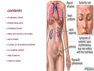
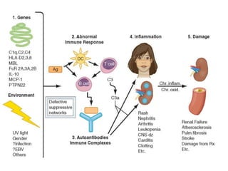
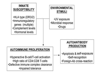
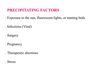
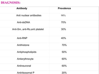
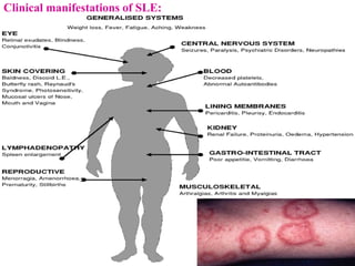
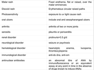
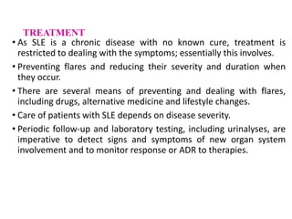
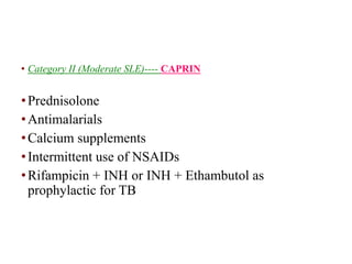
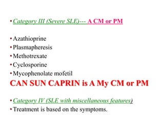
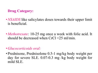
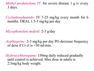
Systemic
Lupus
Erythematosus
contents
• INTRODUCTION
• EPIDEMIOLOGY
• PATHOGENISIS
• PRECIPITATING FACTORS
• DIAGNOSIS
• CLINICAL MANIFESTATIONS
• CLASSIFICATION
• TREATMENT
• CONCLUSION
INTRODUCTION
•SLE was first described in 1828; its name helps to define the
disease.
•Systemic is because the disease can affect organs and tissue
throughout the body.
•Lupus is Latin word for wolf. It refers to the rash that
extends across the bridge of the nose and upper cheekbones
(butter fly) and was thought to resemble a wolf bite.
•Erythematosus is from the Greek word
• for red and refers to the color of the rash
.
SLE can affect any part of the body, but
most often harms the
1. Heart
2. Joints
3. Skin
4. Lungs
5. Blood Vessels
6. Liver
7. Kidneys
8. Nervous System
EPIDEMIOLOGY
•The prevalence of SLE is higher among Asians, Afro-
Americans, Afro-Caribbean’s, Hispanic Americans,
and Asian Indians in Great Britain.
•There is an increased frequency of SLE among
women(10:1) that has been attributed to an estrogen
hormonal effect.
• Prevalence in males was 31.5 and 7.0 per 100 000 in
Asians and Whites respectively; in females the figures
were 69.7 and 31.7 per 100 000.
•SLE is rare in India.
•The reported prevalence of SLE in India is 3.2 per
100,000 population
•In India only 50%-60% survival at10 years.
•The epidemiology of SLE varies substantially between
different sex and age groups and is distributed
unequally among geographical regions; specifically,
SLE occurs more frequently in high-income countries.
•Possible reasons for poor survival in Indian SLE include
delay in diagnosis, referral bias (only the most serious
cases are referred by practitioners), suboptimal health
care facilities and an inherently more severe disease.
PATHOGENISIS
SLE is caused by the interactions between
susceptibility genes and environmental factors,
resulting in abnormal immune responses.
Hyper reactivity of T and B-lymphocytes is indicated
by increased surface expression of molecules such as
HLA-D and CD40L
The end results of this abnormalities is sustained
production of pathogenic auto antibodies and formation
of immune complexes that bind target tissues resulting
in the
1. Sequestration and destruction of Ig coated circulating
cells
2. Fixation and cleaving of complement proteins
3. Release of chemotoxins and vasoactive peptides, and
destructive enzymes into the tissues.
•Many autoantibodies in the persons with SLE are
directed against DNA or RNA complexes such as
nucleosomes, some nucleolar RNA.
• In individual with SLE, phagocytosis and removal of
apoptotic and of immune complexes are impaired.
•Thus in SLE, the antigens are available they are
presented in locations recognized by immune
complexes persists for prolonged periods of time,
allowing tissue damage to accumulate to the point of
clinical illness.
•Female gender is permissive for SLE females make higher
antibody responses than males.
•Estradiol binds to receptors on T and B-lymphocytes,
increasing activation and survival of these cells, thus favoring
prolonged immune responses.
PRECIPITATING FACTORS
 Exposure to the sun, fluorescent lights, or tanning beds
 Infections (Viral)
 Surgery
 Pregnancy
 Therapeutic abortions
 Stress
Antibody Prevalence
Anti nuclear antibodies 98%
Anti-dsDNA 70%
Anti-Sm, anti-Ro,anti platelet 30%
Anti-RNP 40%
Antihistone 70%
Antiphospholipids 50%
Antierythrocyte 60%
Antineuronal 60%
Antiribosomal P 20%
DIAGNOSIS:
Clinical manifestations of SLE:
Malar rash Fixed erethema, flat or raised, over the
malar eminences
Discoid rash Erythematous circular raised paths
Photosensitivity exposure to uv light cause rash
oral ulcers include oral and oesopharangeal ulcers
arthritis arthritis of two or more joints
serositis pleuritis or pericarditis
renal disorder protinuria>0.5 g/d
neurological disorder seizurs or psychosis
hematological disorder heamolytic anemia, lucopenia,
thrombocytopenia
immunological disorder anti-ds dna, anti-sm
antinuclear antibodies an abnormal titre of ANA by
immunoflurescence or an equivalent
assay at any point in time in the absence
of drugs known to induce ANAs
TREATMENT
• As SLE is a chronic disease with no known cure, treatment is
restricted to dealing with the symptoms; essentially this involves.
• Preventing flares and reducing their severity and duration when
they occur.
• There are several means of preventing and dealing with flares,
including drugs, alternative medicine and lifestyle changes.
• Care of patients with SLE depends on disease severity.
• Periodic follow-up and laboratory testing, including urinalyses, are
imperative to detect signs and symptoms of new organ system
involvement and to monitor response or ADR to therapies.
TREATMENT
• Treatment of SLE classified as
Non pharmacologic treatment
Pharmacological treatment
Non Pharmacological Treatment
Diet and nutrition
•Dietary fish oil
•A balanced diet, including carbohydrates, proteins, and
fats
•Active inflammatory disease and fever may require an
increase in caloric intake.
•Steroids (prednisone) increase appetite
•A diet that is low in saturated fat.
•Vitamins
•Calcium
•Herbal supplements
•Exercise
•Immunizations
•Avoiding SLE Triggers
•Reducing Stress
Pharmacological management
•Category I (Mild SLE)-----CAN
Creams and sun blocks
Nonsteroidal anti-inflammatory drugs
Antimalarials drugs
• Category II (Moderate SLE)---- CAPRIN
•Prednisolone
•Antimalarials
•Calcium supplements
•Intermittent use of NSAIDs
•Rifampicin + INH or INH + Ethambutol as
prophylactic for TB
•Category III (Severe SLE)--- A CM or PM
•Azathioprine
•Plasmapheresis
•Methotrexate
•Cyclosporine
•Mycophenolate mofetil
CAN SUN CAPRIN is A My CM or PM
•Category IV (SLE with miscellaneous features)
•Treatment is based on the symptoms.
Drug Category:
•NSAIDS like salicylates doses towards their upper limit
is beneficial.
•Methotrexate: 10-25 mg once a week with folic acid. It
should be decreased when CrCl <25 ml/min.
•Glucocorticoids oral:
•Prednisone, Prednisolone 0.5-1 mg/kg body weight per
day for severe SLE. 0.07-0.3 mg /kg body weight for
mild SLE.
Methyl prednisolone IV: for severe disease 1 g iv every
3 days.
Cyclophosphamide: IV 7-25 mg/kg every month for 6
months. ORAL 1.5-3 mg/kg per day
Mycophenolate mofetil: 2-3 g/day
Azathioprine: 2-3 mg/kg per day PO decrease frequency
of dose if Cr cl is <50 ml/min.
Hydroxychloroquine: 150mg daily reduced gradually
until control is achieved. Max dose in adults is
2.5mg/kg body weight.
NEWER DRUGS
• Hormone Treatments
•Dehydroepiandrosterone (DHEA)
•Prasterone
•Danazol
• Plasmapheresis
Investigational Treatments
•Monoclonal Antibodies (MAbs)
•Epratuzumab
• Belimumab
•Rituximab
•Leflunomide
•Autologous Stem Cell Transplantation:
The procedure first removes the cells from the patient, who then
receives high-dose immunotherapy. The stem cells are then
reintroduced.
UVA-1 Phototherapy
A treatment which uses ultraviolet A-1 (UVA-1) radiation,
which are long UVA wave lengths that do not promote sunburn
and may actually block inflammatory immune factors
CONCLUSION
•Patients with SLE have improved greatly since the
disease was first described.
•Mild disease may require no drugs or only symptomatic
therapy with Paracetamol or an NSAID.
•Hydroxychloroquine often controls mild to moderate
SLE and may keep the disease in remission.
•Patients with more active disease will need treatment
with systemic corticosteroids or cytotoxic drugs such as
azathioprine or cyclophosphamide.
•Many new drugs are under research which will help the
better control over SLE

Systemic Lupus Erythematosus -SLE PT2.ppt

  • 1.
  • 2.
    contents • INTRODUCTION • EPIDEMIOLOGY •PATHOGENISIS • PRECIPITATING FACTORS • DIAGNOSIS • CLINICAL MANIFESTATIONS • CLASSIFICATION • TREATMENT • CONCLUSION
  • 3.
    INTRODUCTION •SLE was firstdescribed in 1828; its name helps to define the disease. •Systemic is because the disease can affect organs and tissue throughout the body. •Lupus is Latin word for wolf. It refers to the rash that extends across the bridge of the nose and upper cheekbones (butter fly) and was thought to resemble a wolf bite. •Erythematosus is from the Greek word • for red and refers to the color of the rash .
  • 4.
    SLE can affectany part of the body, but most often harms the 1. Heart 2. Joints 3. Skin 4. Lungs 5. Blood Vessels 6. Liver 7. Kidneys 8. Nervous System
  • 5.
    EPIDEMIOLOGY •The prevalence ofSLE is higher among Asians, Afro- Americans, Afro-Caribbean’s, Hispanic Americans, and Asian Indians in Great Britain. •There is an increased frequency of SLE among women(10:1) that has been attributed to an estrogen hormonal effect. • Prevalence in males was 31.5 and 7.0 per 100 000 in Asians and Whites respectively; in females the figures were 69.7 and 31.7 per 100 000.
  • 6.
    •SLE is rarein India. •The reported prevalence of SLE in India is 3.2 per 100,000 population •In India only 50%-60% survival at10 years. •The epidemiology of SLE varies substantially between different sex and age groups and is distributed unequally among geographical regions; specifically, SLE occurs more frequently in high-income countries. •Possible reasons for poor survival in Indian SLE include delay in diagnosis, referral bias (only the most serious cases are referred by practitioners), suboptimal health care facilities and an inherently more severe disease.
  • 7.
    PATHOGENISIS SLE is causedby the interactions between susceptibility genes and environmental factors, resulting in abnormal immune responses. Hyper reactivity of T and B-lymphocytes is indicated by increased surface expression of molecules such as HLA-D and CD40L
  • 8.
    The end resultsof this abnormalities is sustained production of pathogenic auto antibodies and formation of immune complexes that bind target tissues resulting in the 1. Sequestration and destruction of Ig coated circulating cells 2. Fixation and cleaving of complement proteins 3. Release of chemotoxins and vasoactive peptides, and destructive enzymes into the tissues.
  • 9.
    •Many autoantibodies inthe persons with SLE are directed against DNA or RNA complexes such as nucleosomes, some nucleolar RNA. • In individual with SLE, phagocytosis and removal of apoptotic and of immune complexes are impaired. •Thus in SLE, the antigens are available they are presented in locations recognized by immune complexes persists for prolonged periods of time, allowing tissue damage to accumulate to the point of clinical illness.
  • 10.
    •Female gender ispermissive for SLE females make higher antibody responses than males. •Estradiol binds to receptors on T and B-lymphocytes, increasing activation and survival of these cells, thus favoring prolonged immune responses.
  • 13.
    PRECIPITATING FACTORS  Exposureto the sun, fluorescent lights, or tanning beds  Infections (Viral)  Surgery  Pregnancy  Therapeutic abortions  Stress
  • 14.
    Antibody Prevalence Anti nuclearantibodies 98% Anti-dsDNA 70% Anti-Sm, anti-Ro,anti platelet 30% Anti-RNP 40% Antihistone 70% Antiphospholipids 50% Antierythrocyte 60% Antineuronal 60% Antiribosomal P 20% DIAGNOSIS:
  • 15.
  • 16.
    Malar rash Fixederethema, flat or raised, over the malar eminences Discoid rash Erythematous circular raised paths Photosensitivity exposure to uv light cause rash oral ulcers include oral and oesopharangeal ulcers arthritis arthritis of two or more joints serositis pleuritis or pericarditis renal disorder protinuria>0.5 g/d neurological disorder seizurs or psychosis hematological disorder heamolytic anemia, lucopenia, thrombocytopenia immunological disorder anti-ds dna, anti-sm antinuclear antibodies an abnormal titre of ANA by immunoflurescence or an equivalent assay at any point in time in the absence of drugs known to induce ANAs
  • 17.
    TREATMENT • As SLEis a chronic disease with no known cure, treatment is restricted to dealing with the symptoms; essentially this involves. • Preventing flares and reducing their severity and duration when they occur. • There are several means of preventing and dealing with flares, including drugs, alternative medicine and lifestyle changes. • Care of patients with SLE depends on disease severity. • Periodic follow-up and laboratory testing, including urinalyses, are imperative to detect signs and symptoms of new organ system involvement and to monitor response or ADR to therapies.
  • 18.
    TREATMENT • Treatment ofSLE classified as Non pharmacologic treatment Pharmacological treatment
  • 19.
    Non Pharmacological Treatment Dietand nutrition •Dietary fish oil •A balanced diet, including carbohydrates, proteins, and fats •Active inflammatory disease and fever may require an increase in caloric intake.
  • 20.
    •Steroids (prednisone) increaseappetite •A diet that is low in saturated fat. •Vitamins •Calcium •Herbal supplements
  • 21.
  • 22.
    Pharmacological management •Category I(Mild SLE)-----CAN Creams and sun blocks Nonsteroidal anti-inflammatory drugs Antimalarials drugs
  • 23.
    • Category II(Moderate SLE)---- CAPRIN •Prednisolone •Antimalarials •Calcium supplements •Intermittent use of NSAIDs •Rifampicin + INH or INH + Ethambutol as prophylactic for TB
  • 24.
    •Category III (SevereSLE)--- A CM or PM •Azathioprine •Plasmapheresis •Methotrexate •Cyclosporine •Mycophenolate mofetil CAN SUN CAPRIN is A My CM or PM •Category IV (SLE with miscellaneous features) •Treatment is based on the symptoms.
  • 25.
    Drug Category: •NSAIDS likesalicylates doses towards their upper limit is beneficial. •Methotrexate: 10-25 mg once a week with folic acid. It should be decreased when CrCl <25 ml/min. •Glucocorticoids oral: •Prednisone, Prednisolone 0.5-1 mg/kg body weight per day for severe SLE. 0.07-0.3 mg /kg body weight for mild SLE.
  • 26.
    Methyl prednisolone IV:for severe disease 1 g iv every 3 days. Cyclophosphamide: IV 7-25 mg/kg every month for 6 months. ORAL 1.5-3 mg/kg per day Mycophenolate mofetil: 2-3 g/day Azathioprine: 2-3 mg/kg per day PO decrease frequency of dose if Cr cl is <50 ml/min. Hydroxychloroquine: 150mg daily reduced gradually until control is achieved. Max dose in adults is 2.5mg/kg body weight.
  • 27.
    NEWER DRUGS • HormoneTreatments •Dehydroepiandrosterone (DHEA) •Prasterone •Danazol • Plasmapheresis
  • 28.
    Investigational Treatments •Monoclonal Antibodies(MAbs) •Epratuzumab • Belimumab •Rituximab •Leflunomide
  • 29.
    •Autologous Stem CellTransplantation: The procedure first removes the cells from the patient, who then receives high-dose immunotherapy. The stem cells are then reintroduced. UVA-1 Phototherapy A treatment which uses ultraviolet A-1 (UVA-1) radiation, which are long UVA wave lengths that do not promote sunburn and may actually block inflammatory immune factors
  • 30.
    CONCLUSION •Patients with SLEhave improved greatly since the disease was first described. •Mild disease may require no drugs or only symptomatic therapy with Paracetamol or an NSAID. •Hydroxychloroquine often controls mild to moderate SLE and may keep the disease in remission. •Patients with more active disease will need treatment with systemic corticosteroids or cytotoxic drugs such as azathioprine or cyclophosphamide. •Many new drugs are under research which will help the better control over SLE