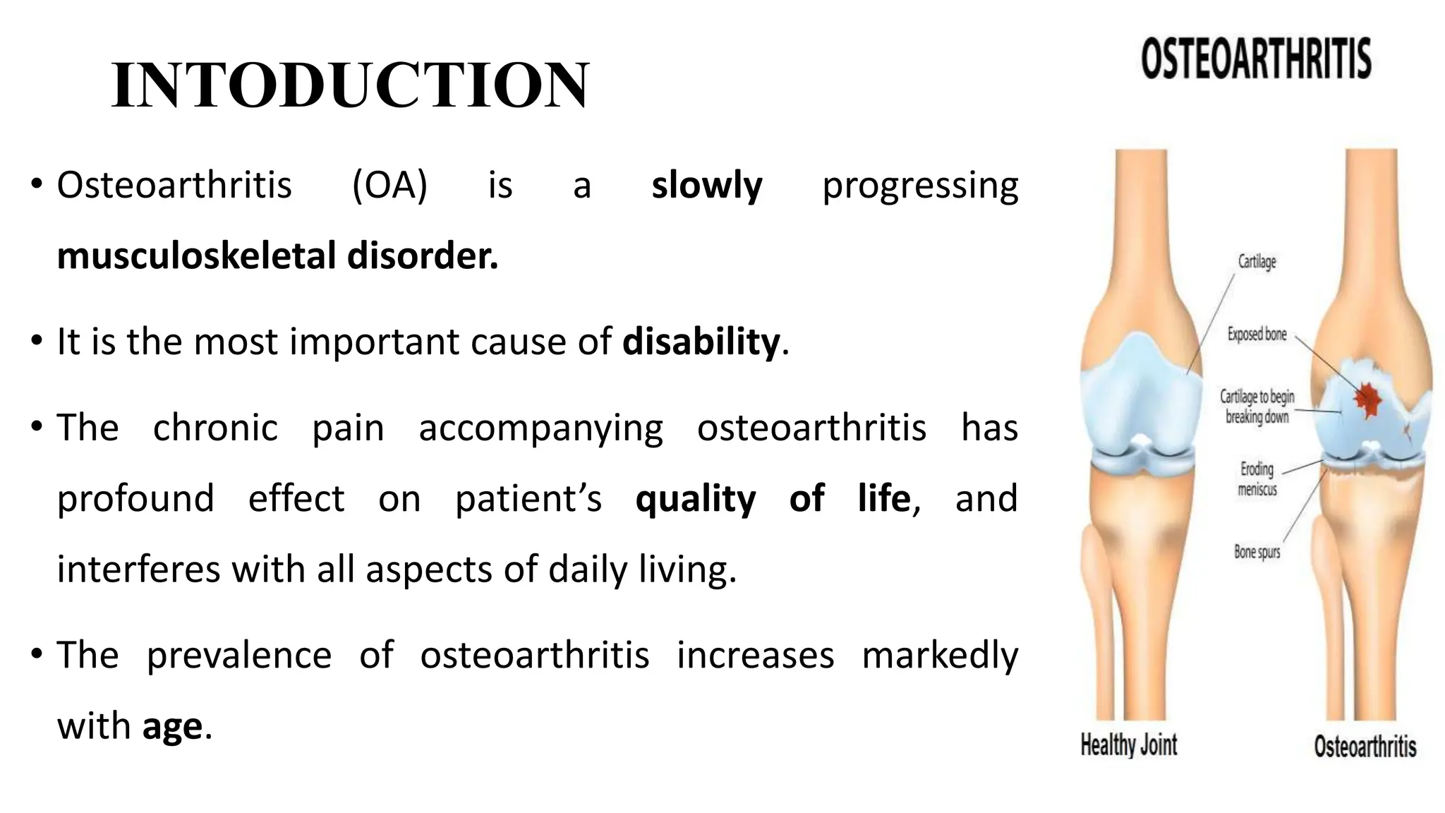
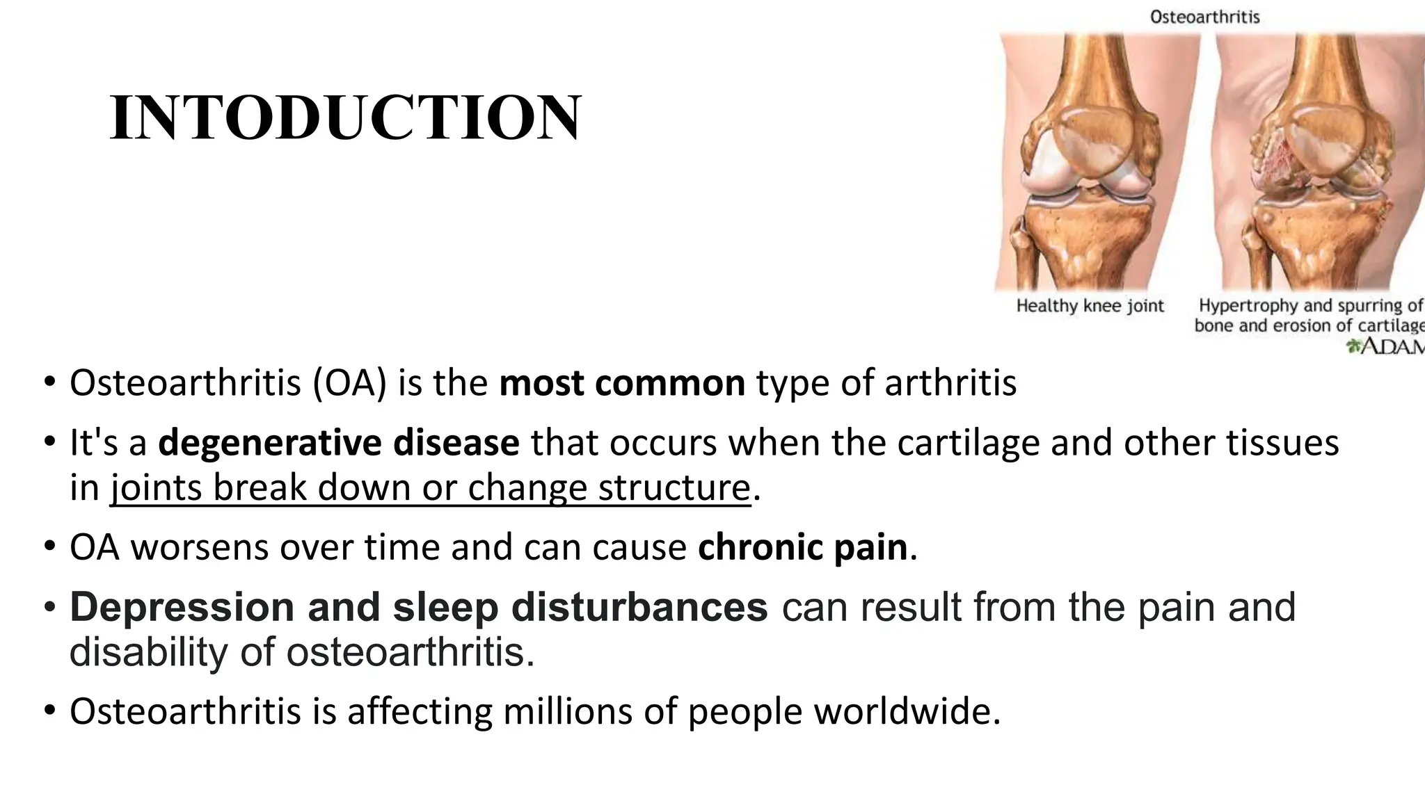
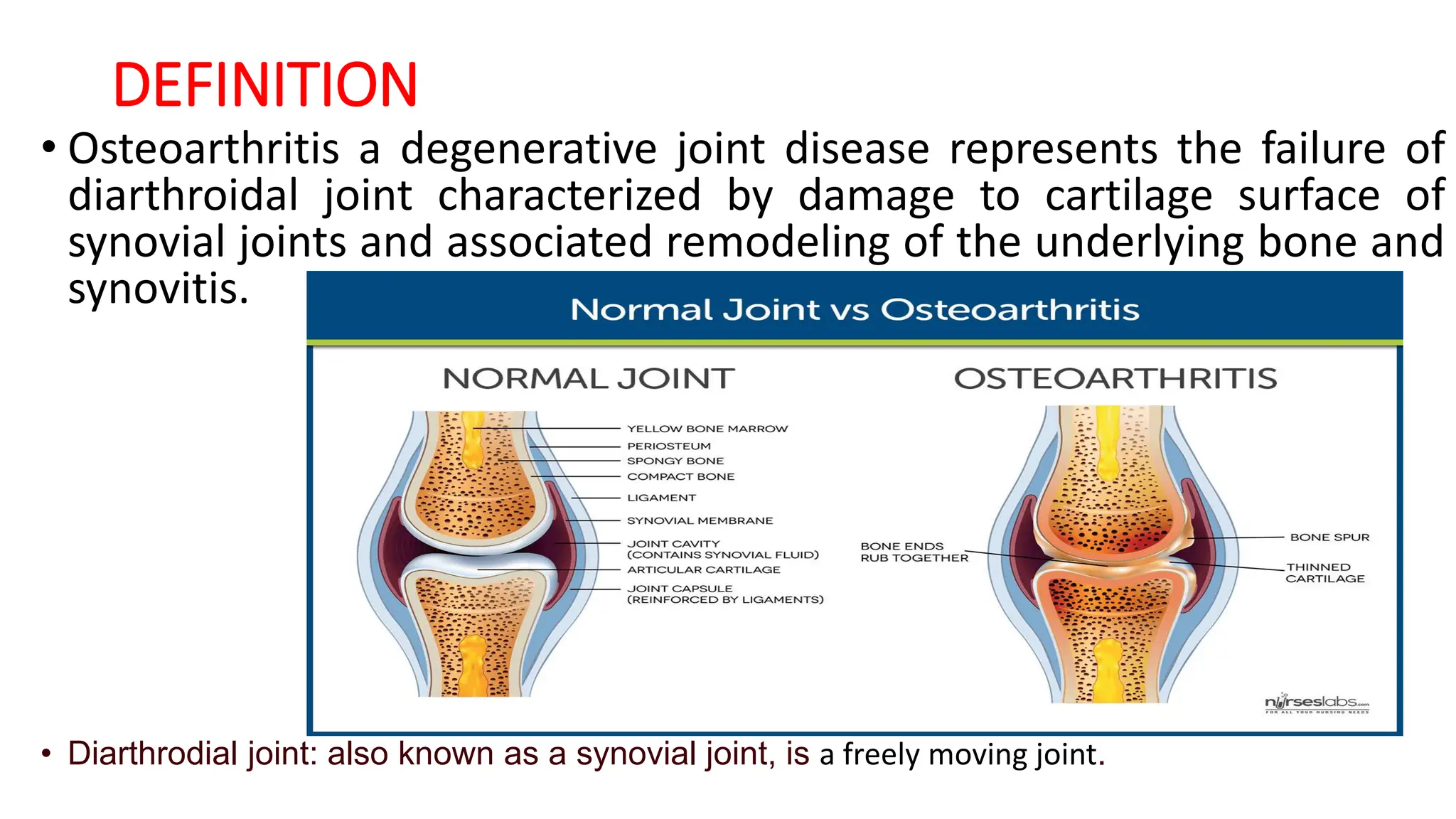
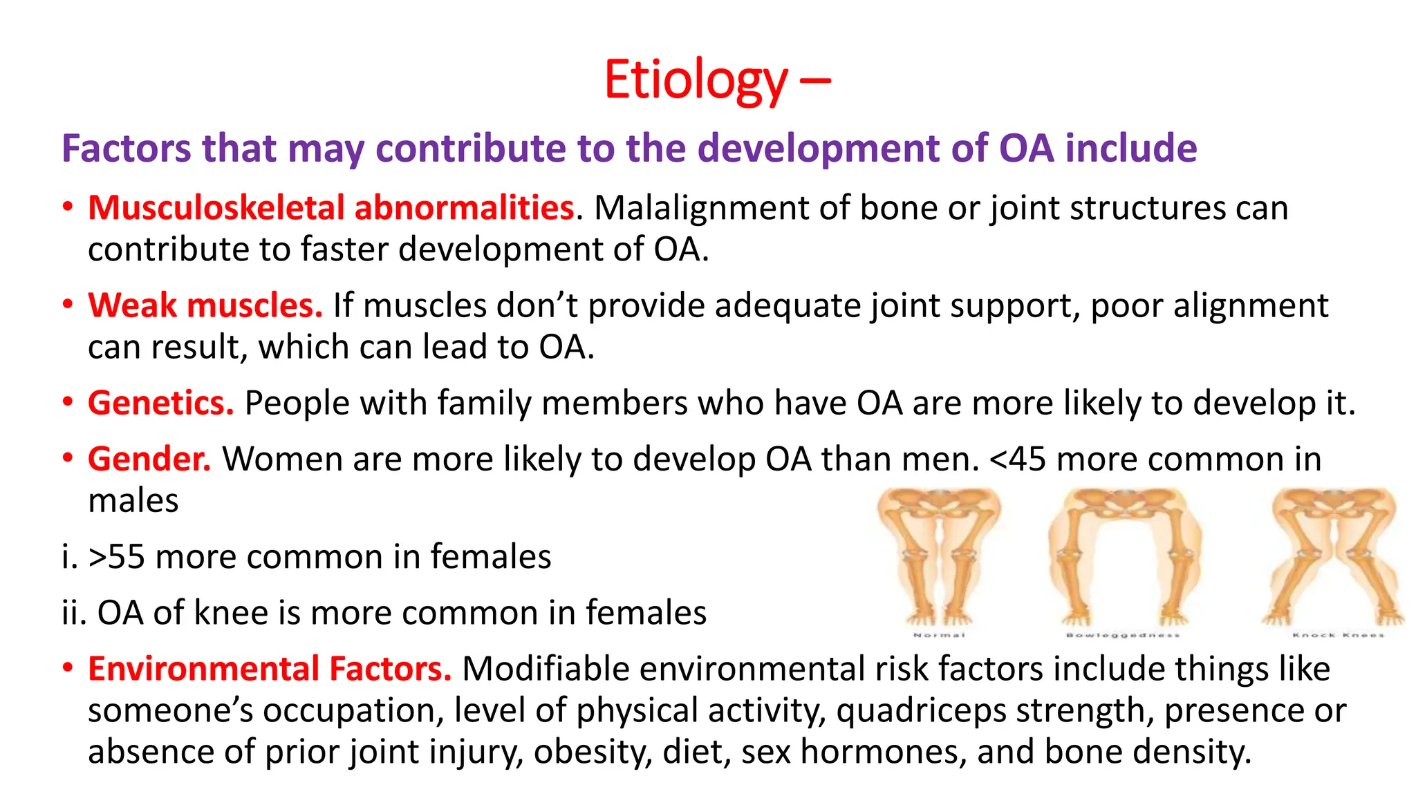
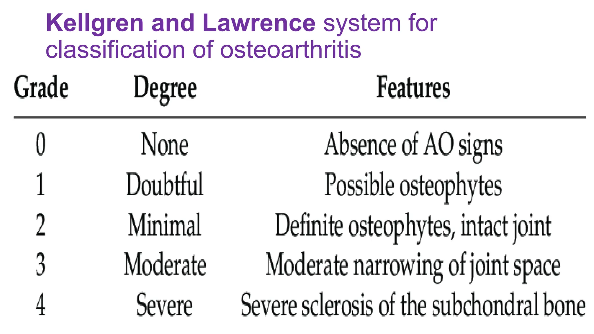
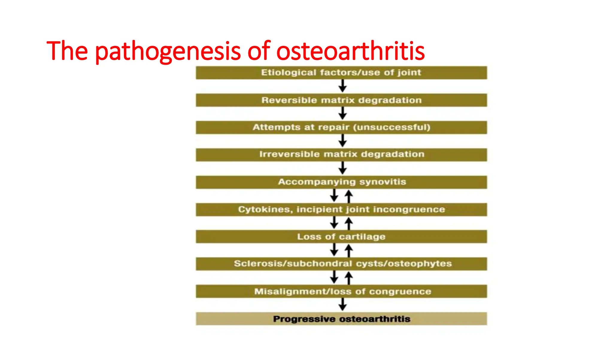
Artifacts in Nuclear Medicine with Identifying and resolving artifacts.
Musculoskeletal disorders: Osteoarthritis,.pptx
1. Osteoarthritis
Mr. Ravinandan A P
Asst. Prof.
Department of Pharmacy Practice
Sree Siddaganga College of Pharmacy
Tumkur, Karnataka
2. INTODUCTION
• Osteoarthritis (OA) is a slowly progressing
musculoskeletal disorder.
• It is the most important cause of disability.
• The chronic pain accompanying osteoarthritis has
profound effect on patient’s quality of life, and
interferes with all aspects of daily living.
• The prevalence of osteoarthritis increases markedly
with age.
3. INTODUCTION
• Osteoarthritis (OA) is the most common type of arthritis
• It's a degenerative disease that occurs when the cartilage and other tissues
in joints break down or change structure.
• OA worsens over time and can cause chronic pain.
• Depression and sleep disturbances can result from the pain and
disability of osteoarthritis.
• Osteoarthritis is affecting millions of people worldwide.
4. • It occurs when the protective cartilage that cushions the ends of the bones
wears down over time.
• Although osteoarthritis can damage any joint, the disorder most
commonly affects joints in your hands, knees, hips and spine.
• Osteoarthritis symptoms can usually be managed, although the damage to
joints can't be reversed.
• Staying active, maintaining a healthy weight and receiving certain
treatments might slow progression of the disease and help improve pain
and joint function.
• Historically, OA was known as a “wear and tear” condition, generally
associated with aging.
• But we know now that it is a disease of the entire joint, including bone,
cartilage, ligaments, fat and the tissues lining the joint (the synovium).
• OA can degrade cartilage, change bone shape and cause inflammation,
resulting in pain, stiffness and loss of mobility.
5. • Its signs and symptoms typically show up more often in individuals
over age 50, but OA can affect much younger people, too, especially
those who have had a prior joint injury.
• There is no cure for OA, but there are ways to manage OA to minimize
pain, continue physical activities, maintain a good quality of life and
remain mobile.
6. DEFINITION
• Osteoarthritis a degenerative joint disease represents the failure of
diarthroidal joint characterized by damage to cartilage surface of
synovial joints and associated remodeling of the underlying bone and
synovitis.
• Diarthrodial joint: also known as a synovial joint, is a freely moving joint.
7.
8. EPIDEMIOLOGY
• OA is the most common and important cause of pain and disability in adult affecting
nearly 50%of those over 65 years of age and almost all individuals of age over75.
• OA stands second to cardiovascular disease in causing disability.
• Hip OA is more common in men and OA of interphalangeal joint and thumb is more
common in women.
• According to the World Health Organization (WHO), 73% of people with
osteoarthritis are over 55 years old, and 60% are female. 43% of people with
osteoarthritis are 65 or older, and 88% are 45 or older. 62% of people with
osteoarthritis are women.
• According to NICE, 9 out of 1,000 at-risk adults are diagnosed with osteoarthritis of
any joint each year.
• In India, osteoarthritis is the most frequent joint disease with a prevalence of 22% to
39%
9. Etiology –
Factors that may contribute to the development of OA include
• Age. The risk of developing OA increases with age and symptoms generally,
but not always, appear in people over 50.
• Joint injury. A bone fracture or cartilage or ligament tear can lead to OA,
sometimes more quickly than in cases where there is not an obvious injury.
• Overuse. Using the same joints over and over in a job or sport can result in
OA.
• Obesity. Excess weight adds stress and pressure on a joint, plus fats cells
promote inflammation.
• Genetic Predisposition-Genetic links are also seen in the development of
OA.
10. Etiology –
Factors that may contribute to the development of OA include
• Musculoskeletal abnormalities. Malalignment of bone or joint structures can
contribute to faster development of OA.
• Weak muscles. If muscles don’t provide adequate joint support, poor alignment
can result, which can lead to OA.
• Genetics. People with family members who have OA are more likely to develop it.
• Gender. Women are more likely to develop OA than men. <45 more common in
males
i. >55 more common in females
ii. OA of knee is more common in females
• Environmental Factors. Modifiable environmental risk factors include things like
someone’s occupation, level of physical activity, quadriceps strength, presence or
absence of prior joint injury, obesity, diet, sex hormones, and bone density.
11. CLASSIFICATION
OA is classified into two major etiologic classes.
• Primary OA (Idiopathic OA) - It is the one in which the underlying cause of the disease is not known-
Common, old age, weight bearing joint like knee and hip
• Secondary OA- it is the one in which the causative factor is known.
Occur at any age after adolescent, occurs mainly in the hip.
Based on the no of sites affected OA is further classified as
• Localized 0A is the one in which symptoms are seen in one or two site
• Generalized OA is the one in which symptoms occur in three or more sites.
• Based on stage:
OA has four stages:
Early,
Mild,
Moderate,
Severe.
13. CLINICAL PRESENTATION
• Depending on the duration of the disease, the joints affected and severity of
joint involvement, the signs and symptoms varies. The common signs and
symptoms are,
• Pain- Localized deep aching pain associated with affected joint (initially pain
occurs when joint is first used and relieved by rest, but on later course pain
occurs with minimal motion and not relieved by rest)
• Joint stiffness – Relatively short episodes occurring during awakening and
resolves with 30 minutes. This may also worsen with weather and barometric
pressure
• Crepitus - Occurs when joint is moved and mainly occurs in OA of knee.
(Related to irregularity that develop on joint surface and loss of cartilage)
• Joint deformity – Herbedens and Bouchards nodes, osteophytes can be seen
as joint deformities
• Synovitis and swelling of joints are also present.
14.
15. The pathogenesis of osteoarthritis
• Pathogenesis of OA includes inflammatory biochemical and immunological changes
occurring in articular cartilage, adjacent bones and synovium.
• The initial biochemical change occurs, as there is an increase in the water content of
the cartilage and resulted thickening of the cartilage.
• Owing to changes in cartilage matrix, the matrix metalloproteins (MMP) are
released which degrade the proteoglycans.
• As a reparative response to this increased degradation, chondrocytes are stimulated
and there is an increased matrix synthesis.
• But as degradation proceeds faster than synthesis there will be a net loss of
proteoglycan despite increased matrix synthesis by chondrocytes.
• Net result of this biochemical process is failure of cartilage to repair itself resulting in
loss of cartilage.
Matrix metalloproteinases (MMPs) are a group of enzymes that break down proteins found
in the spaces between cells in tissues. They are also known as matrix metallopeptidases or
matrixins
16. • As cartilage is destroyed there will be pathologic changes occurring in
underlying bones also.
• Fibrillation of noncalcified cartilage exposes the underlying bones and
causes the abrasion with in the joint.
• As the protective cartilage is eroded away dense smooth bone is exposed.
• The grinding motion stimulates osteoblast activity followed by bone
resorption and vascular changes.
• In the absence of cartilage there won’t be equal distribution of weight
across the bone leading to microfractures and cysts in the subchondral
plate.
• New bone (osteophyte) forms at the joint margin in an attempt to stabilize
the joints.
17. • Joint capsule and synovium changes occurs secondary to OA.
• The release of matrix components as well as bony formation leads to
inflammation with in the synovium (occurs due to PG secreted by the
chondrocyte)
• Pain occurs due to distention of the synovial capsule by increased
joint fluid, microfracture, periosteal irritation, damage to ligaments,
stretched nerve ending around osteophytes.
20. DIAGNOSIS
• Diagnosis of OA is based on clinical and radiographic features. The
most important diagnostic features are as follows.
• X-ray – Initial X-ray of the affected joint will be normal, but as
articular cartilage is lost joint space narrowing and other changes like
subchondral bone sclerosis, cyst, osteophyte formation is visible in
the X-ray.
• Synovial fluid analysis will indicate presence of mild leukocytosis
(<2000WBC/mm3) and increased viscosity
• ESR, which is a nonspecific marker of inflammation, will be slightly
elevated.
21. • American council of Rheumatology has given the following criteria for
the diagnosis of hip and knee OA as follows.
• For hip OA -Patient must have hip pain + two of the following (ESR
<20mm/hr, radiographic femoral or ace tabular osteophyte,
radiographic joint space narrowing )
• For knee OA ----- patient must have knee pain and radiographic
osteophytes + one of the following (age >50 years, morning stiffness
of 30 minutes or less duration, crepitus on motion)
22. MANAGEMENT OF OSTEOARTHRITIS
The primary goals of the therapy are as follows
1. Relieve pain and symptoms
2. To improve joint mobility
3. To reduce functional impairment
4. To maintain and improve quality of life.
23. Treatment
Treatment for OA patient depends on the severity of
joint involvement, co morbid disease states,
concomitant medications and allergies. It is mainly
done by three ways
Non Pharmacological Therapy
Pharmacological therapy
Surgical treatment
24. Non pharmacological treatment
Non pharmacological method of treatment mainly includes the
following
• Patient education - The first step is educating the patient about the
disease process, the extend of OA, prognosis and treatment options.
• Patient should be also taught about self managing of disease like non-
joint stress exercise, importance of avoiding excessive loading of joint,
weight control, use of supportive device.
• Patient education is found to provide additional benefit of about 20-
to30% in the treatment of OA and also helps to manage the patient
expectation.
25. a) Reduction of excessive joint loading - Excessive loading on the joint has to be
avoided eg: patient with hip/knee joint should avoid prolonged standing,
kneeling etc. Obese patient should be counseled to reduce weight. Sufficient
rest also to be taken to avoid further damage.
• Patellar taping - Medial taping may help to decrease severe pain in case of
OA of patellar femoral compartment.
a) Thermal modalities - Application of heat to OA joint decrease pain and
stiffness
b)Exercises - Appropriate non-joint stress exercises, which would strengthen the
periarticular muscles, should be initiated. Exercise programs help to maintain
and restore range motion and reduce pain and muscle spasm.
• Tidal irrigation of the OA knee through large bore needles can be done for the
patient whose pain is refractory analgesics, NSAIDs and intra articular
injections, which help to flush out fibrin, cartilage shards and debris.
26.
27. Pharmacological Treatment
• Drug treatment for OA is palliative or for relieving the symptoms.
Analgesics( acetaminophen)
• Simple analgesics are usually indicted as the first line drug for pain management in
OA (ACR Recommendation) due to their relative safety, efficacy, and low cost
compared to NSAIDs.
• It is used as drug of choice in early and mild osteoarthritis. adverse effects observed
are hepatotoxicity and renal toxicity on long-term use.
• The usual dose of acetaminophen used is 1g QID.
• The common adverse effects observed are hepatotoxicity and renal toxicity on long-
term use.
• It should be used with caution in patients with liver disease or those who chronically
abuse alcohol.
• The important drug interaction of is that with isoniazid there will be increased
hepatotoxicity and with warfarin there is an increased bleeding . Patient should be
warned about the potential toxicity in consuming other prescription and non
prescription products containing acetaminophen.
28. • Nonselective NSAIDs inhibit both COX-1 and COX-2 enzymes to a
significant degree.
• Selective NSAIDs – Selective NSAIDs inhibit COX-2, an enzyme
found at sites of inflammation, more than COX-1, the type that is
normally found in the stomach, blood platelets, and blood vessels.
29. NSAID’S
I. Non selective NSAIDs
II. Selective NSAID’S (Cox – II inhibitors)
Drug and Dose
Acetylated salicylates
Aspirin: 325 – 600mg every 4-6 hours for pain and anti-inflammatory dose
start at 3600 mg/day in divided doses.
Nonacetylated salicylates
Salsalate- 500-1000mg 2-3 times a day
Diflunisal- 500-1000mg 2 times a day
30. • Acetic acids:
• Diclofenac – 100-150 mg/day in
divided dose.
• Indomethacin– 25mg 2-3 times a
day/75mg SR OD
• Etodolac – 800-1200mg/day in divided
dose
• Ketorolac – 10mg every 4-6 hrs.
• Propionic acid
• Fenoprofen – 300-600mg 3-4 times a
day
• Flurbiprofen – 200-30mg in 2-4 divided
dose
• Ibuprofen – 1200-3200mg in 3-4
divided dose
• Ketoprofen – 150-300mg/day
• Naproxen – 250-300mg twice a day
• Oxaprozen – 600-1200 mg OD
• Fenomates
• Meclofenamate – 200-400mg/day in 3-
4 divided
• Mefenamic acid – 250mg every 6
hours.
• Oxicams
• Piroxicam – 10-20mg OD
• Meloxicam – 7.5mgOD
• Coxib
• Celecoxib – 100mg twice daily
• Valadecoxib – 10mg daily
31. Selection of NSAID’S
• About 60% of patient will respond to any NSAID’S, those who do not respond to one
will respond to another.
• If appropriate response not observed within 3 weeks try different NSAID.
• There is no rational in using more than one NSAID.
• It may be combined with paracetamol or an opioid in management of severe pain.
• Measures complete blood count, and liver function before starting the treatment
and repeat at least once a year during continued therapy.
• All NSAID’S (Selective and non selective) should be used with extreme care in patient
with history to GI bleeding .
• To reduce GI complication use paracetamol as an alternative analgesic or prescribe
NSAID with lower history of GI complication (eg: diclofenac and refecoxin) or use a
proton pump inhibitor or a mucosal cytoprotective such as misoprostol in high-risk
patient.
32. Choice of drug and efficacy-
• Non-selective NSAID can be used for patient whom acetaminophen
proves ineffective and who has inflammatory OA.
• Analgesic effect begins with in an hour where as anti-inflammatory
benefit may require 2 to 3 weeks of continuous therapy.
• COX 2 inhibitor can be for initial treatment of patient who are at the high
risk of gastrointestinal side effects and for aspirin sensitive asthma
patient.
• COX-2 selective nonsteroidal anti-inflammatory drugs (c2s NSAIDs), and
aspirin. NSAIDs include ibuprofen, naproxen, ketorolac, and
indomethacin. C2s NSAIDs only include celecoxib.
33. Use of NSAID’S in-patient with other Co-morbidities-
• OA has a greater prevalence in aged people. Hence the initiation of NSAID should be
done with much caution as they may be suffering from other co-morbidities.
1. In asthma patients there is a increased chance of bronchospasm
2. In patients with coagulation disorders non-selective NSAIDs increase the risk of
bleeding and selective NSAID’S increase risk of thrombosis.
3. Use of NSAID in Crohn’s disease may lead to the exacerbation of the disease.
4. Heart failure and hypertension may be exacerbated by sodium and fluid retention
caused by NSAID induced reduction in GFR and renal blood flow.
5. In high cardiovascular risk patients selective NSAIDs may increase risk of
thrombosis events.
6. In renally impaired patients Celecoxib, Parecoxib are not recommended.
34. Adverse effects- Adverse effects associated
with NSAIDs are as follows
• Non-selective NSAIDs- Mainly cause GI side effects and renal side effects
• GI side effects include minor symptoms like nausea, dyspepsia, anorexia,
abdominal pain, flatulence and diarrhoea (10-60%patient). (To minimize NSAID
to be taken with food.) and major symptoms like GI ulcers, perforation,
bleeding etc (To prevent this NSAID to be given with- H2 antagonist or
misoprostol)
• Renal side effects include kidney disease like renal insufficiency tubulo
interstitial nephropathy, hyperkalaemia and renal papillary necrosis
(Acetaminophen is the drug of choice for patient with RF and if NSAID are
used close monitoring of serum creatinine to be done)
• Other side effects include drug induced hepatitis (diclofenac, sulindac)
hypersensitivity, rashes, dizziness, headache, depression, and tinnitus.
35. • COX II inhibitors: - Have same adverse effects as nonselective except GI symptoms.
This may increase prothrombotic activity leading to increase in cardio vascular risk
• Interactions- The common interactions of NSAIDs are as follows
1. ACE inhibitors: - NSAID reduces, antihypertensive effect, increase renal
impairment and hyperkalemia. Combination not to be used if hypo perfusion or
impairment exists.
2. Other antihypertensive – Sodium and fluid retention caused by NSAID reduce
antihypertensive effect.
3. Cyclosporin/ tacrolimus- NSAID induced decreased renal function may increase
the risk of cyclosporin toxicity.
4. Diuretics – diuretic effect will be decreased in presence of NSAID
5. Lithium- NSAID cause decreased renal lithium clearance and thus toxicity.
6. Warfarin – Nonselective NSAID’S antiplatelet action and adverse GI effects
increase risk of bleeding.
7. Potassium sparing diuretics – hyperkalemia.
36. Viscosupplements
• This are the agents containing hyaluronic acid (HA) and are available as intra-
articular injection for treatment of OA.
• This used as an alternative to oral analgesic for patient who cannot tolerate
the NSAIDs
Mechanism of action – Hyaluronan is a polysaccharide molecule that occurs
naturally in synovial fluid, which helps to create a viscous environment and
maintain the normal smooth movement of the bone.
• There is a decrease in concentration and molecular size of HA in OA.
• The visco supplement act as lubricants and shock absorbers in weight bearing
joints.
• They also have structure modifying capacity.
• They inhibit inflammatory mediators such as cytokines and PG, and stimulate
matrix synthesis of cartilage.
37. Dose and administration: -
• The usual dose is 20mg once a week up to 5 weeks.
• The affected knee is first aspirated and hyaluronan is administrated intra-
atricularly into the knee once weekly.
• Pain relief is up to 6 months.
• Duration of action is longer than that of corticosteroids.
• The side effects associated with this treatment are that it causes joint
swelling, local skin reactions, etc.
38.
39. Viscosupplementation Treatment
In its early stages, arthritis of the knee is treated with nonsurgical
methods. Doctor may recommend a range of treatments, including:
• Changes in activity level
• Weight loss
• Pain relievers, such as acetaminophen or nonsteroidal anti-
inflammatory drugs (NSAIDs) like ibuprofen
• Physical therapy
• Corticosteroid injections
40. Side Effects of Viscosupplements
• Typical side effects of viscosupplement injections are mild — redness,
swelling, skin irritation at the injection site — but serious side effects
are possible.
• Closing of the throat
• Difficulty breathing
• Hives
• Swelling of the lips, tongue, or face
41. Glucosamine and Chondroitin
• Endogenous glucosamine is an aminomonosaccaride synthesized from glucose
and is an integral compound for the biosynthesis of proteoglycans and
glycosaminoglycans, which are building block for cartilage.
• Chondroitin provides substrate for formation of healthy joint matrix and block
enzyme that breakdown old cartilage.
42. • Glucosamine and chondroitin are constituents of cartilage, a
component of the joints.
• Glucosamine is a building block for molecules called
glycosaminoglycans that are part of the structure of cartilage.
• Chondroitin is a component of cartilage that plays a role in its
resistance to compression.
43. Corticosteroids
• Intra articular injection of corticosteroids can be used for treatment of OA.
• Treatment is given as monotherapy or adjuvant therapy along with analgesic or
a nonselective NSAID or Cox -2 inhibitor.
• This treatment is preferred when joints are with effusion , painful and swollen.
• Administration- initially the effusion fluid is aspirated and the drug is injected.
Administration is carried out with a prior use of a local anesthetic 1 to 2%.
Dosage may vary with degree of inflammation and size and location of joint.
Beneficial effect is obtained with in 24 hrs after an injection and reaches a
maximum in 1 week and last for 2-4 weeks. Therapy is limited as 3-4 injections
per year.
44. The usual dose is
• Methyl prednisolone- small joint it is 4-10mg and for large joint it is
20-80mg
• Triamcinolone- small joint it is 2.5-10mg and large joint it is 5-40mg
• Mechanism of action – The drug inhibit polymorphonuclear
leukocyte migration and extravasations along with reduction of
vascular permeability resulting in diminished inflammatory response
and secondary pain prevention.
45. Narcotic Analgesics
• Low dose narcotic analgesics are useful in patients who experience no relief
with acetaminophen, NSAID’S, intraarticular injection or topical therapy.
• Particularly useful in patient who cannot take NSAID due to RF, or for patient
whom all treatment options failed and who are at high surgical risk.
• The most commonly given drug is tramadol, which has less abuse potential.
46. Topical Preparations
• Topical preparations containing NSAIDs, analgesics or those having
rubefacient properties can also be used with oral analgesics.
• These preparations should be used in regular basis and usual course is four
times a day.
• One of the most commonly used preparations is capsaicin cream.
• It acts by depleting the local sensory nerve ending of substance P. (it is
implicated in pain transmission in arthritis).
• It effectively reduces joint pain and tenderness.
• Clinical evidence- in four controlled studies carried out to study the effect of
capsaicin it was found that it effectively reduced the pain than placebo
47.
48. Surgical Treatment
• Surgical treatment may provide the most immediate improvement in-
patient with OA.
• Arthroscopic procedures focus on joint debridement to prevent further
destruction of joint surface and to repair by remaining healthy tissue.
• Surgery can be very costly and carries the risk of morbidity and mortality.
• Surgical treatment of OA is generally reserved for severe patients for whom
the potential benefits overweigh the potential risk.
• Debridement= Surgical removal of foreign material and dead tissue from a
wound in order to prevent infection and promote healing
49.
50. NEWER DEVOLEPMENTS IN TREATMENT OF OSTEOARTHRITIS
Vitamin E
• It was found that vitamin E can be used for the treatment of OA as it
fights inflammation by neutralizing the biochemical’s that are
produced during inflammation.
• These biochemical’s released by immune cells contains free radicals
unstable molecules which can grab electrons damaging the body’s
normal tissue.
• Vitamin offers up its own electrons and thus protects the cells.
• Dose- 600 milligram/day PO for 10 days
51. S –adenosyl methionine
• Dose-1200mg/day
• Because of its anti-inflammatory properties it can be used for its
treatment in OA
52. Trolamine salicylate
• It is a topical analgesic used for temporary relief of minor pain associated
with arthritis, simple backache, muscle strains, & sprains.
• Dose – 10 % Trolamine salicylate topical application cream
• Trolamine causes no irritation and offers an alternative to patients with
arthritis who cannot orally tolerate NSAIDs
Orgotenin
• Dose- 4mg once weekly intraarticular injection
• This is a super oxide dismutase enzyme analogue.
• This because of its antioxidant properties lowers inflammatory messengers.
• Having anti-inflammatory effect.
53. CONCLUSION
• OA is painful, progressive condition that may cause
significant disability and compromised quality of life.
• Existing pharmacologic therapy is aimed primarily at relief of
pain and inflammation.
• Newer developments are directed towards identification of
risk factors and specific markers of cartilage destruction and
in the development of disease modifying osteoarthritic
drugs.