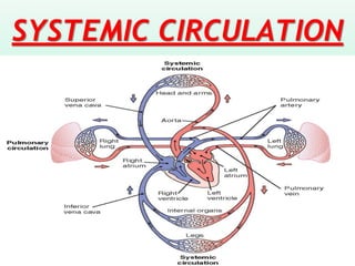
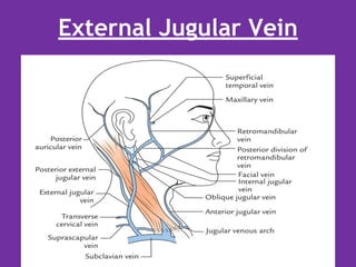
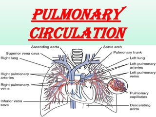
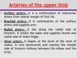
The document summarizes the major arteries of the systemic and pulmonary circulations. It describes the branching patterns of the aorta and its major divisions including the ascending aorta, arch of aorta, descending aorta, abdominal aorta, and their tributaries. It also discusses the pulmonary arteries and veins. Venous drainage of the head, neck, upper limbs, thorax, and abdominal regions are outlined. Common sites used for intravenous injections are noted.
The pericardium is a fibroserous sac that surrounds the heart and roots of the great vessels. It has two layers - an outer fibrous layer and inner serous layer. The serous layer further divides into the parietal layer lining the fibrous sac and visceral layer adhered to the heart. Between these layers is the pericardial cavity containing fluid. The pericardium functions to support and protect the heart while allowing movement. Conditions like pericarditis or excess fluid accumulation can lead to cardiac tamponade.
The document provides an overview of heart anatomy. It describes the heart's location in the thorax and its dimensions. The four chambers - right and left atria and ventricles - are introduced. Each chamber's function is summarized as receiving or pumping blood. The three layers of the heart - epicardium, myocardium, and endocardium - are defined. The cardiac skeleton is described as a fibrous structure that partitions the chambers and anchors the heart valves.
The heart wall consists of three main layers - the endocardium, myocardium and epicardium. The endocardium lines the inner chambers, the myocardium is the thick middle layer of muscle responsible for contractions. The epicardium is the outer layer. Damage to layers can cause conditions like endocarditis (endocardium inflammation), myocarditis (myocardium inflammation) and angina or heart attack due to reduced blood flow from blocked arteries.
The document describes the structure and features of the heart chambers. It states that the heart is composed of 4 chambers - the right atrium, right ventricle, left atrium, and left ventricle. Blood enters the atria and is then pumped into the ventricles. From the left ventricle, blood passes into the aorta for systemic circulation, and from the right it enters the pulmonary circulation via the pulmonary arteries. Each chamber has distinct internal and external features and relations to other cardiac structures. The septa divide the atrial and ventricular chambers.
The document summarizes the internal structure of the heart, including its four chambers, septa that divide them, and valves that regulate blood flow. It describes the conduction system that controls heart contractions, including the sinoatrial node that initiates impulses, the atrioventricular node that relays them to ventricles, and Purkinje fibers that conduct impulses through the ventricles. It also reviews the heart's blood supply, innervation by the autonomic nervous system, and roles of the sympathetic and parasympathetic fibers.
The heart has four chambers - two atria that collect blood and two ventricles that pump blood. The right atrium receives deoxygenated blood from the vena cava and passes it to the right ventricle, which then pumps the blood to the lungs. The left atrium receives oxygenated blood from the lungs and passes it to the left ventricle, which has the thickest walls and pumps oxygenated blood out to the rest of the body via the aorta. Septa divide the atria and ventricles.
The heart is pyramidal in shape with an apex pointing leftward and downward. It has four chambers - two atria which receive blood and two ventricles which pump blood. The right atrium receives deoxygenated blood from the body and pumps it to the right ventricle to be sent to the lungs. The left atrium receives oxygenated blood from the lungs and pumps it to the left ventricle to be sent to the body. Blood flows through the heart via the tricuspid, bicuspid/mitral, pulmonary and aortic valves. The heart's conduction system coordinates contractions and is composed of the sinoatrial node, atrioventricular node and bundle of His.
The aorta is the largest artery that carries oxygenated blood from the left ventricle throughout the body. It can be divided into four sections - the ascending aorta, aortic arch, thoracic aorta, and abdominal aorta. The aortic arch arises from the ascending aorta and curves superiorly, posteriorly and to the left before descending as the thoracic aorta. It gives off three branches - the brachiocephalic trunk, left common carotid artery, and left subclavian artery. The thoracic aorta continues down through the thoracic cavity and passes through the aortic hiatus in the diaphragm to become the abdominal aorta.
Internal feature of right and left atriafarranajwa
This document provides information about the anatomy and structures of the right and left atria of the heart. It discusses the key internal features of each atrium, including chambers, valves, veins and arteries, muscle structures, and other anatomical landmarks. It also briefly explains some clinical significance of atrial structures, mentioning how abnormalities like atrial septal defects or atrial fibrillation can impact blood flow and cardiac output. The document aims to educate the reader on the basic internal structures of the atria and their relationships to clinical functions.
The document describes the muscles and nerves of the thoracic wall. It discusses the three layers of intercostal muscles - external, internal, and innermost. It also describes the intercostal nerves, noting the typical arrangement from T3-T6, and variations in other regions. Finally, it discusses the diaphragm muscle, its origins along the ribs and vertebrae, openings, nerve supply, and role in respiration.
Anatomy of Blood vessels of abdomen pelvic cavities. Portacaval & Cavacaval A...Eneutron
1. The abdominal aorta
a. the parietal branches
b. the visceral branches
2. The common iliac arteries and veins
3. The external iliac artery and veins
4. The internal iliac artery and veins
5. The inferior vena cava
6. The portal vein
7. The cavacacal Anastomoses
8. The portacaval Anastomoses
9. The Fetal Circulation
The heart is a hollow, muscular organ located slightly left of center in the chest. It has four chambers - the right and left atria receive blood, and the right and left ventricles pump it out. The heart is surrounded by membranes and has three layers - the outer fibrous pericardium, middle muscular myocardium, and inner endothelial endocardium. It uses valves to ensure one-way blood flow, pumping deoxygenated blood to the lungs and oxygenated blood throughout the body in continuous circulation. The coronary arteries supply the heart with oxygenated blood.
The document provides details on the anatomy of the abdominal cavity and its contents. It describes the peritoneal cavity and mesenteries that support the intestines. It summarizes the liver, gallbladder and associated ducts. It outlines the locations and blood supply of the stomach, spleen, pancreas, small intestine and large intestine. It also reviews the anatomy of the kidneys, ureters, urinary bladder, and suprarenal glands.
right ventricle internal and external features-
interior is divided into inflowing and outflowing parts (infundibulum)
inflowing part is rough due to trabeculae corneae, - ridges, bridges, pillars. Chordae tendineae- are attached to pillars and cusps of tricuspid valve.
outflowing part is smooth, semilunar valve guards opening of pulmonary valve
This document provides an anatomical overview of the right atrium, right ventricle, pulmonary valve, and tricuspid valve. It describes the external features and internal structures of the right atrium, including its tributaries that receive venous blood. It notes the smooth posterior sinus venarum and rough anterior pectinate part. It also describes the components, surface markings, and location of the tricuspid and pulmonary valves. Finally, it outlines the triangular shape of the right ventricle and notes its inflowing and outflow parts, as well as the muscular ridges within.
The document discusses the anatomy of the intercostal spaces, including the muscles, nerves, arteries, veins and other structures located within the spaces between the ribs. It specifically names the external and internal intercostal muscles, intercostal nerves and their branches, intercostal arteries and veins, as well as related structures like the azygos system. The document also covers applied clinical aspects involving the intercostal spaces like pleural taps, thoracotomies, herpes zoster infections, and other conditions.
This document contains questions from several lectures related to thoracic anatomy:
1. Questions cover the anatomy of structures like the thoracic outlet, internal thoracic artery, intercostal arteries and veins, muscles of respiration, lungs and pleura, heart, mediastinum, large blood vessels, nerves of the thorax, esophagus, and azygos system.
2. The questions test knowledge of the origins, courses, relations, branches and clinical importance of these various thoracic structures.
3. Incomplete statements about the anatomy are also included to be filled in, relating to topics like the root of the lung, bronchial and pulmonary arterial supply, coronary arterial distribution, and veins drain
The spleen is a lymphatic organ located in the left upper abdomen under the diaphragm and ribs. It is oval shaped, 7-14cm long and weighs 150-200g. The spleen has several surfaces - a diaphragmatic surface facing upward, a visceral surface divided into gastric and renal regions, and borders. It filters blood and recycles iron, and contains red and white pulp. The spleen receives blood from the splenic artery and drains into the splenic vein. Diseases can cause an enlarged or absent spleen.
Right Atrium of human heart
This PPT help to understand the external and internal structures of right atrium.
sulcus terminalis on external surface of rt atrium,
crista terminalis on internal side of rt. atrium,
interior is divided into rough anterior part and smooth posterior part ( sinus venarum)
superior and inferior venae cavae drains deoxygenated blood into rt. atrim
there is Eustachian valve to guard the opening of IVC and Thebesian valve to guard the opening of coronary sinus
septal wall presents fossa ovalis with its border limbus fossa ovalis
Surface Anatomy of the abdominal viscera and abdominal regions.Saleh Alorainy
This document discusses surface anatomy of the abdominal region. It aims to define landmarks, planes, and regions; visualize structures at lumbar vertebral levels; locate major blood vessels, viscera, kidneys and spleen; and define surface areas where gut pain is referred. Key points include how to find the superficial inguinal ring and determine lumbar levels, visualizing structures at L1, using quadrants to locate organs, and defining referral regions for pain from the gut.
Anatomy & Physiology Question Paper Revision TutorialSathish Rajamani
This document provides information about anatomy and physiology in three parts. The first part discusses the blood supply of the heart, types of muscles, and cranial nerves. It explains that the heart receives blood from the coronary arteries and veins. It describes the three main types of muscles - cardiac, skeletal, and smooth muscles - in terms of their structure and function. It also lists and briefly describes the 12 pairs of cranial nerves and their functions.
The pericardium is a fibroserous sac that surrounds the heart and roots of the great vessels. It consists of the fibrous pericardium and serous pericardium. The fibrous pericardium is a conical sac made of fibrous tissue that protects the heart. The serous pericardium is a double-layered membrane that forms the pericardial sac, with fluid allowing the heart to move smoothly. The pericardium contains the heart, vessels, and parts of the great vessels, and has transverse and oblique sinuses.
Aorta is the main artery of systemic circulation.
Aorta is divided into 4 parts - 1) Ascending aorta
2) Arch of aorta 3) Descending thoracic aorta 4) Abdominal aorta
1) Ascending aorta - branches - Right and Left coronary arteries . At the level of sternal angle it is continuous with arch of aorta.
2) Arch of aorta - branches - Brachiocephalic artery, Left common carotid artery, Left subclavian artery. Ligamentum arteriosus is attached to arch of aorta and pulmonary trunk. for details about ligamentum arteriosus please go through the video of fetal circulation
https://youtu.be/kBR6p7-GmaE
3) Descending thoracic aorta - is continuation of arch of aorta from the level of T4 vertebra. it descends downwards by giving branches - 9 pairs of Posterior intercostal arteries, esophageal artery, left bronchial artery, pericardial branches, superior thoracic artery.
4) Abdominal aorta - at the level of T12 vertebra thoracic aorta enters into abdomen through aortic opening of diaphragm. Abdominal aorta divides into its terminal branches Left and Right common iliac arteries at the level of L4 vertebra.
The cardiac cycle consists of systole and diastole. During systole, the heart contracts and pumps blood out of the ventricles. During diastole, the heart relaxes and fills with blood. The cycle involves coordinated events in the atria and ventricles. It can be analyzed using a Wiggers diagram which plots various cardiac parameters over time, revealing phases like isovolumic contraction, ejection, isovolumic relaxation, and filling. Precisely measuring time intervals within the cycle using Doppler echocardiography provides clinical insights into cardiac function and timing.
anatomy of spleen, surfaces of spleen, peritoneal an visceral relations, blood supply an lymphatic drainage, hilum of spleen , function of spleen, splenomegaly,
The document summarizes the major arteries and veins of the systemic and pulmonary circulations. It describes the path of blood flow from the aorta and its major branches that supply the head, neck, upper limbs, thorax, abdomen, pelvis and lower limbs. It also details the pulmonary circulation from the pulmonary trunk to the lungs and back to the heart via the pulmonary veins.
The carotid arteries are the primary vessels supplying blood to the brain and face. The right common carotid artery (RCCA) originates in the neck from the brachiocephalic artery while the left common carotid artery (LCCA) arises in the thorax from the arch of the aorta.
The document summarizes the major arteries of the head and neck, including their origins, courses, and branches. It begins with an overview of the aorta and its branches that supply the head and neck (brachiocephalic artery, common carotid artery, subclavian artery). It then describes the external and internal carotid arteries in detail, outlining their branches which include the facial, lingual, occipital, and maxillary arteries. Throughout, clinical correlations and examples of surgical/traumatic implications are provided.
The head and neck receive their primary blood supply from the carotid and vertebral arteries. The common carotid arteries divide into external and internal carotids. The external carotid supplies neck and face structures, branching into the superior thyroid, lingual, facial, occipital, and maxillary arteries. The internal carotid supplies the brain, branching into the anterior and middle cerebral arteries. Venous drainage is performed by the internal and external jugular veins, which drain into the subclavian vein.
The major blood vessels include the arteries, which carry oxygen-rich blood away from the heart, and veins, which carry deoxygenated blood back to the heart. The circulatory system is divided into the pulmonary and systemic circuits. The pulmonary circuit carries deoxygenated blood to the lungs and returns oxygenated blood to the heart. The systemic circuit pumps oxygenated blood to the entire body through progressively smaller arteries and returns deoxygenated blood to the heart via veins. The major arteries include the aorta, carotid, brachial and iliac arteries. The major veins include the jugular, subclavian, brachial and iliac veins.
arteries of human body including head and neck, upper extremities, lower extremities
different between the veins and arteries. common carotid arteries, internal carotid arteries, external carotid arteries, subclavian arteries
circle of Willis
The heart is a hollow muscular organ located in the middle mediastinum. It is approximately the size of a fist and weighs 250-300 grams. The heart has four chambers - two upper atria and two lower ventricles. It is surrounded by a double-walled sac called the pericardium. The heart pumps blood through two circuits - the pulmonary circulation and the systemic circulation - using a series of valves to ensure one-way blood flow.
The document summarizes the anatomy and flow patterns of the major veins in the human body. It describes the three major systemic veins that return deoxygenated blood to the heart - the coronary sinus, inferior vena cava (IVC), and superior vena cava (SVC). It then provides details on the venous drainage of specific regions including the head and neck, upper limbs, thorax, abdomen, and pelvis. Key veins discussed include the internal and external jugular veins, azygos vein system, hepatic portal vein, common iliac veins, and renal veins.
Vascular structure head and neck Almas khan , Khorfakkhan hospitalalmasmkm
This document discusses the vascular structure of the head and neck. It begins by explaining the components of the circulatory system, including the cardiovascular and lymphatic systems. It then describes the three main circuits of circulation: coronary, pulmonary, and systemic. The document focuses on the head and neck vasculature, detailing arteries like the aorta, carotid, vertebral and various branches. It also discusses venous drainage and the circle of Willis. Common clinical indications involving the head and neck vessels are presented. In conclusion, the circulatory system of the head and neck is reviewed to aid in angiography identification and understanding normal and abnormal vascular distributions.
The cardiovascular system summary is as follows:
1. The heart is located in the chest behind the sternum and is about the size of a fist. It has four chambers - two atria for receiving blood and two ventricles for pumping blood out.
2. The pericardium is a sac that surrounds and protects the heart. It has two layers - an outer fibrous layer and an inner serous layer that secretes fluid.
3. Blood enters the right atrium from the body and is pumped into the pulmonary artery to the lungs. Oxygenated blood returns to the left atrium and is pumped by the left ventricle through the aorta to the body.
MEDISTINUM and The HEART.pptx1111111111111111111marrahmohamed33
The document discusses the mediastinum and structures contained within it, including the heart. It provides details on the different compartments of the mediastinum (superior, anterior, middle, posterior), and structures found in each. It describes the pericardium and heart in detail. The pericardium surrounds and protects the heart. It has fibrous and serous layers. Conditions like pericarditis and pericardial effusion are discussed. The heart is located in the middle mediastinum and has an apex pointing left and downward and a base facing posteriorly.
The heart has four chambers. The two superior receiving chambers are the atria (= entry halls or chambers), and the two inferior pumping chambers are the ventricles (= little bellies).
On the anterior surface of each atrium is a wrinkled pouchlike structure called an auricle
Be the first to comment
Anatomy And Physiology of Human Heart
1. ANATOMY OF THE HEART By: Dr Mohammed Faez
2. The Heart The heart is a chambered muscular organ that pumps blood received from the veins into the arteries, thereby maintaining the flow of blood through the entire circulatory system.
3. The Heart • The heart is surrounded by membrane called Pericardium.
4. The Pericardium • The pericardium is a fibroserous sac that encloses the heart and the roots of the great vessels. • The pericardium lies within the middle mediastinum.
5. The Pericardium
6. The Pericardium • Its function is to restrict excessive movements of the heart as a whole and to serve as a lubricated container in which the different parts of the heart can contract.
1) The document describes the major arteries of the systemic circulation, including branches off the aorta such as the coronary, carotid, and brachial arteries.
2) It also details important veins that drain blood from the head, neck, arms and abdomen, including the internal and external jugular veins, brachial veins, and hepatic portal vein.
3) The hepatic portal system is summarized as carrying nutrient-rich blood from the abdominal viscera to the liver through the hepatic portal vein and its tributaries, before the blood continues to the inferior vena cava and heart.
The document summarizes the major arteries and veins supplying the head and neck region. It begins by listing the objectives which are to describe the major arteries including the common carotid arteries, external carotid artery, internal carotid artery, and subclavian artery. It then provides details on the branches and relations of these arteries. Next, it discusses the major veins including the external jugular vein, internal jugular vein, and their tributaries. It concludes by describing the formation of the brachiocephalic veins and superior vena cava which drain the head and neck veins.
The document describes the structure and function of the heart. It discusses the location of the heart in the mediastinum and its external and internal anatomy. The four chambers of the heart (right and left atria and ventricles) are described along with the valves that regulate blood flow. The circulations of blood through the pulmonary system and systemic circulation are also summarized. Key details about the layers of the heart wall, coronary circulation and blood flow through arteries, capillaries and veins are provided.
Anatomy & physiology of cardiovascular systemvinayanerurkar
This document provides an overview of the anatomy and physiology of the cardiovascular system. It describes the location and structure of the heart, including its chambers and layers. It explains the circulation of blood through the heart, into the pulmonary circulation to oxygenate blood and into the systemic circulation to distribute oxygenated blood to the body. It details the coronary circulation which provides blood supply to the heart muscle.
The document provides an overview of the anatomy of the heart and its associated vessels. It describes the internal and external structures of the heart including the chambers, valves, arteries and veins. Key points include:
- The heart is located in the middle mediastinum and is surrounded by the pericardium. It has four chambers - right and left atria on top which receive blood and right and left ventricles on bottom which pump blood out.
- Blood flows from the vena cava and pulmonary veins into the atria, through valves into the ventricles, and out through the pulmonary artery and aorta into the lungs and body.
- Coronary arteries supply blood to the heart muscle and coronary
Similar to systemic and pulmonary circulation.pdf (20)
Retinopathy of prematurity.pdf ROP newbornPooja Rani
Retinopathy of prematurity (ROP) is a retinal disorder that affects premature infants, caused by abnormal blood vessel growth in the retina. It develops due to retinal immaturity and hyperoxia from high oxygen therapy in preterm infants less than 37 weeks gestational age. Early stages involve mild abnormal vessel growth, while later stages involve retinal detachment. Treatment depends on the stage, and may include laser therapy, scleral buckling surgery, or prevention through careful oxygen management for preterm infants.
The Braden Scale and Norton Scale are tools used to assess risk for pressure ulcer development. The Braden Scale consists of 6 subscales that are scored from 1-4 or 1-3, with lower total scores indicating higher risk. The Norton Scale consists of 5 subscales scored from 1-4, with total scores below 14 indicating higher risk. Both scales provide structured assessments of factors like sensory perception, mobility, nutrition, and moisture that are used to determine a patient's pressure ulcer risk level.
Neonatal Intensive Care Unit. NICU. neonatePooja Rani
The document discusses the organization and management of neonatal intensive care units (NICU) in India. It outlines a 3-tier system of neonatal care based on weight and gestational age. Level I care is for healthy newborns, Level II care is for preterm or low birth weight babies, and Level III care is for very preterm and low birth weight babies requiring advanced equipment. It provides details on NICU unit size, location, design, equipment, staffing, and infection control practices to optimize care for sick newborns.
Kangaroo Mother Care . neonate. newborncarePooja Rani
Kangaroo Mother Care (KMC) is a technique for caring for low birth weight babies that provides skin-to-skin contact between the mother and baby to promote thermal control, breastfeeding, infection prevention, and bonding. Key components of KMC include prolonged, continuous skin-to-skin contact; exclusive breastfeeding; and early discharge from the hospital with regular follow-up care. KMC has benefits like increased breastfeeding rates, better temperature control for the baby, earlier discharge from the hospital, and lower morbidity for the infant.
methods of data collection in nursing researchPooja Rani
This document discusses various tools and methods used for data collection in research studies. It describes primary and secondary sources of data, as well as common data collection methods like interviews, questionnaires, attitude scales, and observations. Interviews can be structured, semi-structured, or unstructured, while questionnaires contain open-ended or closed-ended questions. Attitude scales assess perspectives using tools like Likert scales or semantic differentials. Observations are structured, unstructured, participant, or non-participant. Proper planning is required to select the most appropriate data collection methods and tools for different research objectives.
Nose anatomy & physiology sensory systemPooja Rani
The nose consists of the external nose and nasal cavity divided by a septum. The external nose has two nostrils separated by the nasal septum. The framework of the external nose is made up of nasal bones, frontal processes of maxillae, and nasal part of frontal bone. The nose contains 10-100 million olfactory receptors for smell within the olfactory epithelium in the nasal cavity. Olfactory receptors are neurons that transmit signals from inhaled chemicals to the olfactory bulb.
fertilization and implantation-converted.pdfPooja Rani
Fertilization occurs when a sperm fuses with an egg in the fallopian tube, forming a zygote. Thousands of sperm are ejaculated into the vagina during sex and travel to meet the egg released from the ovary. Within 24 hours, a single sperm will fertilize the egg. The zygote then begins dividing rapidly as it travels down the fallopian tube and into the uterus over the next few days. By 8 weeks, it is called an embryo and implants in the uterine wall, beginning pregnancy.
moving and lifting of patient-converted.pdfPooja Rani
This document provides guidelines for moving and lifting patients, including indications, contraindications, instructions, and nursing responsibilities. It describes how to transfer patients from bed to wheelchair or stretcher safely with assistance. Key points include planning the movement, supporting the head/shoulders/hips/legs, using lifting devices like gait belts, and positioning patients with pillows when rolling or logrolling them. Nursing assessments and preparations are outlined to ensure transfers are performed correctly.
The document discusses the structure and function of the tongue. It describes how taste is sensed on the tongue and transmitted through cranial nerves to the brainstem and cortex. Taste pathways are discussed for the front, middle, and back of the tongue. Factors influencing taste sensation and abnormalities of taste are also summarized, along with pathological terms related to the tongue and changes in tongue color.
The cardiac cycle describes the repeating sequence of events in the heart from one heartbeat to the next. It involves systole, when the heart contracts to pump blood, and diastole, when the heart relaxes and refills with blood. The normal cardiac cycle lasts 0.8 seconds and includes atrial systole, ventricular systole, and relaxation. An electrocardiogram (ECG) records and measures the heart's electrical activity through electrodes placed on the skin, detecting the P wave, QRS complex, ST segment, and T wave to evaluate the heart's rhythm and electrical conduction.
This document discusses the nutritional needs of patients from a nursing perspective. It defines nutrients and their importance for energy, tissue maintenance, and bodily functions. Nutritional needs are affected by various factors like age, activity level, medical conditions, and more. A nutritional assessment evaluates intake, lifestyle, medical history, and includes anthropometric measurements, biochemical tests, clinical exams, and dietary analyses. Nurses play an important role in meeting patients' nutritional needs by considering preferences, assisting with meals, discussing diet importance, and more.
There are three main types of joints in the body: fibrous or fixed joints which do not allow movement; cartilaginous or slightly movable joints which allow limited movement; and synovial or freely movable joints which allow the greatest range of motion. Synovial joints contain synovial fluid that lubricates and nourishes the joint space. The six types of synovial joints each allow different ranges and axes of movement and include ball-and-socket, hinge, pivot, gliding, saddle, and condyloid joints.
The lymphatic system removes excess fluid from tissues, absorbs fat and transports white blood cells and antigens. It comprises a network of lymphatic vessels that carry lymph fluid towards the heart. Lymph is filtered through lymph nodes which contain lymphocytes and phagocytes that help fight infection and disease. The major components are lymph, lymph vessels, lymphoid tissues and lymphocytes.
The document summarizes the ABO and Rh blood group systems. It explains that the ABO system categorizes blood into four main groups - A, B, AB, and O - based on the presence or absence of antigens A and B on red blood cells. The Rh system further distinguishes blood as Rh positive or negative based on the presence of the D antigen. The document outlines Karl Landsteiner's discovery of the ABO system in 1901 and the discovery of the Rh system in 1940. It notes that the ABO system is important for blood transfusions and describes the potential for Rh incompatibility between mother and fetus.
Hemostasis is the process by which bleeding is stopped. It involves four key mechanisms:
1) Vasoconstriction of blood vessels to reduce blood flow from the injury site.
2) Formation of a platelet plug through adhesion, activation, and aggregation of platelets at the injury.
3) Coagulation of blood to form a fibrin clot from fibrinogen.
4) Later lysis of the clot through fibrinolysis to restore blood flow.
The eye is a spherical organ that allows for vision. It has three layers - an outer fibrous coat, middle vascular coat, and inner nervous coat containing photoreceptor cells. Light enters through the cornea and is focused by the lens onto the retina. Photoreceptor cells in the retina convert light into neural signals via the optic nerve. Accessory structures like the eyelids and lacrimal system help protect and lubricate the eye. Common refractive errors that impact vision like myopia, hyperopia, and astigmatism can often be corrected using lenses.
The kidneys are located in the back of the abdomen and are bean-shaped organs that filter waste from the blood to produce urine. The kidneys regulate blood volume, pressure, pH, and electrolyte levels. They remove waste and toxins from the body. The kidneys receive blood through the renal artery and filter it through nephrons to remove waste, which is collected by minor and major calyces and exits through the ureters as urine.
The nephron is the basic structural and functional unit of the kidney that filters blood and forms urine. Each nephron consists of 10 parts including the glomerulus, Bowman's capsule, proximal convoluted tubule, loop of Henle, distal convoluted tubule, and collecting duct. The glomerulus filters blood in the renal corpuscle which is composed of the glomerulus and Bowman's capsule. Filtrate then passes through the renal tubule where reabsorption and secretion occur to regulate water, salt, and waste removal before final collection in the ureters. Each kidney contains about 1 million nephrons to perform these vital filtration and regulatory functions.
This document discusses anti-tubercular drugs, classifying them into first and second line drugs. It describes the mechanism of action, dosage, contraindications, adverse effects and drug interactions of major first line drugs - isoniazid, rifampicin, pyrazinamide, ethambutol and streptomycin. It also summarizes the same details for several second line drugs used when tuberculosis is resistant to first line treatment or these cannot be tolerated, including para-amino salicylate, ethionamide, cycloserine, thiacetazone, fluoroquinolones, clarithromycin and aminoglycosides like kanamycin, amikacin and capreomycin.
Osteoporosis - Definition , Evaluation and Management .pdfJim Jacob Roy
Osteoporosis is an increasing cause of morbidity among the elderly.
In this document , a brief outline of osteoporosis is given , including the risk factors of osteoporosis fractures , the indications for testing bone mineral density and the management of osteoporosis
Promoting Wellbeing - Applied Social Psychology - Psychology SuperNotesPsychoTech Services
A proprietary approach developed by bringing together the best of learning theories from Psychology, design principles from the world of visualization, and pedagogical methods from over a decade of training experience, that enables you to: Learn better, faster!
ABDOMINAL TRAUMA in pediatrics part one.drhasanrajab
Abdominal trauma in pediatrics refers to injuries or damage to the abdominal organs in children. It can occur due to various causes such as falls, motor vehicle accidents, sports-related injuries, and physical abuse. Children are more vulnerable to abdominal trauma due to their unique anatomical and physiological characteristics. Signs and symptoms include abdominal pain, tenderness, distension, vomiting, and signs of shock. Diagnosis involves physical examination, imaging studies, and laboratory tests. Management depends on the severity and may involve conservative treatment or surgical intervention. Prevention is crucial in reducing the incidence of abdominal trauma in children.
Adhd Medication Shortage Uk - trinexpharmacy.comreignlana06
The UK is currently facing a Adhd Medication Shortage Uk, which has left many patients and their families grappling with uncertainty and frustration. ADHD, or Attention Deficit Hyperactivity Disorder, is a chronic condition that requires consistent medication to manage effectively. This shortage has highlighted the critical role these medications play in the daily lives of those affected by ADHD. Contact : +1 (747) 209 – 3649 E-mail : sales@trinexpharmacy.com
Rasamanikya is a excellent preparation in the field of Rasashastra, it is used in various Kushtha Roga, Shwasa, Vicharchika, Bhagandara, Vatarakta, and Phiranga Roga. In this article Preparation& Comparative analytical profile for both Formulationon i.e Rasamanikya prepared by Kushmanda swarasa & Churnodhaka Shodita Haratala. The study aims to provide insights into the comparative efficacy and analytical aspects of these formulations for enhanced therapeutic outcomes.
Basavarajeeyam is a Sreshta Sangraha grantha (Compiled book ), written by Neelkanta kotturu Basavaraja Virachita. It contains 25 Prakaranas, First 24 Chapters related to Rogas& 25th to Rasadravyas.
2. Systemic Arteries:
• It resembles a tree trunk that continue to branch
and re-branch forming arterioles and capillaries.
• Aorta is the major artery that serves as main trunk.
It consist of ascending aorta, arch of aorta and
descending aorta.
• The descending aorta when it pass through thoracic
cavity and abdominal cavity it is called as thoracic
aorta and abdominal aorta respectively.
• The branches of arch of the aorta is different in
right and left.
4. THE AORTA
• The Ascending
aorta:
• Short section of
aorta, 5 cms –
located behind
the sternum.
• Branches are the
R and L Coronary
Arteries.
• They arise at the
level of the aortic
valve
5. The Arch of Aorta:
• Lies behind the Manubrium of the sternum.
Runs upwards, backwards and to the left in
front of the trachea.
• 3 Main Branches of the Aortic Arch deliver
blood to head and neck:
1. Brachio-cephalic artery
2. Left common carotid artery
3. Left subclavian artery
7. • The Brachiocephalic Artery 5 cms long - at
the level of the sterno-clavicular joint it
divides into Branches to form:
– Right subclavian artery
– Right common carotid artery
• The Subclavian Arteries branches within
thoracic cavity:
– Internal thoracic artery
– Vertebral artery
– Thyro-cervical trunk
9. All the blood supply to head and neck arise
from arch of aorta.
The major branches of arch of aorta are;
1. Brachiocephalic artery (innominate)
a. Right Common Carotid (right internal carotid
and right external carotid arteries)
b. Right Subclavian (right vertebral, right axillary
and right brachial.)
2. Left Common carotid artery (left internal carotid
and left external carotid)
3. Left Subclavian artery (left vertebral, left axillary
and left brachial.)
10. The Vertebral Arteries
▪ Also supply brain with
blood
• Left and right vertebral
arteries:
– arise from subclavian
arteries
– enter cranium through
foramen magnum
– fuse to form basilar
artery
11.
12. The Common Carotid Arteries
• Carry blood to head and neck (Left and right
common carotid artery)
• At the upper border of the Thyroid gland -
common carotid divides into:
– External carotid artery- Supplies Neck,
lower jaw, face.
- Internal carotid artery- Enters skull and
divides into: ophthalmic artery and
cerebral artery.
13.
14. External Carotid Artery
It supplies superficial tissues of the neck and
face. Branches are
• Superior thyroid artery
• Ascending pharyngeal artery
• Lingual artery
• Facial artery
• Occipital artery
• Posterior auricular
• Maxillary artery
• Superficial temporal artery
15. Internal carotid artery
• Major contributor of the Circle of
Willis.
• It enters skull and divides into:
ophthalmic artery and cerebral
artery
17. The Circulus Arteriosus
The Circle of Willis is the
joining area of several
arteries at the bottom
(inferior) side of the brain.
At the Circle of Willis, the
internal carotid arteries
branch into smaller
arteries that supply
oxygenated blood to over
80% of the cerebrum.
18.
19. Carotid Body/ Carotid Sinus
• The carotid body is a
chemoreceptor located in the
bifurcation of the common
carotid artery and senses for
pCO2, and pO2.
• The carotid sinus is a
baroreceptor that senses
changes in systemic blood
pressure and is located in the
carotid bulb of the internal
carotid artery.
20. Veins of the Head and neck
• Venous drainage from the face is entirely
superficial and it drains to external jugular veins
which further joins to subclavian vein.
• Venous drainage from the head and neck
terminate in the internal jugular vein which join
the subclavian vein to form the brachio-cephalic
vein.
• Two brachio-cephalic veins unite to form superior
vena cava.
22. Internal Jugular Vein
➢ The internal jugular vein receives blood
from the head & neck.
➢ It descends through the neck in the carotid
sheath & unites with the subclavian vein to
form the brachiocephalic vein.
➢ The main branches of internal jugular veins
are Pharyngeal vein, lingual veins, Facial
veins and veins of larynx and thyroid.
24. External Jugular Vein
➢It drains mainly scalp and face.
➢It is formed by the union of posterior
division of the retro-mandibular vein with
the posterior auricular vein.
➢It descends obliquely superficial to the
sterno-cleidomastoid (muscle lateral to
neck) to the root of the neck and then it
ends in the subclavian vein.
29. The Pulmonary Circuit
• Deoxygenated blood arrives at heart from
systemic circuit:
– passes through right atrium and ventricle
– enters pulmonary trunk
• At the lungs:
– CO2 is removed
– O2 is added
• Oxygenated blood:
– returns to the heart via pulmonary veins and
distributed to systemic circuit
30. Pulmonary Vessels
• Pulmonary arteries: Carry deoxygenated blood
• The Pulmonary trunk branches to left and right
pulmonary arteries.
• The Pulmonary arteries branch into pulmonary
arterioles and further into capillary networks
that surround alveoli.
• Pulmonary veins carry oxygenated blood to the
heart. Capillary networks around alveoli join to
form venules. Venules join to form 4 pulmonary
veins. Pulmonary veins empty into left atrium.
33. • The Subclavian Arteries Branches in thoracic
cavity:
– internal thoracic artery or mammary artery.
– vertebral artery
– thyrocervical trunk (cervical artery & thyroid
artery)
• Leaving the thoracic cavity:
– become axillary artery in arm and brachial
artery distally
– Further divides into radial artery and ulnar
artery and then Superficial Palmar arch is
formed
34.
35.
36. Arteries of the upper limb
• Axillary artery- it is a continuation of subclavian
artery from lateral margin of first rib.
• Brachial artery- it is continuation of the axillary
artery and supplies arm.
• Radial artery- it lies along the radial side of
forearm. It enters the palm and supplies thumb and
radial side of index finger.
• Ulnar artery- it begins at the level of the neck of
radius. It runs downwards and reaches the medial
side of forearm midway between the elbow and the
wrist.
37. Arteries of the upper limb
Arterial arcs of the palm-
1. Deep palmer arch- it is mainly formed by radial
artery, completed by the deep branch of the ulnar
artery.
2. Superficial palmer arch- it is mainly formed by the
ulnar artery and completed by the superficial
branch of radial artery.
38. ARTERIES OF THE TRUNK
Descending Aorta –
is divided by
diaphragm into:
thoracic aorta and
abdominal aorta
39. Arteries of the Thorax
Thoracic Aorta branches are anatomically grouped
into visceral and parietal arteries.
Visceral arteries Supply visceral organs:
– Two bronchial arteries: blood supply to
bronchial tree and surrounding lung tissues.
– Two pericardial arteries: blood supply to
pericardial sac.
– Two esophageal arteries: Blood supply to all
tissues of esophagus.
– Two mediastinal arteries: Blood supply to
connective tissues in mediastinum
40. Arteries of the Thorax
Parietal arteries:
- Posterior intercostal arteries: 3rd to 11th
intercoastal spaces
- Subcostal arteries: below 12th rib
- Superior phrenic arteries: supply the diaphragm
41. Veins of the Upper Limb
The veins of the upper limb is divided into deep
veins and superficial veins.
1. Deep veins
a. Subclavian veins: skin, muscles bones, shoulder
and neck.
b. Axillary veins: skin, muscles bones, shoulder and
axilla.
c. Brachial veins: muscles and bones of elbow and
brachial regions.
d. Ulnar vein: muscles and bones of medial aspects
of forearm.
e. Radial vein: muscles and bones of lateral aspects
of forearm.
42. Veins of the Upper Limb
2. Superficial veins
a. Cephalic veins: superficial aspects of upper
limb.
b. Basilic vein: skin and superficial aspects of
upper limb.
c. Median ante brachial veins: drain from palmar
venous plexus and palmar digital veins.
46. Veins of the Thorax
• Brachiocephalic veins: It is the union of
subclavian and internal jugular veins. This
ultimately forms superior vena cava. It drains
head, neck, upper limbs, mammary glands and
thorax.
• Azygos vein: It is present anterior to vertebral
column. It includes esophageal, mediastinal,
pericardial and bronchial veins.
• Hemiazygos vein: Present anterior to vertebral
column and joins with Azygos vein.
49. The Abdominal Aorta
• The abdominal aorta begins at the level of
the diaphragm, crossing it via the aortic
hiatus.
• It runs parallel to the inferior vena cava,
which is located just to the right of the
abdominal aorta.
• It becomes smaller in diameter as it gives
off branches.
50.
51. Branches of the Abdominal
Aorta
The abdominal aorta is branched into two
categories such as
1. Unpaired branches
2. Paired branches
52. Unpaired Branches
1. Celiac artery: The major branch called as
celiac trunk/artery arise from abdominal aorta
anteriorly which further divides into three as
follows;
❖ Left gastric artery: supplies stomach
❖ Splenic artery: largest branch of celiac trunk.
Supplies stomach, spleen and pancreas
❖ Common hepatic artery: Three sub branches:
proper hepatic artery (supplies liver and gall
bladder), right gastric artery (supplies
stomach), gastro-duodenal artery (stomach
and duodenum).
53. Cont..
2. Superior mesenteric arteries: arteries
pertaining to intestine. It mainly posses five
branches
❖ Inferior pancreatic-duodenal artery: Pancreas
& Duodenum
❖ Jejunal artery: Jejunum, ileum
❖ Ileocolic artery: ileum, cecum, appendix,
ascending colon
❖ Right colic artery: ascending colon, transverse
colon
❖ Middle colic artery: tarnsverse colon
58. Paired Branches
1.Inferior Phrenic arteries: Diaphragm
2.Suprarenal arteries: suprarenal areas such as
adrenal glands.
3.Renal arteries: supplies Kidneys
4.Gonadal arteries: testicular arteries(males:
testis, epididymis, ductus deferens, ureters),
ovarian arteries (female: ovaries, fallopian
tubes and ureters) and uterine arteries
(uterus).
5.Lumbar arteries: Lumbar vertebrae
59.
60. Arteries of Pelvis and lower
limbs
The abdominal aorta ends by dividing into left
and right common iliac arteries. It give rise
two branches such as internal iliac arteries and
external iliac arteries.
1. Internal iliac arteries: it supplies all the
pelvic viscera, buttocks, external genitals,
and muscles of thighs. In females it give rise
to uterine and vaginal arteries.
2. External iliac arteries: larger than internal
iliac arteries. It starts at sacroiliac joint and
passes to form femoral artery.
61. Arteries of lower limbs.
❖Femoral arteries: continuation of external
iliac arteries. Muscles of thigh, femur,
ligaments and tendons around the knee joint.
❖Popliteal arteries: continuation of femoral
arteries pass through popliteal fossa (space
behind knee joint). Knee, tibia, fibula, skin
and muscles.
❖Further it divides into anterior tibial and
posterior tibial arteries. This further forms
plantar arch and metatarsal arteries &
plantar digital arteries. The dorsal arteries at
ankle is also called as dorsalis pedis.
62.
63. Veins of the Abdomen and
Pelvis
The venous supply of the abdomen and pelvis
reaches the heart through inferior vena cava. It
has the following branches
1. Inferior Phrenic veins: inferior diaphragm and
adjacent peritoneum.
2. Hepatic veins: mainly three branches:
superior mesenteric vein, the inferior
mesenteric vein, the splenic vein and gastric
vein.
64. The Hepatic Portal Circulation
• The hepatic portal system is the venous
system that returns blood from the digestive tract
and spleen to the liver (where raw nutrients in
blood are processed before the blood returns to
the heart).
• It consists of the hepatic portal vein and other
veins that drain into the hepatic portal vein, i.e.,
the superior mesenteric vein, the inferior
mesenteric vein, the splenic vein and gastric
vein.
65.
66. Cont..
• Since blood received from the hepatic
portal vein may be contaminated with
pathogens such as bacteria, the liver is rich
in specialized immune cells called Kupffer
cells that detect and destroy foreign
organisms.
• Following processing, blood collects into
the hepatic vein and finally through
inferior vena cava to the right atria of the
heart.
67. Veins of the Abdomen and
Pelvis
3. Lumbar veins: lumbar vertebrae, spinal cord
4. Suprarenal veins: adrenal glands
5. Renal veins: kidneys.
6. Gonadal veins: testes, ovaries, ureters.
7. Common iliac veins: internal (pelvis,
gluteal, external genitalia) and external
(lower limbs, lower abdominal wall)
68. The veins of lower limbs
a. Deep veins: They are branches of external
iliac veins. The main branches are;
1. Femoral veins: skin, bones and muscles of the
thigh.
2. Popliteal veins: skin, muscle, and bones of
the knee joint.
3. Posterior tibial veins: posterior and lateral
aspects of leg.
4. Anterior tibial veins: anterior aspects of leg
such as foot, ankle, tibio-fibular joint.
69. The veins of lower limbs
b. Superficial veins:
1. Great Saphenous veins: longest vein in the
body. It starts from metatarsal veins to groin
region. It drains blood from muscles of lower
limbs, groin and lower abdomen.
2. Small Saphenous vein: It begins from the
venous arch of the foot and ends in popliteal
veins. Posterior aspects of leg is also drained
with this vein.