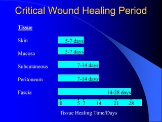
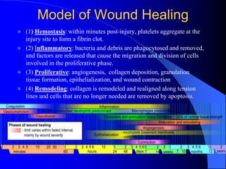
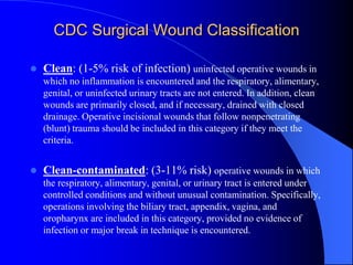
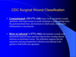
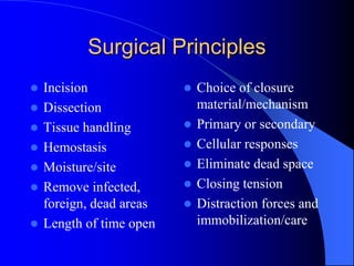
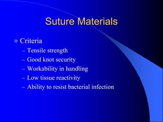
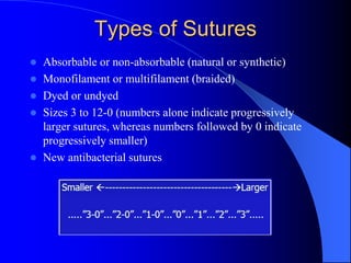
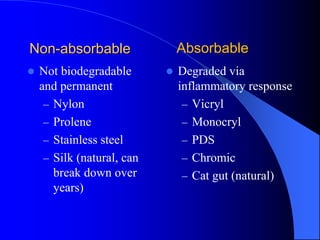
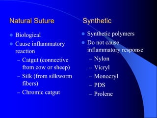
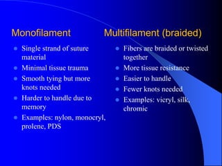
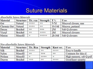
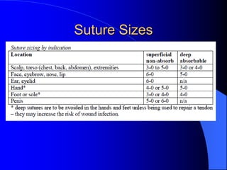
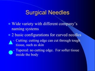
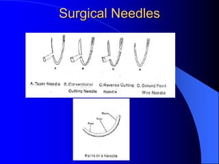
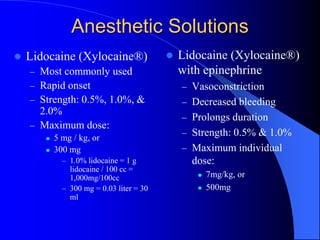
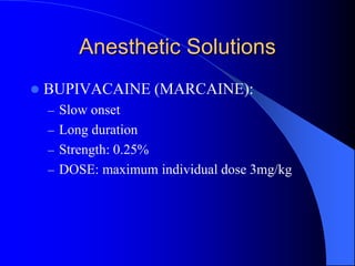
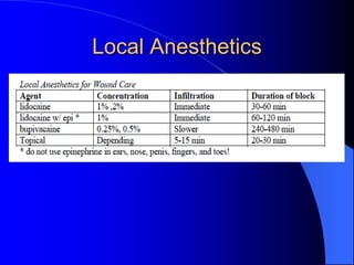
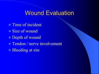
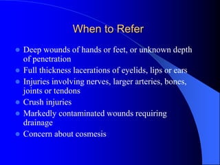
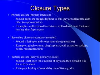
Sutures and Suturing
Critical Wound Healing Period
Tissue
Skin
Mucosa
Subcutaneous
Peritoneum
Fascia
5-7 days
5-7 days
7-14 days
7-14 days
14-28 days
0 5 7 14 21 28
Tissue Healing Time/Days
Model of Wound Healing
 (1) Hemostasis: within minutes post-injury, platelets aggregate at the
injury site to form a fibrin clot.
 (2) Inflammatory: bacteria and debris are phagocytosed and removed,
and factors are released that cause the migration and division of cells
involved in the proliferative phase.
 (3) Proliferative: angiogenesis, collagen deposition, granulation
tissue formation, epithelialization, and wound contraction
 (4) Remodeling: collagen is remodeled and realigned along tension
lines and cells that are no longer needed are removed by apoptosis.
Wound Healing Concepts
 Patient factors
 Wound classification
 Mechanism of injury
 Tetanus/antibiotics/local anesthetics
 Surgical principles and wound prep
 Suture/needle/stitch choice
 Management/care/follow-up
Common Patient Factors
 Age
 Blood supply to the
area
 Nutritional status
 Tissue quality
 Revision/infection
 Compliance
 Weight
 Dehydration
 Chronic disease
 Immune response
 Radiation therapy
CDC Surgical Wound Classification
 Clean: (1-5% risk of infection) uninfected operative wounds in
which no inflammation is encountered and the respiratory, alimentary,
genital, or uninfected urinary tracts are not entered. In addition, clean
wounds are primarily closed, and if necessary, drained with closed
drainage. Operative incisional wounds that follow nonpenetrating
(blunt) trauma should be included in this category if they meet the
criteria.
 Clean-contaminated: (3-11% risk) operative wounds in which
the respiratory, alimentary, genital, or urinary tract is entered under
controlled conditions and without unusual contamination. Specifically,
operations involving the biliary tract, appendix, vagina, and
oropharynx are included in this category, provided no evidence of
infection or major break in technique is encountered.
CDC Surgical Wound Classification
 Contaminated: (10-17% risk) open, fresh, accidental wounds,
operations with major breaks in sterile technique or gross spillage from
the gastrointestinal tract, and incisions in which acute, nonpurulent
inflammation is encountered.
 Dirty or infected: (>27% risk) old traumatic wounds with
retained devitalized tissue and those that involve existing clinical
infection or perforated viscera. This definition suggests that the
organisms causing postoperative infection were present in the
operative field before the operation.
Surgical Principles
 Incision
 Dissection
 Tissue handling
 Hemostasis
 Moisture/site
 Remove infected,
foreign, dead areas
 Length of time open
 Choice of closure
material/mechanism
 Primary or secondary
 Cellular responses
 Eliminate dead space
 Closing tension
 Distraction forces and
immobilization/care
Suture Materials
 Criteria
– Tensile strength
– Good knot security
– Workability in handling
– Low tissue reactivity
– Ability to resist bacterial infection
Types of Sutures
 Absorbable or non-absorbable (natural or synthetic)
 Monofilament or multifilament (braided)
 Dyed or undyed
 Sizes 3 to 12-0 (numbers alone indicate progressively
larger sutures, whereas numbers followed by 0 indicate
progressively smaller)
 New antibacterial sutures
Non-absorbable
 Not biodegradable
and permanent
– Nylon
– Prolene
– Stainless steel
– Silk (natural, can
break down over
years)
 Degraded via
inflammatory response
– Vicryl
– Monocryl
– PDS
– Chromic
– Cat gut (natural)
Absorbable
Natural Suture
 Biological
 Cause inflammatory
reaction
– Catgut (connective
from cow or sheep)
– Silk (from silkworm
fibers)
– Chromic catgut
Synthetic
 Synthetic polymers
 Do not cause
inflammatory response
– Nylon
– Vicryl
– Monocryl
– PDS
– Prolene
Monofilament
 Single strand of suture
material
 Minimal tissue trauma
 Smooth tying but more
knots needed
 Harder to handle due to
memory
 Examples: nylon, monocryl,
prolene, PDS
Multifilament (braided)
 Fibers are braided or twisted
together
 More tissue resistance
 Easier to handle
 Fewer knots needed
 Examples: vicryl, silk,
chromic
Suture Materials
Suture Selection
 Do not use dyed sutures on the skin
 Use monofilament on the skin as multifilament
harbor BACTERIA
 Non-absorbable cause less scarring but must be
removed
 Plus sutures (staph, monocryl for E. coli,
Klebsiella)
 Location and layer, patient factors, strength,
healing, site and availability
Suture Selection
 Absorbable for GI, urinary or biliary
 Non-absorbable or extended for up to 6 mos
for skin, tendons, fascia
 Cosmetics = monofilament or subcuticular
 Ligatures usually absorbable
Suture Sizes
Surgical Needles
 Wide variety with different company’s
naming systems
 2 basic configurations for curved needles
– Cutting: cutting edge can cut through tough
tissue, such as skin
– Tapered: no cutting edge. For softer tissue
inside the body
Surgical Needles
Anesthetic Solutions
 Lidocaine (Xylocaine®)
– Most commonly used
– Rapid onset
– Strength: 0.5%, 1.0%, &
2.0%
– Maximum dose:
 5 mg / kg, or
 300 mg
– 1.0% lidocaine = 1 g
lidocaine / 100 cc =
1,000mg/100cc
– 300 mg = 0.03 liter = 30
ml
 Lidocaine (Xylocaine®)
with epinephrine
– Vasoconstriction
– Decreased bleeding
– Prolongs duration
– Strength: 0.5% & 1.0%
– Maximum individual
dose:
 7mg/kg, or
 500mg
Anesthetic Solutions
 CAUTIONS: due to its vasoconstriction
properties never use Lidocaine with epinephrine
on:
– Eyes, Ears, Nose
– Fingers, Toes
– Penis, Scrotum
Anesthetic Solutions
 BUPIVACAINE (MARCAINE):
– Slow onset
– Long duration
– Strength: 0.25%
– DOSE: maximum individual dose 3mg/kg
Local Anesthetics
Wound Evaluation
 Time of incident
 Size of wound
 Depth of wound
 Tendon / nerve involvement
 Bleeding at site
When to Refer
 Deep wounds of hands or feet, or unknown depth
of penetration
 Full thickness lacerations of eyelids, lips or ears
 Injuries involving nerves, larger arteries, bones,
joints or tendons
 Crush injuries
 Markedly contaminated wounds requiring
drainage
 Concern about cosmesis
Contraindications to Suturing
 Redness
 Edema of the wound margins
 Infection
 Fever
 Puncture wounds
 Animal bites
 Tendon, verve, or vessel involvement
 Wound more than 12 hours old (body) and 24 hrs
(face)
Closure Types
 Primary closure (primary intention)
– Wound edges are brought together so that they are adjacent to each
other (re-approximated)
– Examples: well-repaired lacerations, well reduced bone fractures,
healing after flap surgery
 Secondary closure (secondary intention)
– Wound is left open and closes naturally (granulation)
– Examples: gingivectomy, gingivoplasty,tooth extraction sockets,
poorly reduced fractures
 Tertiary closure (delayed primary closure)
– Wound is left open for a number of days and then closed if it is
found to be clean
– Examples: healing of wounds by use of tissue grafts.
Wound Preparation
 Most important step for reducing the risk of wound
infection.
 Remove all contaminants and devitalized tissue before
wound closure.
– IRRIGATE w/ NS or TAP WATER (AVOID H2O2,
POVIDONE-IODINE)
– CUT OUT DEAD, FRAGMENTED TISSUE
 If not, the risk of infection and of a cosmetically poor scar
are greatly increased
 Personal Precautions
Suturing - before you go…
 Need for tetanus globulin and/or vaccine?
– Dirty (playground nail) vs clean (kitchen knife)
– Immunization history (>10 yrs need booster or >5 yrs if
contaminated)
 Tell pt to return in one day for recheck, for signs of infection
(redness, heat, pain, puss, etc), inadequate analgesia, or suture
complications (suture strangulation or knot failure with possible
wound dehiscence)
 It should be emphasized to patients that they return at the
appropriate time for suture removal or complications may arise
leading to further scarring or subsequent surgical removal of
buried sutures.
Patient instructions and follow up care
 Wound care
– After the first 24-48 hours, patients should gently wash
the wound with soap and water, dry it carefully, apply
topical antibiotic ointment, and replace the
dressing/bandages.
– Facial wounds generally only need topical antibiotic
ointment without bandaging.
– Eschar or scab formation should be avoided.
– Sunscreen spf 30 should be applied to the wound to
prevent subsequent hyperpigmentation.
Suture Removal
 Average time frame is 7 – 10 days
– FACE: 3 – 5 d
– NECK: 5 – 7 d
– SCALP: 7 – 12 days
– UPPER EXTREMITY, TRUNK: 10 – 14 days
– LOWER EXTREMITY: 14 – 28 days
– SOLES, PALMS, BACK OR OVER JOINTS: 10 days
 Any suture with pus or signs of infections should be
removed immediately.
Suture Removal
 Clean with hydrogen peroxide to remove any
crusting or dried blood
 Using the tweezers, grasp the knot and snip the
suture below the knot, close to the skin
 Pull the suture line through the tissue- in the
direction that keeps the wound closed - and place
on a 4x4. Count them.
 Most wounds have < 15% of final wound
strength after 2 wks, so steri-strips should be
applied afterwards.
Topical Adhesives
 Indications: selection of approximated, superficial, clean
wounds especially face, torso, limbs. May be used in
conjunction with deep sutures
 Benefits: Cosmetic, seals out bacteria, apply in 3 min,
holds 7 days (5-10 to slough), seal moisture, faster, clear,
convenient, less supplies, no removal, less expensive
 Contraindicated with infection, gangrene, mucosal, damp
or hairy areas, allergy to formaldehyde or cryanoacrylate,
or high tension areas
Dermabond®
 A sterile, liquid topical skin
adhesive
 Reacts with moisture on skin
surface to form a strong, flexible
bond
 Only for easily approximated
skin edges of wounds
– punctures from minimally
invasive surgery
– simple, thoroughly cleansed,
lacerations
References
 http://depts.washington.edu/uwemig/media_files/EMIG%20Suture%20Handout.pdf
 Thomsen, T. Basic Laceration Repair. The New England Journal of Medicine. Oct.
355: 17.
 Edgerton, M. The Art of Surgical Technique. Baltimore, Williams & Wilkins, 1988.
 www.uptodateonline.com; 2009, topic lacerations, etc.
 http://dermnetnz.org/procedures/pdf/suturing-dermnetnz.pdf
 http://www.mnpa.us/handouts/Session%2005%20%20-
%20%20Basic%20Suturing%20%202010%20MNPA.pdf
 http://www.practicalplasticsurgery.org/docs/Practical_01.pdf
 http://health.usf.edu/NR/rdonlyres/ABB54A41-80A1-4E2B-8AE8-
7EB5D06CE8DF/0/wound_healing_manual.pdf
 Jackson, E. Wound Care – Suture, Laceration, Dressing: Essentials for Family
Physicians. AAFP Scientific Assembly. 2010.
http://www.aafp.org/online/etc/medialib/aafp_org/documents/cme/courses/conf/asse
mbly/2010handouts/071.Par.0001.File.tmp/071-072.pdf

Sutures.ppt

  • 1.
  • 2.
    Critical Wound HealingPeriod Tissue Skin Mucosa Subcutaneous Peritoneum Fascia 5-7 days 5-7 days 7-14 days 7-14 days 14-28 days 0 5 7 14 21 28 Tissue Healing Time/Days
  • 3.
    Model of WoundHealing  (1) Hemostasis: within minutes post-injury, platelets aggregate at the injury site to form a fibrin clot.  (2) Inflammatory: bacteria and debris are phagocytosed and removed, and factors are released that cause the migration and division of cells involved in the proliferative phase.  (3) Proliferative: angiogenesis, collagen deposition, granulation tissue formation, epithelialization, and wound contraction  (4) Remodeling: collagen is remodeled and realigned along tension lines and cells that are no longer needed are removed by apoptosis.
  • 4.
    Wound Healing Concepts Patient factors  Wound classification  Mechanism of injury  Tetanus/antibiotics/local anesthetics  Surgical principles and wound prep  Suture/needle/stitch choice  Management/care/follow-up
  • 5.
    Common Patient Factors Age  Blood supply to the area  Nutritional status  Tissue quality  Revision/infection  Compliance  Weight  Dehydration  Chronic disease  Immune response  Radiation therapy
  • 6.
    CDC Surgical WoundClassification  Clean: (1-5% risk of infection) uninfected operative wounds in which no inflammation is encountered and the respiratory, alimentary, genital, or uninfected urinary tracts are not entered. In addition, clean wounds are primarily closed, and if necessary, drained with closed drainage. Operative incisional wounds that follow nonpenetrating (blunt) trauma should be included in this category if they meet the criteria.  Clean-contaminated: (3-11% risk) operative wounds in which the respiratory, alimentary, genital, or urinary tract is entered under controlled conditions and without unusual contamination. Specifically, operations involving the biliary tract, appendix, vagina, and oropharynx are included in this category, provided no evidence of infection or major break in technique is encountered.
  • 7.
    CDC Surgical WoundClassification  Contaminated: (10-17% risk) open, fresh, accidental wounds, operations with major breaks in sterile technique or gross spillage from the gastrointestinal tract, and incisions in which acute, nonpurulent inflammation is encountered.  Dirty or infected: (>27% risk) old traumatic wounds with retained devitalized tissue and those that involve existing clinical infection or perforated viscera. This definition suggests that the organisms causing postoperative infection were present in the operative field before the operation.
  • 8.
    Surgical Principles  Incision Dissection  Tissue handling  Hemostasis  Moisture/site  Remove infected, foreign, dead areas  Length of time open  Choice of closure material/mechanism  Primary or secondary  Cellular responses  Eliminate dead space  Closing tension  Distraction forces and immobilization/care
  • 9.
    Suture Materials  Criteria –Tensile strength – Good knot security – Workability in handling – Low tissue reactivity – Ability to resist bacterial infection
  • 10.
    Types of Sutures Absorbable or non-absorbable (natural or synthetic)  Monofilament or multifilament (braided)  Dyed or undyed  Sizes 3 to 12-0 (numbers alone indicate progressively larger sutures, whereas numbers followed by 0 indicate progressively smaller)  New antibacterial sutures
  • 11.
    Non-absorbable  Not biodegradable andpermanent – Nylon – Prolene – Stainless steel – Silk (natural, can break down over years)  Degraded via inflammatory response – Vicryl – Monocryl – PDS – Chromic – Cat gut (natural) Absorbable
  • 12.
    Natural Suture  Biological Cause inflammatory reaction – Catgut (connective from cow or sheep) – Silk (from silkworm fibers) – Chromic catgut Synthetic  Synthetic polymers  Do not cause inflammatory response – Nylon – Vicryl – Monocryl – PDS – Prolene
  • 13.
    Monofilament  Single strandof suture material  Minimal tissue trauma  Smooth tying but more knots needed  Harder to handle due to memory  Examples: nylon, monocryl, prolene, PDS Multifilament (braided)  Fibers are braided or twisted together  More tissue resistance  Easier to handle  Fewer knots needed  Examples: vicryl, silk, chromic
  • 14.
  • 15.
    Suture Selection  Donot use dyed sutures on the skin  Use monofilament on the skin as multifilament harbor BACTERIA  Non-absorbable cause less scarring but must be removed  Plus sutures (staph, monocryl for E. coli, Klebsiella)  Location and layer, patient factors, strength, healing, site and availability
  • 16.
    Suture Selection  Absorbablefor GI, urinary or biliary  Non-absorbable or extended for up to 6 mos for skin, tendons, fascia  Cosmetics = monofilament or subcuticular  Ligatures usually absorbable
  • 17.
  • 18.
    Surgical Needles  Widevariety with different company’s naming systems  2 basic configurations for curved needles – Cutting: cutting edge can cut through tough tissue, such as skin – Tapered: no cutting edge. For softer tissue inside the body
  • 19.
  • 20.
    Anesthetic Solutions  Lidocaine(Xylocaine®) – Most commonly used – Rapid onset – Strength: 0.5%, 1.0%, & 2.0% – Maximum dose:  5 mg / kg, or  300 mg – 1.0% lidocaine = 1 g lidocaine / 100 cc = 1,000mg/100cc – 300 mg = 0.03 liter = 30 ml  Lidocaine (Xylocaine®) with epinephrine – Vasoconstriction – Decreased bleeding – Prolongs duration – Strength: 0.5% & 1.0% – Maximum individual dose:  7mg/kg, or  500mg
  • 21.
    Anesthetic Solutions  CAUTIONS:due to its vasoconstriction properties never use Lidocaine with epinephrine on: – Eyes, Ears, Nose – Fingers, Toes – Penis, Scrotum
  • 22.
    Anesthetic Solutions  BUPIVACAINE(MARCAINE): – Slow onset – Long duration – Strength: 0.25% – DOSE: maximum individual dose 3mg/kg
  • 23.
  • 24.
    Wound Evaluation  Timeof incident  Size of wound  Depth of wound  Tendon / nerve involvement  Bleeding at site
  • 25.
    When to Refer Deep wounds of hands or feet, or unknown depth of penetration  Full thickness lacerations of eyelids, lips or ears  Injuries involving nerves, larger arteries, bones, joints or tendons  Crush injuries  Markedly contaminated wounds requiring drainage  Concern about cosmesis
  • 26.
    Contraindications to Suturing Redness  Edema of the wound margins  Infection  Fever  Puncture wounds  Animal bites  Tendon, verve, or vessel involvement  Wound more than 12 hours old (body) and 24 hrs (face)
  • 27.
    Closure Types  Primaryclosure (primary intention) – Wound edges are brought together so that they are adjacent to each other (re-approximated) – Examples: well-repaired lacerations, well reduced bone fractures, healing after flap surgery  Secondary closure (secondary intention) – Wound is left open and closes naturally (granulation) – Examples: gingivectomy, gingivoplasty,tooth extraction sockets, poorly reduced fractures  Tertiary closure (delayed primary closure) – Wound is left open for a number of days and then closed if it is found to be clean – Examples: healing of wounds by use of tissue grafts.
  • 28.
    Wound Preparation  Mostimportant step for reducing the risk of wound infection.  Remove all contaminants and devitalized tissue before wound closure. – IRRIGATE w/ NS or TAP WATER (AVOID H2O2, POVIDONE-IODINE) – CUT OUT DEAD, FRAGMENTED TISSUE  If not, the risk of infection and of a cosmetically poor scar are greatly increased  Personal Precautions
  • 29.
    Suturing - beforeyou go…  Need for tetanus globulin and/or vaccine? – Dirty (playground nail) vs clean (kitchen knife) – Immunization history (>10 yrs need booster or >5 yrs if contaminated)  Tell pt to return in one day for recheck, for signs of infection (redness, heat, pain, puss, etc), inadequate analgesia, or suture complications (suture strangulation or knot failure with possible wound dehiscence)  It should be emphasized to patients that they return at the appropriate time for suture removal or complications may arise leading to further scarring or subsequent surgical removal of buried sutures.
  • 30.
    Patient instructions andfollow up care  Wound care – After the first 24-48 hours, patients should gently wash the wound with soap and water, dry it carefully, apply topical antibiotic ointment, and replace the dressing/bandages. – Facial wounds generally only need topical antibiotic ointment without bandaging. – Eschar or scab formation should be avoided. – Sunscreen spf 30 should be applied to the wound to prevent subsequent hyperpigmentation.
  • 31.
    Suture Removal  Averagetime frame is 7 – 10 days – FACE: 3 – 5 d – NECK: 5 – 7 d – SCALP: 7 – 12 days – UPPER EXTREMITY, TRUNK: 10 – 14 days – LOWER EXTREMITY: 14 – 28 days – SOLES, PALMS, BACK OR OVER JOINTS: 10 days  Any suture with pus or signs of infections should be removed immediately.
  • 32.
    Suture Removal  Cleanwith hydrogen peroxide to remove any crusting or dried blood  Using the tweezers, grasp the knot and snip the suture below the knot, close to the skin  Pull the suture line through the tissue- in the direction that keeps the wound closed - and place on a 4x4. Count them.  Most wounds have < 15% of final wound strength after 2 wks, so steri-strips should be applied afterwards.
  • 33.
    Topical Adhesives  Indications:selection of approximated, superficial, clean wounds especially face, torso, limbs. May be used in conjunction with deep sutures  Benefits: Cosmetic, seals out bacteria, apply in 3 min, holds 7 days (5-10 to slough), seal moisture, faster, clear, convenient, less supplies, no removal, less expensive  Contraindicated with infection, gangrene, mucosal, damp or hairy areas, allergy to formaldehyde or cryanoacrylate, or high tension areas
  • 34.
    Dermabond®  A sterile,liquid topical skin adhesive  Reacts with moisture on skin surface to form a strong, flexible bond  Only for easily approximated skin edges of wounds – punctures from minimally invasive surgery – simple, thoroughly cleansed, lacerations
  • 35.
    References  http://depts.washington.edu/uwemig/media_files/EMIG%20Suture%20Handout.pdf  Thomsen,T. Basic Laceration Repair. The New England Journal of Medicine. Oct. 355: 17.  Edgerton, M. The Art of Surgical Technique. Baltimore, Williams & Wilkins, 1988.  www.uptodateonline.com; 2009, topic lacerations, etc.  http://dermnetnz.org/procedures/pdf/suturing-dermnetnz.pdf  http://www.mnpa.us/handouts/Session%2005%20%20- %20%20Basic%20Suturing%20%202010%20MNPA.pdf  http://www.practicalplasticsurgery.org/docs/Practical_01.pdf  http://health.usf.edu/NR/rdonlyres/ABB54A41-80A1-4E2B-8AE8- 7EB5D06CE8DF/0/wound_healing_manual.pdf  Jackson, E. Wound Care – Suture, Laceration, Dressing: Essentials for Family Physicians. AAFP Scientific Assembly. 2010. http://www.aafp.org/online/etc/medialib/aafp_org/documents/cme/courses/conf/asse mbly/2010handouts/071.Par.0001.File.tmp/071-072.pdf

Editor's Notes

  • #4 Phases are sequential, yet overlap
  • #8 This classification scheme has been shown in numerous studies to predict the relative probability that a wound will become infected. Clean wounds have a 1%-5% risk of infection; clean-contaminated, 3%-11%; contaminated, 10%-17%; and dirty, over 27% (2,3,7). These infection rates were affected by many appropriate prevention measures taken during the studies, such as use of prophylactic antimicrobials, and would have been higher if no prevention measures had been taken.
  • #12 Nylon (Ethilon®): of all the non-absorbable suture materials, monofilament nylon is the most commonly used in surface closures.  Polypropylene (Prolene®): appears to be stronger then nylon and has better overall wound security. BRAIDED: includes cotton, silk, braided nylon and multifilament dacron. Before the advent of synthetic fibers, silk was the mainstay of wound closure. It is the most workable and has excellent knot security. Disadvantages: high reactivity and infection due to the absorption of body fluids by the braided fibers.