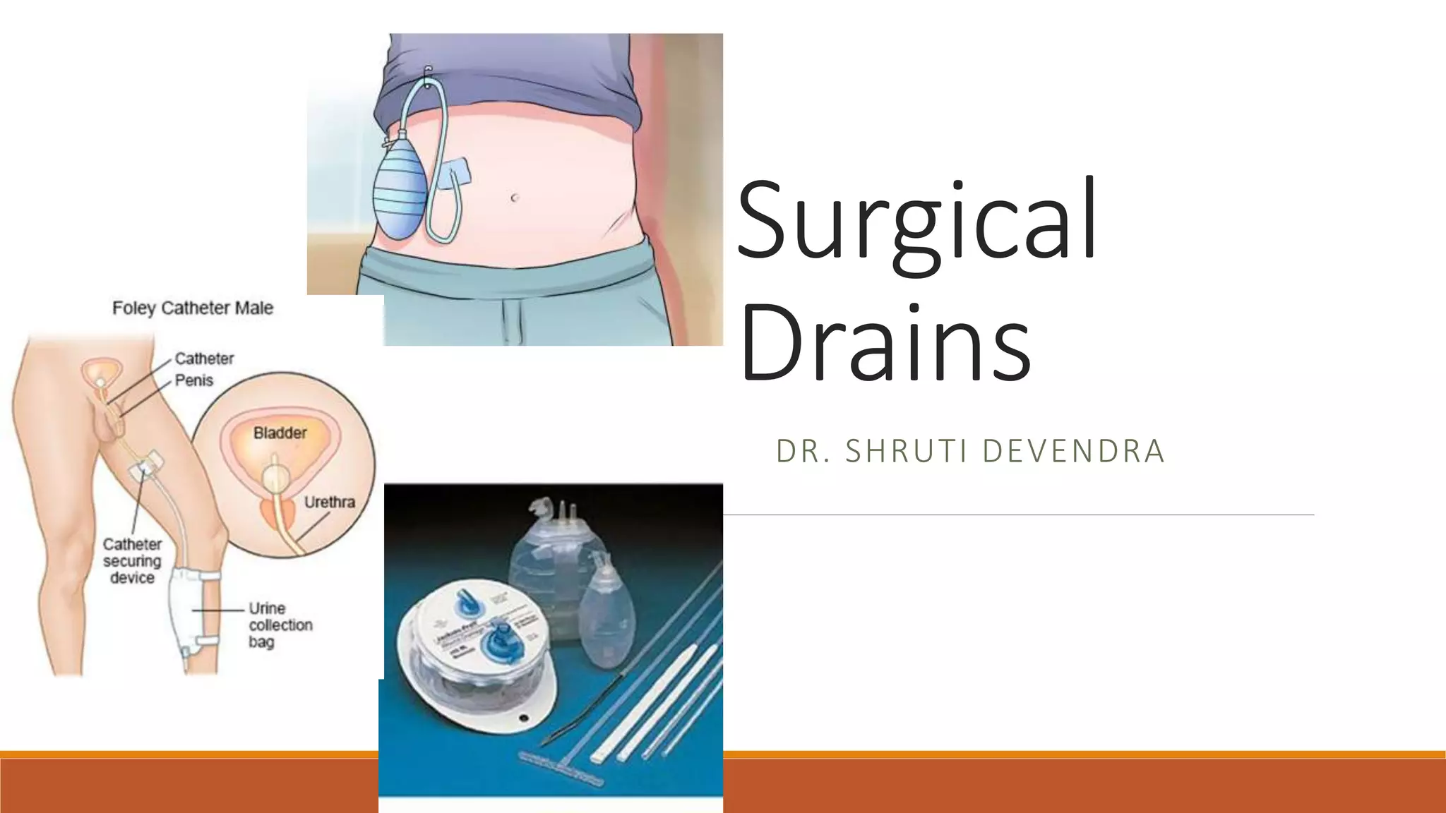
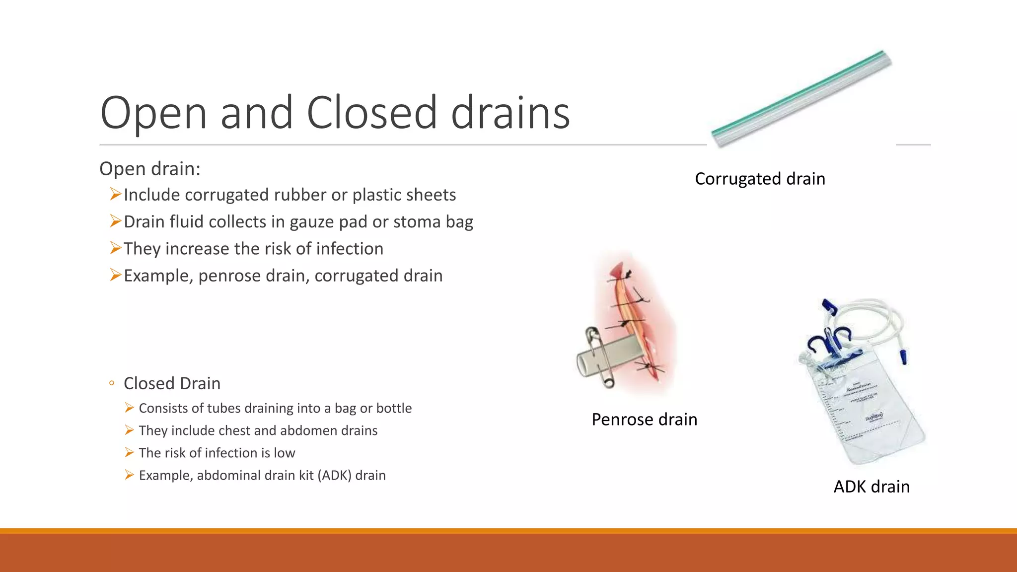
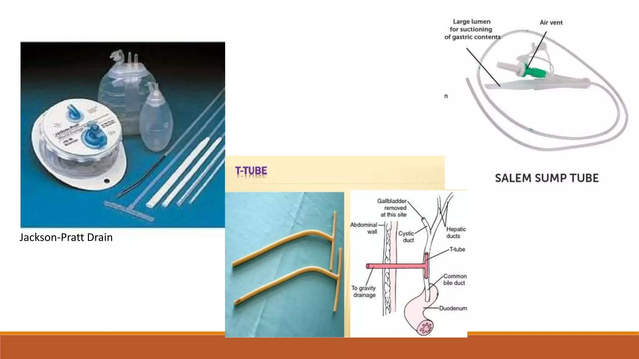
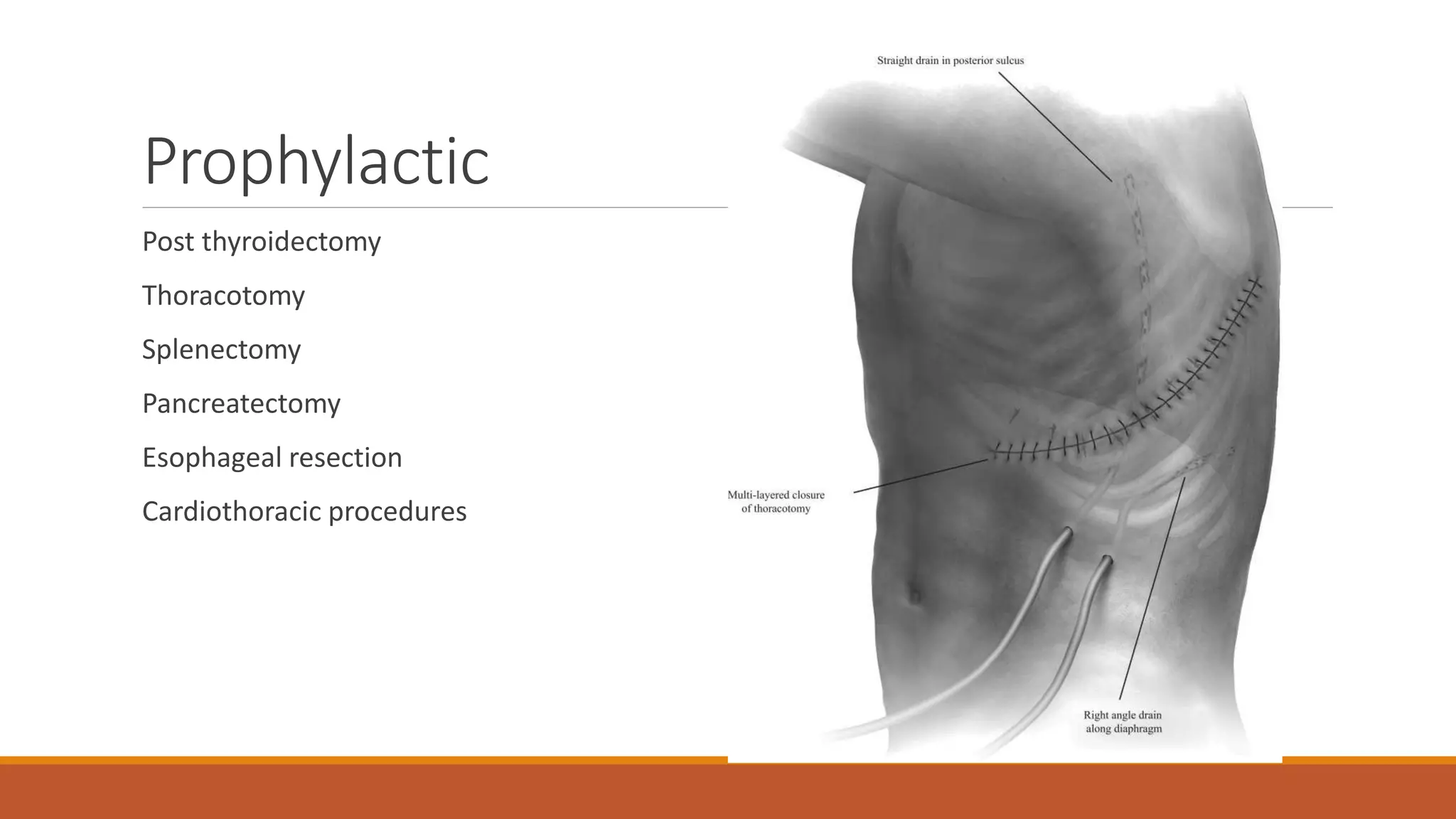
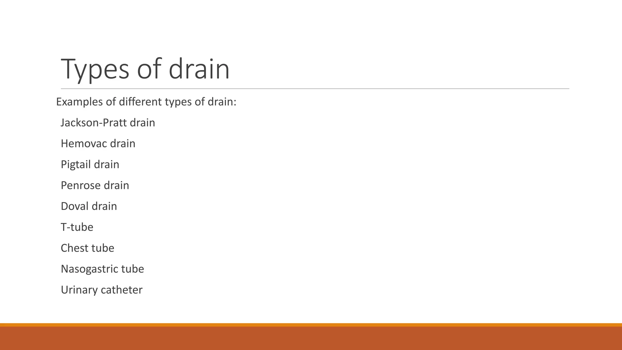
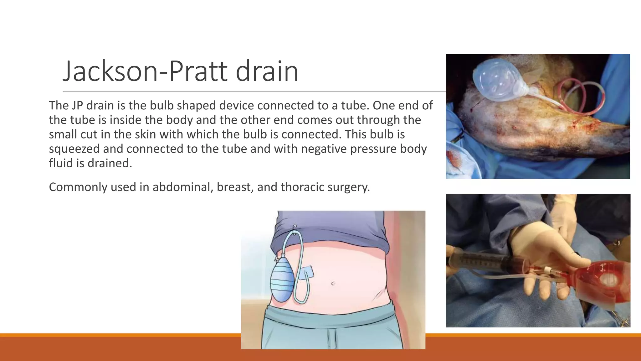
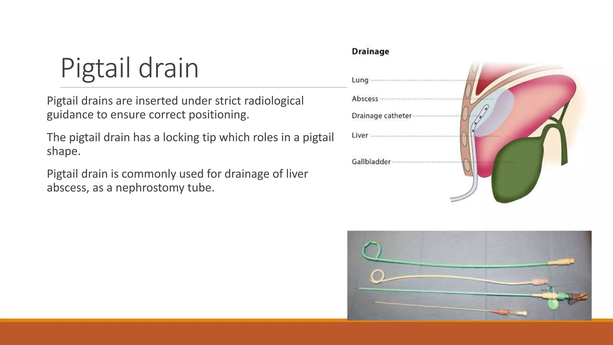
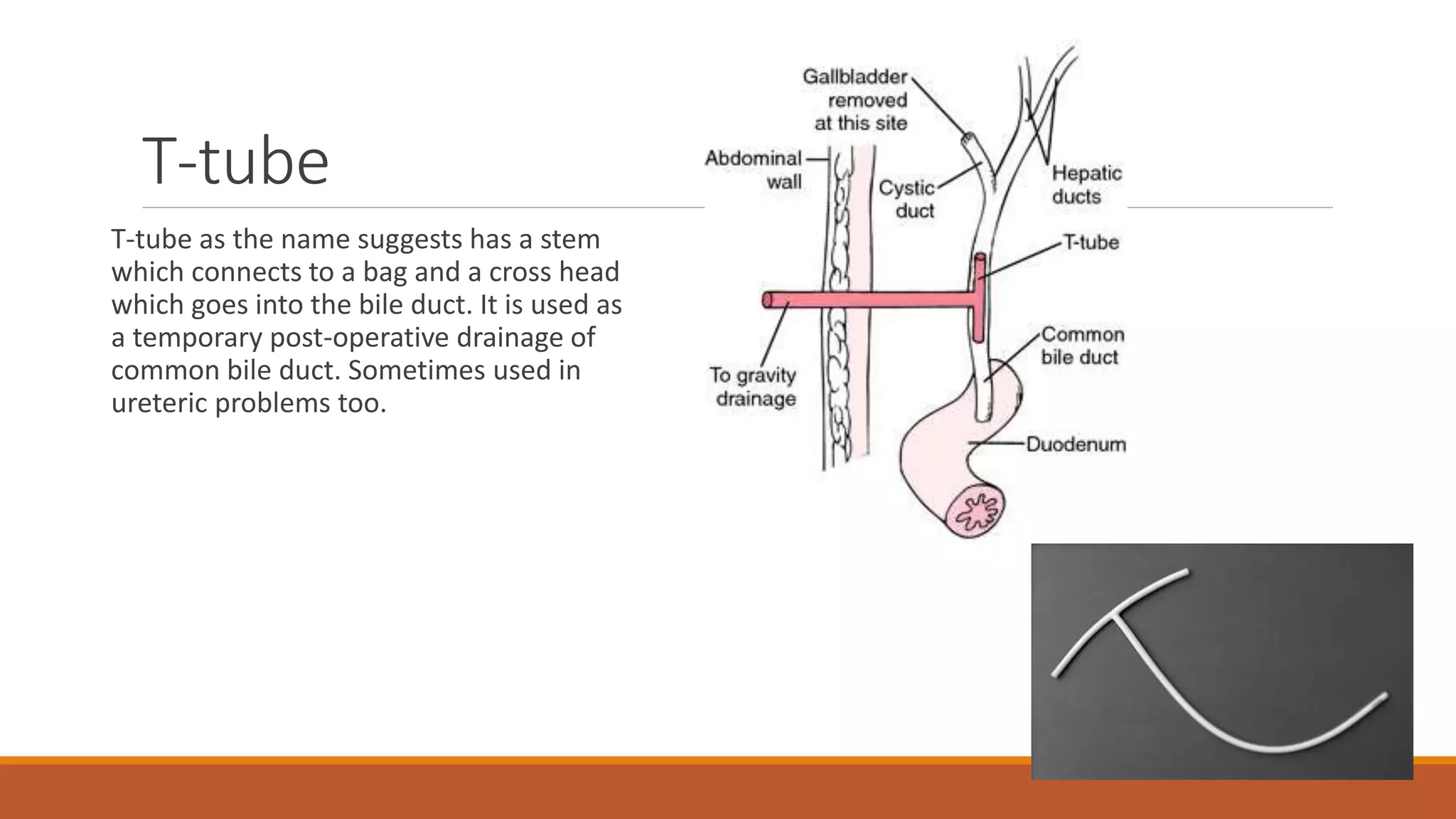
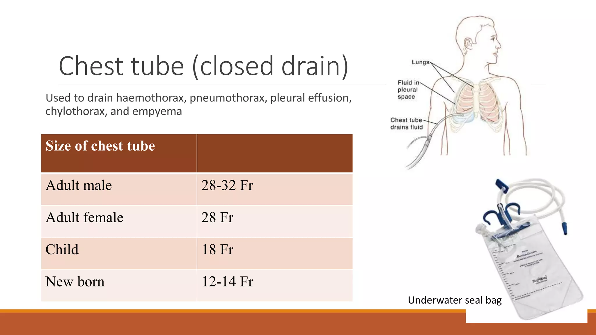
Surgical
Drains
DR. SHRUTI DEVENDRA
Outline
Introduction
Ideal drain
Classification
Indications
Types
Assessment
Removal
Complications
conclusion
Introduction
Drains are the devices that acts as a channel to drain established or potential collection of blood,
pus, body fluids or air.
Drains are available in various types and different sizes.
Drains are often inserted after surgery to drain body fluids that may accumulate and may
become focus of infection.
They are hooked up wall suction, a portable suction device, or they may be left to drain by
gravity depending on the location and need.
Recording of the quantity of drainage as well as the content is vital to ensure proper healing.
Depending on the amount of drainage it may be kept in place from few days to weeks.
Ideal drain
Firm, not too rigid
It should not be too soft as it may twist or kink or become blocked
Smooth
It should be resistant to decomposition and disintegration
Wide and patent enough to prevent easy blockage
It should be non electrolytic, non carcinogenic, and non thrombogenic when used in vascular
surgery.
Classification
Open or closed
Active or passive
Open and Closed drains
Open drain:
Include corrugated rubber or plastic sheets
Drain fluid collects in gauze pad or stoma bag
They increase the risk of infection
Example, penrose drain, corrugated drain
◦ Closed Drain
 Consists of tubes draining into a bag or bottle
 They include chest and abdomen drains
 The risk of infection is low
 Example, abdominal drain kit (ADK) drain
Corrugated drain
Penrose drain
ADK drain
Active and Passive drain
Active drain
Maintained under suction
They can be under low or high pressure
Closed e.g., Jackson-Pratt, hemovac drain
Open e.g., sump drain
Low risk of infection, can be placed anywhere
Disadvantage is clogging and tissue injury due to
negative pressure
◦ Passive drain
 They have no suction
 Drains by means of pressure differentials, overflow, and gravity
 Disadvantage is gravity dependent and easily clogged
Jackson-Pratt Drain
Indications
Therapeutic
Diagnostic
Prophylactic
Monitoring
Palliative
Therapeutic
Tension pneumothorax
Pleural fluid
Abscess cavity
Seroma
Acute urinary retention
Acute supportive arthritis
Infected cyst
Diagnostic
T-tube cholangiogram for retained gall stones in common bile duct
Biliary fistula
Prophylactic
Post thyroidectomy
Thoracotomy
Splenectomy
Pancreatectomy
Esophageal resection
Cardiothoracic procedures
Monitoring and Palliative
Monitoring
GI bleeding
Urethral catheterization
◦ Palliative
 Advanced carcinoma esophagus
 hydrocephalus
Types of drain
Examples of different types of drain:
Jackson-Pratt drain
Hemovac drain
Pigtail drain
Penrose drain
Doval drain
T-tube
Chest tube
Nasogastric tube
Urinary catheter
Jackson-Pratt drain
The JP drain is the bulb shaped device connected to a tube. One end of
the tube is inside the body and the other end comes out through the
small cut in the skin with which the bulb is connected. This bulb is
squeezed and connected to the tube and with negative pressure body
fluid is drained.
Commonly used in abdominal, breast, and thoracic surgery.
Hemovac drain
One end of the tube with multiple holes goes
into the body and the other end is connected
to the hemovac bag which is squeezed before
connecting the tube.
Pigtail drain
Pigtail drains are inserted under strict radiological
guidance to ensure correct positioning.
The pigtail drain has a locking tip which roles in a pigtail
shape.
Pigtail drain is commonly used for drainage of liver
abscess, as a nephrostomy tube.
Penrose drain
A penrose drain is soft and flexible. This drain
doesn’t have a collection devise. It empties into
absorptive dressing material, it promotes
drainage passively. With the drainage moving
from the area of greater pressure in the wound or
surgical site to the area of less pressure.
Doval drain
This suction device has a rubber bulb on top of the
drain that acts as a pump. To inflate the balloon in
the drainage bottle rubber bulb is squeezed
repeatedly in a pumping motion until the balloon
in the drainage bottle is inflated. Then quickly the
plug in the drain is replaced before the balloon
deflates. The inflated balloon in the bottle creates
suction.
T-tube
T-tube as the name suggests has a stem
which connects to a bag and a cross head
which goes into the bile duct. It is used as
a temporary post-operative drainage of
common bile duct. Sometimes used in
ureteric problems too.
Chest tube (closed drain)
Used to drain haemothorax, pneumothorax, pleural effusion,
chylothorax, and empyema
Size of chest tube
Adult male 28-32 Fr
Adult female 28 Fr
Child 18 Fr
New born 12-14 Fr
Underwater seal bag
Nasogastric tube
A tube passes form nostril to stomach
Indications
Aspiration of gastric juices
Lavage: in case of poisoning or drug
overdose
Feeding
Complications
Epistaxis
Aspiration
Erosion in the nasal cavity and nasopharynx
For adults 16-18 Fr
Urinary catheters
Indications
Urinary retention
Intra and post operative period
Incontinence
Drain assessment
Assess drain insertion site for signs of leakage, redness or signs of ooze
Assess the patency of the drain kinks, knots or clogging
Monitor the patients signs of sepsis
Ensure the suction is maintained in negative pressure drains
Drainage documented every 4 hourly and if high output then more frequently
Drain should be removed as soon as practicable, the longer the drain remains in situ, higher the
risk of infection and granulation tissue formation at the drain site which will increase pain and
trauma upon removal
Drain removal
Once the drainage has stopped or output less than 25 ml/day or when the drain has stopped
serving the desired function.
In case reactionary body fluid suspected in the drain then intermittent clamping trial is given to
see if the daily output has reduced before removing the drain
Drain can be shortened by 2 cm per day allowing the site to heal gradually
Before the drain is pulled patient is asked to take a deep breath. Once the patient inhales drain
is withdrawn steadily and swiftly
Once the drain is removed, sterile dry dressing is placed at the site of drain minimum for 24
hours. If the drain site shows discharge beyond 24 hours this means new drain need to be placed
Complications
Immediate
Pain
Irritation
Bleeding
perforation
Early
Occlusion
Leaking around drain
Displacement
Infection
Loss of fluid electrolytes and protein
Late
Pressure/suction necrosis
Fistula
Scar
hernia
Complications during removal
Pain
Infection (cellulitis/abscess)
Injury to adjacent structures
Retained or fragmentation of tube
Conclusion
The use of drains in surgical practice has been contentious over the years
The essential questions a surgeon needs to answer when deciding on the value of surgical drain
are
What purpose would drain serve if placed?
What type of drain should be used?
How long should the drain be left in place?
Once these questions are carefully and adequately answered each time a drain is used, the
effectiveness and advantage can be maximized with minimal problems.
Thank you!

Surgical Drains

  • 1.
  • 2.
  • 3.
    Introduction Drains are thedevices that acts as a channel to drain established or potential collection of blood, pus, body fluids or air. Drains are available in various types and different sizes. Drains are often inserted after surgery to drain body fluids that may accumulate and may become focus of infection. They are hooked up wall suction, a portable suction device, or they may be left to drain by gravity depending on the location and need. Recording of the quantity of drainage as well as the content is vital to ensure proper healing. Depending on the amount of drainage it may be kept in place from few days to weeks.
  • 4.
    Ideal drain Firm, nottoo rigid It should not be too soft as it may twist or kink or become blocked Smooth It should be resistant to decomposition and disintegration Wide and patent enough to prevent easy blockage It should be non electrolytic, non carcinogenic, and non thrombogenic when used in vascular surgery.
  • 5.
  • 6.
    Open and Closeddrains Open drain: Include corrugated rubber or plastic sheets Drain fluid collects in gauze pad or stoma bag They increase the risk of infection Example, penrose drain, corrugated drain ◦ Closed Drain  Consists of tubes draining into a bag or bottle  They include chest and abdomen drains  The risk of infection is low  Example, abdominal drain kit (ADK) drain Corrugated drain Penrose drain ADK drain
  • 7.
    Active and Passivedrain Active drain Maintained under suction They can be under low or high pressure Closed e.g., Jackson-Pratt, hemovac drain Open e.g., sump drain Low risk of infection, can be placed anywhere Disadvantage is clogging and tissue injury due to negative pressure ◦ Passive drain  They have no suction  Drains by means of pressure differentials, overflow, and gravity  Disadvantage is gravity dependent and easily clogged
  • 8.
  • 9.
  • 10.
    Therapeutic Tension pneumothorax Pleural fluid Abscesscavity Seroma Acute urinary retention Acute supportive arthritis Infected cyst
  • 11.
    Diagnostic T-tube cholangiogram forretained gall stones in common bile duct Biliary fistula
  • 12.
  • 13.
    Monitoring and Palliative Monitoring GIbleeding Urethral catheterization ◦ Palliative  Advanced carcinoma esophagus  hydrocephalus
  • 14.
    Types of drain Examplesof different types of drain: Jackson-Pratt drain Hemovac drain Pigtail drain Penrose drain Doval drain T-tube Chest tube Nasogastric tube Urinary catheter
  • 15.
    Jackson-Pratt drain The JPdrain is the bulb shaped device connected to a tube. One end of the tube is inside the body and the other end comes out through the small cut in the skin with which the bulb is connected. This bulb is squeezed and connected to the tube and with negative pressure body fluid is drained. Commonly used in abdominal, breast, and thoracic surgery.
  • 16.
    Hemovac drain One endof the tube with multiple holes goes into the body and the other end is connected to the hemovac bag which is squeezed before connecting the tube.
  • 17.
    Pigtail drain Pigtail drainsare inserted under strict radiological guidance to ensure correct positioning. The pigtail drain has a locking tip which roles in a pigtail shape. Pigtail drain is commonly used for drainage of liver abscess, as a nephrostomy tube.
  • 18.
    Penrose drain A penrosedrain is soft and flexible. This drain doesn’t have a collection devise. It empties into absorptive dressing material, it promotes drainage passively. With the drainage moving from the area of greater pressure in the wound or surgical site to the area of less pressure.
  • 19.
    Doval drain This suctiondevice has a rubber bulb on top of the drain that acts as a pump. To inflate the balloon in the drainage bottle rubber bulb is squeezed repeatedly in a pumping motion until the balloon in the drainage bottle is inflated. Then quickly the plug in the drain is replaced before the balloon deflates. The inflated balloon in the bottle creates suction.
  • 20.
    T-tube T-tube as thename suggests has a stem which connects to a bag and a cross head which goes into the bile duct. It is used as a temporary post-operative drainage of common bile duct. Sometimes used in ureteric problems too.
  • 21.
    Chest tube (closeddrain) Used to drain haemothorax, pneumothorax, pleural effusion, chylothorax, and empyema Size of chest tube Adult male 28-32 Fr Adult female 28 Fr Child 18 Fr New born 12-14 Fr Underwater seal bag
  • 22.
    Nasogastric tube A tubepasses form nostril to stomach Indications Aspiration of gastric juices Lavage: in case of poisoning or drug overdose Feeding Complications Epistaxis Aspiration Erosion in the nasal cavity and nasopharynx For adults 16-18 Fr
  • 23.
    Urinary catheters Indications Urinary retention Intraand post operative period Incontinence
  • 24.
    Drain assessment Assess draininsertion site for signs of leakage, redness or signs of ooze Assess the patency of the drain kinks, knots or clogging Monitor the patients signs of sepsis Ensure the suction is maintained in negative pressure drains Drainage documented every 4 hourly and if high output then more frequently Drain should be removed as soon as practicable, the longer the drain remains in situ, higher the risk of infection and granulation tissue formation at the drain site which will increase pain and trauma upon removal
  • 25.
    Drain removal Once thedrainage has stopped or output less than 25 ml/day or when the drain has stopped serving the desired function. In case reactionary body fluid suspected in the drain then intermittent clamping trial is given to see if the daily output has reduced before removing the drain Drain can be shortened by 2 cm per day allowing the site to heal gradually Before the drain is pulled patient is asked to take a deep breath. Once the patient inhales drain is withdrawn steadily and swiftly Once the drain is removed, sterile dry dressing is placed at the site of drain minimum for 24 hours. If the drain site shows discharge beyond 24 hours this means new drain need to be placed
  • 26.
    Complications Immediate Pain Irritation Bleeding perforation Early Occlusion Leaking around drain Displacement Infection Lossof fluid electrolytes and protein Late Pressure/suction necrosis Fistula Scar hernia Complications during removal Pain Infection (cellulitis/abscess) Injury to adjacent structures Retained or fragmentation of tube
  • 27.
    Conclusion The use ofdrains in surgical practice has been contentious over the years The essential questions a surgeon needs to answer when deciding on the value of surgical drain are What purpose would drain serve if placed? What type of drain should be used? How long should the drain be left in place? Once these questions are carefully and adequately answered each time a drain is used, the effectiveness and advantage can be maximized with minimal problems.
  • 28.