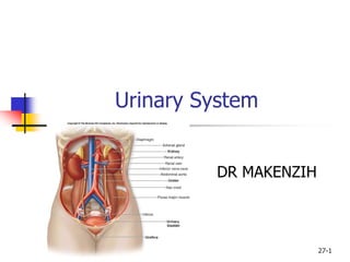
27-1
Urinary System
DR MAKENZIH
27-2
General Structure and Functions
of the Urinary System
 General Concept:
 Waste products accumulate in blood
 Are toxic
 Must be removed to maintain
homeostasis
 Urinary System organs
 remove waste products from the blood
 then from the body
 Major homeostatic system
27-3
General Structure and Functions
of the Urinary System
 Organs of the Urinary System:
 Kidneys
 Ureters
 Urinary Bladder
 Urethra
 Primary organs: kidneys
 filter waste products from the bloodstream
 convert the filtrate into urine.
 The Urinary Tract:
 Includes:
 ureters
 urinary bladder
 urethra
 Because they transport the urine out of the body.
4
5
6
27-7
Functions of the Urinary System
 Removing waste products from the bloodstream.
 Storage of urine.
 the urinary bladder is an expandable, muscular sac that can
store as much as 1 liter of urine
 Excretion of urine.
 Blood volume regulation.
 the kidneys control the volume of interstitial fluid and blood
under the direction of certain hormones
 Regulation of erythrocyte production.
 as the kidneys filter the blood, they are also indirectly
measuring the oxygen level in the blood
 Erythropoietin (EPO): hormone produced by kidney
 Released if blood oxygen levels fall
 Stimulates RBC production in red bone marrow
27-8
Kidneys: Gross and Sectional
Anatomy
 Retroperitoneal
 Anterior surface covered with peritoneum
 Posterior surface against posterior
abdominal wall
 Superior pole: T-12
 Inferior pole: L-3
 Right kidney ~ 2cm lower than left
 Adrenal gland on superior pole
9
27-10
Kidneys: Gross and Sectional
Anatomy
 Hilum: concave medial border
 Renal sinus: internal space
 Houses blood vessels, lymphatic vessels,
nerves
 Houses renal pelvis, renal calyces
 Also fat
27-11
Kidneys: Gross and Sectional
Anatomy
 Surrounding tissues, from deep to superficial:
 Fibrous capsule (renal capsule)
 Dense irregular CT
 Covers outer surface
 Perinephric fat (adipose capsule)
 Also called perirenal fat
 Completely surrounds kidney
 Cushioning and insulation
 Renal fascia
 Dense irregular CT
 Anchors kidney to posterior wall and peritoneum
 Paranephric fat
 Between renal fascia and peritoneum
12
27-13
Kidneys: Gross and Sectional
Anatomy
 Sectioned on a coronal plane:
 Renal Cortex
 Renal arches
 Renal columns
 Renal Medulla
 Divided into renal pyramids
 8 to 15 per kidney
 Base against cortex
 Apex called renal papilla
27-14
Kidneys: Gross and Sectional
Anatomy
 Minor calyx:
 Funnel shaped
 Receives renal papilla
 8 to 15 per kidney, one per pyramid
 Major calyx
 Fusion of minor calyces
 2 to 3 per kidney
 Major calyces merge to form renal pelvis
 Renal Lobe
 Pyramid plus some cortical tissue
 8 to 15 per kidney
15
16
27-17
Blood Supply to the Kidney
 About 20 to 25% of cardiac output to
kidneys
 Path:
 Renal artery to segmental arteries to
interlobar arteries to arcuate arteries to
interlobular arteries to:
 Afferent arteriole to glomerulus to efferent
arteriole to peritubular capilaries and vasa
recta
18
27-19
Blood Supply to the Kidney
 Blood plasma is filtered across the glomerulus
into the glomerular space.
 Once the blood plasma is filtered
 blood leaves the glomerulus
 enters an efferent arteriole.
 efferent arteriole is still carrying oxygenated
blood
 a gas and nutrient exchange with the kidney
tissues has not yet occurred.
27-20
Blood Supply to the Kidney
 The efferent arterioles branch into one of two
types of capillary networks:
 peritubular capillaries
 vasa recta
 these capillary networks are responsible for
the actual exchange of gases and nutrients
 Peritubular capillaries: primarily in cortex
 Vasa recta: surround the thin tubes that
project into the medulla.
27-21
Blood Supply to the Kidney
 Path for veins:
 Interlobar veins to arcuate veins to
interlobar veins to the renal vein
22
27-23
Nephrons
 The functional filtration unit in the kidney.
 Consists of the following:
 Renal corpuscle
 Glomerulus
 Glomerular capsule (Bowman’s capsule)
 Proximal convoluted tubule (PCT)
 Nephron loop (loop of Henle)
 Ascending loop of Henle
 Descending loop of Henle
 Distal convoluted tubule (DCT)
 collectively called the renal tubule
 In both kidneys: approximately 2.5 million nephrons.
 Are microscopic: measure about 5 centimeters in
length.
27-24
Nephrons
 Cortical Nephrons
 Near peripheral edge of cortex
 Short nephron loops
 Have peritubular capillaries
 Juxtamedullary nephrons
 Near corticomedullary border
 Long nephron loops
 Have vasa recta
25
27-26
Urine Formation
 Three processes
 Filtration
 Renal corpuscle: forms filtrate
 From blood to tubule
 Reabsorption
 Mostly PCT
 Water and salt: rest of nephron
 From tubule to blood
 Secretion
 From blood to tubule
27-27
Renal Corpuscle
 Vascular pole
 Afferent and efferent arterioles
 Tubular pole
 Connects to PCT
 Two structures:
 Glomerulus and glomerular capsule
 Glomerulus
 Capillary bed
 High pressure
 fenestrations
27-28
Renal Corpuscle
 Glomerular Capsule
 Parietal layer
 Simple squamous epithelium
 Visceral layer
 Podocytes
 Pedicels
 Filtration slits
 Capsular space (Bowman’s capsule): location of
filtrate
 Filtration membrane
 Fenestrations
 Filtration slits
29
30
27-31
Proximal Convoluted Tubule
 Begins at tubular pole of the renal corpuscle.
 Cells: simple cuboidal epithelium
 actively reabsorb from the filtrate:
 almost all nutrients (glucose and amino acids)
 electrolytes
 plasma proteins
 Osmosis: reabsorption of 60% to 65% of the
water in filtrate.
 Have microvilli
 Solutes and water:
 moved into blood plasma
 via the peritubular capillaries.
27-32
Nephron Loop (loop of Henle)
 originates at end of proximal convoluted tubule
 projects toward and/or into the medulla.
 Each loop has two limbs.
 descending limb:
 from cortex toward and/or into the medulla
 ascending limb:
 returns back to the renal cortex
33
34
27-35
Distal Convoluted Tubule
 begins at the end of the thick ascending limb of the
nephron loop
 adjacent to the afferent arteriole (important physiologically)
 Juxtaglomerular apparatus.
 primary function:
 Secretion
 From blood plasma to filtrate.
 secretes ions
 potassium (K+)
 acid (H+)
 Reabsorption of water also occurs:
 influenced by two hormones
 Aldosterone
 antidiuretic hormone (ADH).
27-36
Collecting Collecting Ducts
 Function in a well hydrated person:
 transport the tubular fluid into the papillary duct and then
into the minor calyx.
 Function in a dehydrated person:
 water conservation
 more-concentrated urine is produced.
 ADH can act on the collecting duct epithelium
 Cells become permeable to water
 Water moves from filtrate into blood plasma
 Involves vasa recta.
27-37
Innervation of the Kidney
 innervated by a mass of autonomic nervous system
fibers
 called the renal plexus.
 The renal plexus
 accompanies each renal artery
 enters the kidney through the hilum.
27-38
Urinary Tract : Ureters
 long, fibromuscular tubes
 conduct urine from the kidneys to the urinary
bladder.
 average 25 centimeters in length
 retroperitoneal.
 ureters originate at the renal pelvis
 extend inferiorly to enter the posterolateral wall of
the base of the urinary bladder.
 wall is composed of three concentric tunics.
 mucosa
 muscularis
 adventitia.
27-39
Urinary Tract – Urinary Bladder
 The urinary bladder:
 expandable, muscular container
 serves as a reservoir for urine
 positioned immediately superior and posterior to the pubic
symphysis.
 in females
 the urinary bladder is in contact with the uterus posterosuperiorly
and with the vagina posteroinferiorly.
 in males
 it is in contact with the rectum posterosuperiorly and is
immediately superior to the prostate gland.
 is a retroperitoneal organ.
 when empty exhibits an upside-down pyramidal shape.
 Filling with urine distends it superiorly until it assumes an oval
shape.
27-40
Urinary Tract – Urinary Bladder
 Trigone
 posteroinferior triangular area of the urinary bladder wall
 formed by imaginary lines
 connect the two posterior ureteral openings
 and the anterior urethral opening.
 The trigone remains immovable as the urinary
bladder fills and evacuates.
 It functions as a funnel
 directs urine into the urethra as the bladder wall contracts
 four tunics
 mucosa
 submucosa
 Muscularis: called the detrusor muscle
 adventitia.
 Internal urethral sphincter (smooth muscle)
41
42
43
27-44
Micturition (Urination)
 The expulsion of urine from the bladder.
 Initiated by a complex sequence of events called the
micturition reflex.
 The bladder is supplied by both parasympathetic and
sympathetic nerve fibers of the autonomic nervous
system.
27-45
Urethra
 Fibromuscular tube
 exits the urinary bladder through the urethral opening
 at anteroinferior surface
 conducts urine to the exterior of the body.
 Tunica mucosa: is a protective mucous membrane
 houses clusters of mucin-producing cells called urethral
glands.
 Tunica muscularis: primarily smooth muscle fibers
 help propel urine to the outside of the body.
 Two urethral sphincters:
 Internal urethral sphincter
 restrict the release of urine until the pressure within the urinary
bladder is high enough
 External urethral sphincter
 and voluntary activities needed to release the urine are
activated.
27-46
Urethra
 The internal urethral sphincter
 involuntary (smooth muscle)
 superior sphincter surrounding the neck of the bladder,
where the urethra originates.
 a circular thickening of the detrusor muscle
 controlled by the autonomic nervous system
 The external urethral sphincter
 inferior to the internal urethral sphincter
 formed by skeletal muscle fibers of the urogenital
diaphragm.
 a voluntary sphincter
 controlled by the somatic nervous system
 this is the muscle children learn to control when they
become “toilet-trained”
27-47
Female Urethra
 Has a single function:
 to transport urine from the urinary bladder to the vestibule,
an external space immediately internal to the labia minora
 3 to 5 centimeters long, and opens to the outside of
the body at the external urethral orifice located in the
female perineum.
48
27-49
Male Urethra
 Urinary and reproductive functions:
 passageway for both urine and semen
 Approximately 18 to 20 centimeters long.
 Partitioned into three segments:
 prostatic urethra is approximately 3 to 4 centimeters long and is the
most dilatable portion of the urethra
 extends through the prostate gland, immediately inferior to the male
bladder, where multiple small prostatic ducts enter it
 membranous urethra is the shortest and least dilatable portion
 extends from the inferior surface of the prostate gland through the
urogenital diaphragm
 spongy urethra is the longest part (15 centimeters)
 encased within a cylinder of erectile tissue in the penis called the
corpus spongiosum
 extends to the external urethral orifice
50
27-51
Aging and the Urinary System
 Changes in the size and functioning of the kidneys begin at 30.
 Gradual reduction in kidney size.
 Reduced blood flow to the kidneys.
 Decrease in the number of functional nephrons.
 Reabsorption and secretion are reduced.
 Diminished ability to filter and cleanse the blood.
 Less aldosterone or antidiuretic hormone.
 Ability to control blood volume and blood pressure is reduced.
 Bladder decreases in size.
 More frequent urination.
 Control of the urethral sphincters—and micturition—may be lost.
52

Urinary_System-1.ppt

  • 1.
  • 2.
    27-2 General Structure andFunctions of the Urinary System  General Concept:  Waste products accumulate in blood  Are toxic  Must be removed to maintain homeostasis  Urinary System organs  remove waste products from the blood  then from the body  Major homeostatic system
  • 3.
    27-3 General Structure andFunctions of the Urinary System  Organs of the Urinary System:  Kidneys  Ureters  Urinary Bladder  Urethra  Primary organs: kidneys  filter waste products from the bloodstream  convert the filtrate into urine.  The Urinary Tract:  Includes:  ureters  urinary bladder  urethra  Because they transport the urine out of the body.
  • 4.
  • 5.
  • 6.
  • 7.
    27-7 Functions of theUrinary System  Removing waste products from the bloodstream.  Storage of urine.  the urinary bladder is an expandable, muscular sac that can store as much as 1 liter of urine  Excretion of urine.  Blood volume regulation.  the kidneys control the volume of interstitial fluid and blood under the direction of certain hormones  Regulation of erythrocyte production.  as the kidneys filter the blood, they are also indirectly measuring the oxygen level in the blood  Erythropoietin (EPO): hormone produced by kidney  Released if blood oxygen levels fall  Stimulates RBC production in red bone marrow
  • 8.
    27-8 Kidneys: Gross andSectional Anatomy  Retroperitoneal  Anterior surface covered with peritoneum  Posterior surface against posterior abdominal wall  Superior pole: T-12  Inferior pole: L-3  Right kidney ~ 2cm lower than left  Adrenal gland on superior pole
  • 9.
  • 10.
    27-10 Kidneys: Gross andSectional Anatomy  Hilum: concave medial border  Renal sinus: internal space  Houses blood vessels, lymphatic vessels, nerves  Houses renal pelvis, renal calyces  Also fat
  • 11.
    27-11 Kidneys: Gross andSectional Anatomy  Surrounding tissues, from deep to superficial:  Fibrous capsule (renal capsule)  Dense irregular CT  Covers outer surface  Perinephric fat (adipose capsule)  Also called perirenal fat  Completely surrounds kidney  Cushioning and insulation  Renal fascia  Dense irregular CT  Anchors kidney to posterior wall and peritoneum  Paranephric fat  Between renal fascia and peritoneum
  • 12.
  • 13.
    27-13 Kidneys: Gross andSectional Anatomy  Sectioned on a coronal plane:  Renal Cortex  Renal arches  Renal columns  Renal Medulla  Divided into renal pyramids  8 to 15 per kidney  Base against cortex  Apex called renal papilla
  • 14.
    27-14 Kidneys: Gross andSectional Anatomy  Minor calyx:  Funnel shaped  Receives renal papilla  8 to 15 per kidney, one per pyramid  Major calyx  Fusion of minor calyces  2 to 3 per kidney  Major calyces merge to form renal pelvis  Renal Lobe  Pyramid plus some cortical tissue  8 to 15 per kidney
  • 15.
  • 16.
  • 17.
    27-17 Blood Supply tothe Kidney  About 20 to 25% of cardiac output to kidneys  Path:  Renal artery to segmental arteries to interlobar arteries to arcuate arteries to interlobular arteries to:  Afferent arteriole to glomerulus to efferent arteriole to peritubular capilaries and vasa recta
  • 18.
  • 19.
    27-19 Blood Supply tothe Kidney  Blood plasma is filtered across the glomerulus into the glomerular space.  Once the blood plasma is filtered  blood leaves the glomerulus  enters an efferent arteriole.  efferent arteriole is still carrying oxygenated blood  a gas and nutrient exchange with the kidney tissues has not yet occurred.
  • 20.
    27-20 Blood Supply tothe Kidney  The efferent arterioles branch into one of two types of capillary networks:  peritubular capillaries  vasa recta  these capillary networks are responsible for the actual exchange of gases and nutrients  Peritubular capillaries: primarily in cortex  Vasa recta: surround the thin tubes that project into the medulla.
  • 21.
    27-21 Blood Supply tothe Kidney  Path for veins:  Interlobar veins to arcuate veins to interlobar veins to the renal vein
  • 22.
  • 23.
    27-23 Nephrons  The functionalfiltration unit in the kidney.  Consists of the following:  Renal corpuscle  Glomerulus  Glomerular capsule (Bowman’s capsule)  Proximal convoluted tubule (PCT)  Nephron loop (loop of Henle)  Ascending loop of Henle  Descending loop of Henle  Distal convoluted tubule (DCT)  collectively called the renal tubule  In both kidneys: approximately 2.5 million nephrons.  Are microscopic: measure about 5 centimeters in length.
  • 24.
    27-24 Nephrons  Cortical Nephrons Near peripheral edge of cortex  Short nephron loops  Have peritubular capillaries  Juxtamedullary nephrons  Near corticomedullary border  Long nephron loops  Have vasa recta
  • 25.
  • 26.
    27-26 Urine Formation  Threeprocesses  Filtration  Renal corpuscle: forms filtrate  From blood to tubule  Reabsorption  Mostly PCT  Water and salt: rest of nephron  From tubule to blood  Secretion  From blood to tubule
  • 27.
    27-27 Renal Corpuscle  Vascularpole  Afferent and efferent arterioles  Tubular pole  Connects to PCT  Two structures:  Glomerulus and glomerular capsule  Glomerulus  Capillary bed  High pressure  fenestrations
  • 28.
    27-28 Renal Corpuscle  GlomerularCapsule  Parietal layer  Simple squamous epithelium  Visceral layer  Podocytes  Pedicels  Filtration slits  Capsular space (Bowman’s capsule): location of filtrate  Filtration membrane  Fenestrations  Filtration slits
  • 29.
  • 30.
  • 31.
    27-31 Proximal Convoluted Tubule Begins at tubular pole of the renal corpuscle.  Cells: simple cuboidal epithelium  actively reabsorb from the filtrate:  almost all nutrients (glucose and amino acids)  electrolytes  plasma proteins  Osmosis: reabsorption of 60% to 65% of the water in filtrate.  Have microvilli  Solutes and water:  moved into blood plasma  via the peritubular capillaries.
  • 32.
    27-32 Nephron Loop (loopof Henle)  originates at end of proximal convoluted tubule  projects toward and/or into the medulla.  Each loop has two limbs.  descending limb:  from cortex toward and/or into the medulla  ascending limb:  returns back to the renal cortex
  • 33.
  • 34.
  • 35.
    27-35 Distal Convoluted Tubule begins at the end of the thick ascending limb of the nephron loop  adjacent to the afferent arteriole (important physiologically)  Juxtaglomerular apparatus.  primary function:  Secretion  From blood plasma to filtrate.  secretes ions  potassium (K+)  acid (H+)  Reabsorption of water also occurs:  influenced by two hormones  Aldosterone  antidiuretic hormone (ADH).
  • 36.
    27-36 Collecting Collecting Ducts Function in a well hydrated person:  transport the tubular fluid into the papillary duct and then into the minor calyx.  Function in a dehydrated person:  water conservation  more-concentrated urine is produced.  ADH can act on the collecting duct epithelium  Cells become permeable to water  Water moves from filtrate into blood plasma  Involves vasa recta.
  • 37.
    27-37 Innervation of theKidney  innervated by a mass of autonomic nervous system fibers  called the renal plexus.  The renal plexus  accompanies each renal artery  enters the kidney through the hilum.
  • 38.
    27-38 Urinary Tract :Ureters  long, fibromuscular tubes  conduct urine from the kidneys to the urinary bladder.  average 25 centimeters in length  retroperitoneal.  ureters originate at the renal pelvis  extend inferiorly to enter the posterolateral wall of the base of the urinary bladder.  wall is composed of three concentric tunics.  mucosa  muscularis  adventitia.
  • 39.
    27-39 Urinary Tract –Urinary Bladder  The urinary bladder:  expandable, muscular container  serves as a reservoir for urine  positioned immediately superior and posterior to the pubic symphysis.  in females  the urinary bladder is in contact with the uterus posterosuperiorly and with the vagina posteroinferiorly.  in males  it is in contact with the rectum posterosuperiorly and is immediately superior to the prostate gland.  is a retroperitoneal organ.  when empty exhibits an upside-down pyramidal shape.  Filling with urine distends it superiorly until it assumes an oval shape.
  • 40.
    27-40 Urinary Tract –Urinary Bladder  Trigone  posteroinferior triangular area of the urinary bladder wall  formed by imaginary lines  connect the two posterior ureteral openings  and the anterior urethral opening.  The trigone remains immovable as the urinary bladder fills and evacuates.  It functions as a funnel  directs urine into the urethra as the bladder wall contracts  four tunics  mucosa  submucosa  Muscularis: called the detrusor muscle  adventitia.  Internal urethral sphincter (smooth muscle)
  • 41.
  • 42.
  • 43.
  • 44.
    27-44 Micturition (Urination)  Theexpulsion of urine from the bladder.  Initiated by a complex sequence of events called the micturition reflex.  The bladder is supplied by both parasympathetic and sympathetic nerve fibers of the autonomic nervous system.
  • 45.
    27-45 Urethra  Fibromuscular tube exits the urinary bladder through the urethral opening  at anteroinferior surface  conducts urine to the exterior of the body.  Tunica mucosa: is a protective mucous membrane  houses clusters of mucin-producing cells called urethral glands.  Tunica muscularis: primarily smooth muscle fibers  help propel urine to the outside of the body.  Two urethral sphincters:  Internal urethral sphincter  restrict the release of urine until the pressure within the urinary bladder is high enough  External urethral sphincter  and voluntary activities needed to release the urine are activated.
  • 46.
    27-46 Urethra  The internalurethral sphincter  involuntary (smooth muscle)  superior sphincter surrounding the neck of the bladder, where the urethra originates.  a circular thickening of the detrusor muscle  controlled by the autonomic nervous system  The external urethral sphincter  inferior to the internal urethral sphincter  formed by skeletal muscle fibers of the urogenital diaphragm.  a voluntary sphincter  controlled by the somatic nervous system  this is the muscle children learn to control when they become “toilet-trained”
  • 47.
    27-47 Female Urethra  Hasa single function:  to transport urine from the urinary bladder to the vestibule, an external space immediately internal to the labia minora  3 to 5 centimeters long, and opens to the outside of the body at the external urethral orifice located in the female perineum.
  • 48.
  • 49.
    27-49 Male Urethra  Urinaryand reproductive functions:  passageway for both urine and semen  Approximately 18 to 20 centimeters long.  Partitioned into three segments:  prostatic urethra is approximately 3 to 4 centimeters long and is the most dilatable portion of the urethra  extends through the prostate gland, immediately inferior to the male bladder, where multiple small prostatic ducts enter it  membranous urethra is the shortest and least dilatable portion  extends from the inferior surface of the prostate gland through the urogenital diaphragm  spongy urethra is the longest part (15 centimeters)  encased within a cylinder of erectile tissue in the penis called the corpus spongiosum  extends to the external urethral orifice
  • 50.
  • 51.
    27-51 Aging and theUrinary System  Changes in the size and functioning of the kidneys begin at 30.  Gradual reduction in kidney size.  Reduced blood flow to the kidneys.  Decrease in the number of functional nephrons.  Reabsorption and secretion are reduced.  Diminished ability to filter and cleanse the blood.  Less aldosterone or antidiuretic hormone.  Ability to control blood volume and blood pressure is reduced.  Bladder decreases in size.  More frequent urination.  Control of the urethral sphincters—and micturition—may be lost.
  • 52.