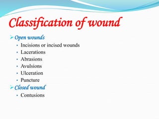
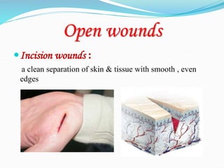
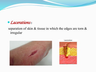
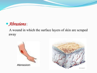
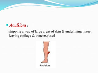
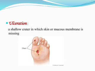
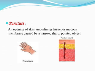
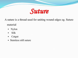
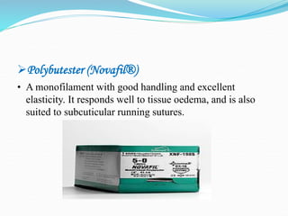
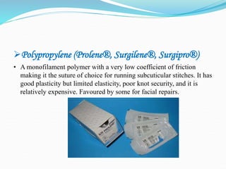
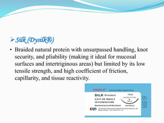
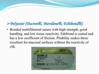
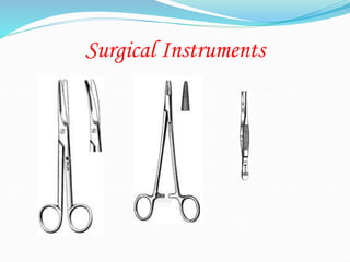
wound
 Wound : damaged skin or soft tissue result from injury
Two basic type of wound :
 Open wound : the surface of the skin or mucous membrane
is no longer intact .
 Closed wound : no opening in the skin or mucous
membrane .
Classification of wound
Open wounds
• Incisions or incised wounds
• Lacerations
• Abrasions
• Avulsions
• Ulceration
• Puncture
Closed wound
• Contusions
Open wounds
Incision wounds :
a clean separation of skin & tissue with smooth , even
edges
Lacerations :
separation of skin & tissue in which the edges are torn &
irregular
Abrasions :
A wound in which the surface layers of skin are scraped
away
Avulsions :
stripping a way of large areas of skin & underlining tissue,
leaving catilage & bone exposed
Ulceration :
a shallow crater in which skin or mucous membrane is
missing
Puncture :
An opening of skin, underlining tissue, or mucous
membrane caused by a narrow, sharp, pointed object
Closed wound
Contusions :
injury to soft tissue underlining the skin from the force of
contact with a hard object sometimes called a bruise
wound management
wound management : involve techniques that promote
wound healing .
 involve using :
 Dressing
 Drain
 Bandage and binder
 Sutures & staples
Suture
A suture is a thread used for uniting wound edges eg. Suture
material
 Nylon
 Silk
 Catgut
 Stainless still suture
Sutures
Goals
1. Provide an adequate tension of wound closure without dead space but
loose enough to obviate tissue ischemia and necrosis
2. Maintain hemostasis.
3. Permit primary intention healing
4. Reduce postoperative pain
5. Provide support for tissue margins until they have healed and the support
no longer needed
6. Prevent bone exposure resulting in delayed healing and unnecessary
resorption
7. Permit proper flap position
Qualities of a suture material
• Adequate tensile strength
• Functional strength
• Non capillary
• Non reactivity
• Flexibility & elasticity
• Easy to handle
• Knotable
• Easily sterlisable
• Uniformity
• Smooth surface
• Monofilament
• Absorbility
Suture Materials
 Absorbable
Those that are absorbed or digested by the body cells and
tissue fluids in which they are embedded during and after the
healing processes.
Non-Absorbable
Those suture materials that can not be absorbed by the body
cells or fluids.
Types of absorbable suture material
 Surgical Gut
• Plain gut loses its strength in 7-10 days and is completely digested by 60
days. It is seldom used now due to poor strength and high tissue reactivity
(due to proteolytic enzyme degradation rather than hydrolysis).
• Chromic gut has been manufactured with chromium salts to reduce
enzyme digestion and therefore maintains strength for 10-14 days making
it useful for mucosal closures.
• Fast-absorbing gut is produced by pre-heating and can be used for
attaching skin grafts, or in areas of low tension where the wound is well
supported by deep sutures, and suture removal would be difficult. It
maintains strength for 3-5 days
 Polyglactin 910 (Vicryl®, Polysorb®)
• A synthetic braided co-polymer which maintains 75% strength at 2
weeks, and 50% at 3 weeks. Absorption is usually complete by 3
months. It handles well, has minimal tissue reactivity, and does not
tear tissue. It may occasionally persist as a small nodule or extrude
(‘spitting’).
 Poliglecaprone 25 (Monocryl®)
• Monofilament maintaining 50-60% strength at 7 days with complete
absorption by 3 months. It offers better handling and knot security
than most other monofilament sutures, with even less tissue reaction
than Vicryl® and is therefore useful where minimal tissue reaction is
essential.
Polydioxanone (PDS II®)
• Monofilament polymer with prolonged tensile strength (70% at 2
weeks, 50% at 4 weeks) and may persist for more than 6 months.
Good for high-tension areas or contaminated wounds, but being a
monofilament it has poor handling and knot security. Its minimal
tissue reaction makes it good for repair of cartilage where
inflammation would lead to significant discomfort.
Polytrimethylene carbonate (Maxon®)
• A monofilament that combines the prolonged strength of
PDS® and the good handling and knotting of Vicryl®.
80% strength at 2 weeks, 60% at 4 weeks, and complete
absorption by 6 months. Minimal tissue reaction.
Glycomer 631 (Biosyn®)
• A monofilament similar to Monocryl® in characteristics
but with prolonged strength akin to Maxon®.
Non-absorbable Sutures
Nylon (Ethilon®, Dermalon®, Surgilon®, Nurolon®, Nylene®)
• Inexpensive monofilament with good tensile strength, and
minimal tissue reactivity. Disadvantages are its handling and
knot security, but it remains one of the most popular non-
absorbable sutures in dermatological surgery. Surgilon® and
Nurolon® handle better but are more expensive.
Polybutester (Novafil®)
• A monofilament with good handling and excellent
elasticity. It responds well to tissue oedema, and is also
suited to subcuticular running sutures.
Polypropylene (Prolene®, Surgilene®, Surgipro®)
• A monofilament polymer with a very low coefficient of friction
making it the suture of choice for running subcuticular stitches. It has
good plasticity but limited elasticity, poor knot security, and it is
relatively expensive. Favoured by some for facial repairs.
Silk (Dysilk®)
• Braided natural protein with unsurpassed handling, knot
security, and pliability (making it ideal for mucosal
surfaces and intertriginous areas) but limited by its low
tensile strength, and high coefficient of friction,
capillarity, and tissue reactivity.
Polyester (Dacron®, Mersilene®, Ethibond®)
• Braided multifilament suture with high strength, good
handling, and low tissue reactivity. Ethibond is coated and
has a low coefficient of friction. Pliability makes these
excellent for mucosal surfaces without the reactivity of
silk.
Selecting the Gauge
Selection of the gauge depends on the strength of the repair required,
the number of sutures to be used, the type of material used and the
cosmetic requirements of the wound. In practice, the selection
depends on experience of the surgeon and his knowledge of the
material.
The gauging may seem confusing to the novice. Long before current
suture materials were available, the finest thread was a 1 gauge with
thicker threads being given a higher number depending on diameter.
When a thread of smaller diameter than a 1 gauge was made, it was
named a 0 gauge (1/0). Subsequent smaller threads were termed 00
gauge (2/0), 000 gauge (3/0), etc as the diameter decreased to the
point where sutures of a 11/0 gauge are now used in microsurgery to
join blood vessels less than a millimeter in diameter.
When we suture a wound on the face, we would look to using a
very fine material with multiple sutures closely placed to obtain
the best cosmetic result, for example a 6/0 monofilament
nonabsorbable suture like Nylon. Closure of the abdominal
wall following laparotomy requires a strong suture such as a 0
gauge monofilarnent. A tendon repair would require a small
diameter suture with an inherently strong nonabsorbable
material such as a polyester (3/0 Ticron).
Needle Characteristics
There are 5 factors which we take into account when we select an
appropriate needle.
• Method of Use: The needle can either be hand-held or instrument
held.
• Shape of the Needle: The needle can either be straight or curved
• Length of the Needle: Needle length ranges from 2 to 60 mm.
• Tissue Penetration Characteristics: The tip of the needle can be
either a round bodied for passing through soft tissues such as fat
or muscle or a cutting point for penetration of tougher tissues
such as skin.
• Attachment of Suture to Needle: The material can either be
threaded through the eye of the needle or could be swaged into
the end of a needle.
Suture needles types
1. straight needles
2. curved needles
• Round needles
• Cutting needles
Surgical Instruments
Techniques of Suturing
The aims of repairing a skin wound is to end up with a fine linear scar
situated in a natural skin crease line without evidence of suture marks. The
final appearance of a scar depends on the use of atraumatie technique, scar
placement, the age of the patient, the region of the body, the skin type and
complicating factors such as infection.
The principals of repair of wounds are as follows:
• Adequate debridement (removal of dead, contaminated tissues and foreign
bodies)
• Atraumatic technique (gentle handling of tissues preventing inadvertent crushing
of wound edges and desiccation)
• Haemostasis
• Closure of the wound in layers under minimal tension (“appose don’t nccrose”)
using fine, high quality instruments
• Early removal of sutures to prevent marks
Suture Procedures
Suturing Techniques
1. Interrupted Pattern
2. Continuous Pattern
Simple interrupted suture
Simple continuous suture
Interrupted horizontal mattress suture
Continuous horizontal mattress suture
Vertical mettress suture
Near and Far suture
Sub cuticular suture
Cruciate suture
Continuous lock stitch
Dressing of the Wound
Do not underestimate the importance of a good, neat dressing on the
wound as patients will judge your work partly on the only bit they
can initially see, the dressing! Poor dressings look sloppy and are
a reflection of your work and lack of attention to detail. The aims
of a dressing are as follows:
• Occlusion of dirt, bacteria and inquisitive fingers
• Absorption of blood and exudate
• Wound immobilisation and pain relief
• Pressure application
• Medication carrier such as antibiotics and antiseptics
• Aesthetic wound covering
Suture Removal
Time frame for removing sutures:
Average time frame is 7-10 days
FACE: 4-5 days
BODY & SCALP: 7 days
SOLES, PALMS, BACK OR OVER JOINTS: 10 days
Any suture with pus or signs of infections should be removed immediately.
1. Clean with hydrogen peroxide to remove any crusting or dried blood
2. Using the tweezers, grasp the knot and snip the suture below the knot,
close to the skin
3. Pull the suture line through the tissue- in the direction that keeps the
wound closed - and place on a 4x4
Once all sutures have been removed, count the sutures
The number of sutures needs to match the number indicated
in the patient's health record
Thank you for listening

closure-material#5.ppt

  • 2.
    wound  Wound :damaged skin or soft tissue result from injury Two basic type of wound :  Open wound : the surface of the skin or mucous membrane is no longer intact .  Closed wound : no opening in the skin or mucous membrane .
  • 3.
    Classification of wound Openwounds • Incisions or incised wounds • Lacerations • Abrasions • Avulsions • Ulceration • Puncture Closed wound • Contusions
  • 4.
    Open wounds Incision wounds: a clean separation of skin & tissue with smooth , even edges
  • 5.
    Lacerations : separation ofskin & tissue in which the edges are torn & irregular
  • 6.
    Abrasions : A woundin which the surface layers of skin are scraped away
  • 7.
    Avulsions : stripping away of large areas of skin & underlining tissue, leaving catilage & bone exposed
  • 8.
    Ulceration : a shallowcrater in which skin or mucous membrane is missing
  • 9.
    Puncture : An openingof skin, underlining tissue, or mucous membrane caused by a narrow, sharp, pointed object
  • 10.
    Closed wound Contusions : injuryto soft tissue underlining the skin from the force of contact with a hard object sometimes called a bruise
  • 11.
    wound management wound management: involve techniques that promote wound healing .  involve using :  Dressing  Drain  Bandage and binder  Sutures & staples
  • 12.
    Suture A suture isa thread used for uniting wound edges eg. Suture material  Nylon  Silk  Catgut  Stainless still suture
  • 13.
    Sutures Goals 1. Provide anadequate tension of wound closure without dead space but loose enough to obviate tissue ischemia and necrosis 2. Maintain hemostasis. 3. Permit primary intention healing 4. Reduce postoperative pain 5. Provide support for tissue margins until they have healed and the support no longer needed 6. Prevent bone exposure resulting in delayed healing and unnecessary resorption 7. Permit proper flap position
  • 14.
    Qualities of asuture material • Adequate tensile strength • Functional strength • Non capillary • Non reactivity • Flexibility & elasticity • Easy to handle • Knotable
  • 15.
    • Easily sterlisable •Uniformity • Smooth surface • Monofilament • Absorbility
  • 16.
    Suture Materials  Absorbable Thosethat are absorbed or digested by the body cells and tissue fluids in which they are embedded during and after the healing processes. Non-Absorbable Those suture materials that can not be absorbed by the body cells or fluids.
  • 17.
    Types of absorbablesuture material  Surgical Gut • Plain gut loses its strength in 7-10 days and is completely digested by 60 days. It is seldom used now due to poor strength and high tissue reactivity (due to proteolytic enzyme degradation rather than hydrolysis). • Chromic gut has been manufactured with chromium salts to reduce enzyme digestion and therefore maintains strength for 10-14 days making it useful for mucosal closures. • Fast-absorbing gut is produced by pre-heating and can be used for attaching skin grafts, or in areas of low tension where the wound is well supported by deep sutures, and suture removal would be difficult. It maintains strength for 3-5 days
  • 18.
     Polyglactin 910(Vicryl®, Polysorb®) • A synthetic braided co-polymer which maintains 75% strength at 2 weeks, and 50% at 3 weeks. Absorption is usually complete by 3 months. It handles well, has minimal tissue reactivity, and does not tear tissue. It may occasionally persist as a small nodule or extrude (‘spitting’).
  • 19.
     Poliglecaprone 25(Monocryl®) • Monofilament maintaining 50-60% strength at 7 days with complete absorption by 3 months. It offers better handling and knot security than most other monofilament sutures, with even less tissue reaction than Vicryl® and is therefore useful where minimal tissue reaction is essential.
  • 20.
    Polydioxanone (PDS II®) •Monofilament polymer with prolonged tensile strength (70% at 2 weeks, 50% at 4 weeks) and may persist for more than 6 months. Good for high-tension areas or contaminated wounds, but being a monofilament it has poor handling and knot security. Its minimal tissue reaction makes it good for repair of cartilage where inflammation would lead to significant discomfort.
  • 21.
    Polytrimethylene carbonate (Maxon®) •A monofilament that combines the prolonged strength of PDS® and the good handling and knotting of Vicryl®. 80% strength at 2 weeks, 60% at 4 weeks, and complete absorption by 6 months. Minimal tissue reaction.
  • 22.
    Glycomer 631 (Biosyn®) •A monofilament similar to Monocryl® in characteristics but with prolonged strength akin to Maxon®.
  • 23.
    Non-absorbable Sutures Nylon (Ethilon®,Dermalon®, Surgilon®, Nurolon®, Nylene®) • Inexpensive monofilament with good tensile strength, and minimal tissue reactivity. Disadvantages are its handling and knot security, but it remains one of the most popular non- absorbable sutures in dermatological surgery. Surgilon® and Nurolon® handle better but are more expensive.
  • 24.
    Polybutester (Novafil®) • Amonofilament with good handling and excellent elasticity. It responds well to tissue oedema, and is also suited to subcuticular running sutures.
  • 25.
    Polypropylene (Prolene®, Surgilene®,Surgipro®) • A monofilament polymer with a very low coefficient of friction making it the suture of choice for running subcuticular stitches. It has good plasticity but limited elasticity, poor knot security, and it is relatively expensive. Favoured by some for facial repairs.
  • 26.
    Silk (Dysilk®) • Braidednatural protein with unsurpassed handling, knot security, and pliability (making it ideal for mucosal surfaces and intertriginous areas) but limited by its low tensile strength, and high coefficient of friction, capillarity, and tissue reactivity.
  • 27.
    Polyester (Dacron®, Mersilene®,Ethibond®) • Braided multifilament suture with high strength, good handling, and low tissue reactivity. Ethibond is coated and has a low coefficient of friction. Pliability makes these excellent for mucosal surfaces without the reactivity of silk.
  • 28.
    Selecting the Gauge Selectionof the gauge depends on the strength of the repair required, the number of sutures to be used, the type of material used and the cosmetic requirements of the wound. In practice, the selection depends on experience of the surgeon and his knowledge of the material. The gauging may seem confusing to the novice. Long before current suture materials were available, the finest thread was a 1 gauge with thicker threads being given a higher number depending on diameter. When a thread of smaller diameter than a 1 gauge was made, it was named a 0 gauge (1/0). Subsequent smaller threads were termed 00 gauge (2/0), 000 gauge (3/0), etc as the diameter decreased to the point where sutures of a 11/0 gauge are now used in microsurgery to join blood vessels less than a millimeter in diameter.
  • 29.
    When we suturea wound on the face, we would look to using a very fine material with multiple sutures closely placed to obtain the best cosmetic result, for example a 6/0 monofilament nonabsorbable suture like Nylon. Closure of the abdominal wall following laparotomy requires a strong suture such as a 0 gauge monofilarnent. A tendon repair would require a small diameter suture with an inherently strong nonabsorbable material such as a polyester (3/0 Ticron).
  • 30.
    Needle Characteristics There are5 factors which we take into account when we select an appropriate needle. • Method of Use: The needle can either be hand-held or instrument held. • Shape of the Needle: The needle can either be straight or curved • Length of the Needle: Needle length ranges from 2 to 60 mm. • Tissue Penetration Characteristics: The tip of the needle can be either a round bodied for passing through soft tissues such as fat or muscle or a cutting point for penetration of tougher tissues such as skin. • Attachment of Suture to Needle: The material can either be threaded through the eye of the needle or could be swaged into the end of a needle.
  • 31.
    Suture needles types 1.straight needles 2. curved needles • Round needles • Cutting needles
  • 32.
  • 33.
    Techniques of Suturing Theaims of repairing a skin wound is to end up with a fine linear scar situated in a natural skin crease line without evidence of suture marks. The final appearance of a scar depends on the use of atraumatie technique, scar placement, the age of the patient, the region of the body, the skin type and complicating factors such as infection. The principals of repair of wounds are as follows: • Adequate debridement (removal of dead, contaminated tissues and foreign bodies) • Atraumatic technique (gentle handling of tissues preventing inadvertent crushing of wound edges and desiccation) • Haemostasis • Closure of the wound in layers under minimal tension (“appose don’t nccrose”) using fine, high quality instruments • Early removal of sutures to prevent marks
  • 34.
  • 35.
    Suturing Techniques 1. InterruptedPattern 2. Continuous Pattern
  • 36.
  • 37.
  • 38.
  • 39.
  • 40.
  • 41.
  • 42.
  • 43.
  • 44.
  • 45.
    Dressing of theWound Do not underestimate the importance of a good, neat dressing on the wound as patients will judge your work partly on the only bit they can initially see, the dressing! Poor dressings look sloppy and are a reflection of your work and lack of attention to detail. The aims of a dressing are as follows: • Occlusion of dirt, bacteria and inquisitive fingers • Absorption of blood and exudate • Wound immobilisation and pain relief • Pressure application • Medication carrier such as antibiotics and antiseptics • Aesthetic wound covering
  • 46.
    Suture Removal Time framefor removing sutures: Average time frame is 7-10 days FACE: 4-5 days BODY & SCALP: 7 days SOLES, PALMS, BACK OR OVER JOINTS: 10 days Any suture with pus or signs of infections should be removed immediately. 1. Clean with hydrogen peroxide to remove any crusting or dried blood 2. Using the tweezers, grasp the knot and snip the suture below the knot, close to the skin 3. Pull the suture line through the tissue- in the direction that keeps the wound closed - and place on a 4x4
  • 47.
    Once all sutureshave been removed, count the sutures The number of sutures needs to match the number indicated in the patient's health record
  • 48.
    Thank you forlistening