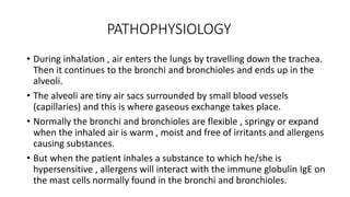
ASTHMA
CHIPO JAMES MAINDA
Nurses Zone(documentary)
DEFINITION
• Asthma is a chronic lower respiratory condition affecting the bronchi
and bronchioles in which there is inflammation of the airways
resulting in hyperresponsiveness (bronchospasms) , mucosal edema
and excessive mucus production mainly caused by allergies and
characterized by cough , dyspnea and wheezing.
PREDISPOSING FACTORS
• Family history of asthma
• Viral respiratory infection
• Allergens such as pollen , perfume , dust , smoke
• Emotional upsets
• Drugs such as NSAIDs like aspirin
• Air pollution
• Cold air
• Vigorous exercises
TYPES OF ASTHMA
INTRINSIC/CRYPTOGENIC ASTHMA
• Attacks tend to increase in severity causing irreversible lung damage.
• No history of asthma in the family
• May occur secondary to other respiratory tract infections
• No causative agent can be identified
• Often starts in middle age
• This type of asthma is not triggered by allergens
• Usually there is no history of childhood allergies.
TYPES OF ASTHMA
EXTRINSIC/ATOPIC ASTHMA
• It occurs in childhood and young adults
• There is usually family history of asthma
• Attacks tend to be less severe
• It is triggered by allergens like dust , feathers , perfume
• A definitive external cause is identified
PATHOPHYSIOLOGY
• During inhalation , air enters the lungs by travelling down the trachea.
Then it continues to the bronchi and bronchioles and ends up in the
alveoli.
• The alveoli are tiny air sacs surrounded by small blood vessels
(capillaries) and this is where gaseous exchange takes place.
• Normally the bronchi and bronchioles are flexible , springy or expand
when the inhaled air is warm , moist and free of irritants and allergens
causing substances.
• But when the patient inhales a substance to which he/she is
hypersensitive , allergens will interact with the immune globulin IgE on
the mast cells normally found in the bronchi and bronchioles.
PATHOPHYSIOLOGY
• This causes the mast cells to rupture releasing chemicals such as
prostaglandins , histamine , bradykinin.
• These mediators causes inflammation lead to bronchoconstriction ,
edema and hyper secretion of mucus inside the bronchi.
• Mucosal thickening , swelling and production of mucus interferes with
airflow hence the patient will present with dyspnea , wheezing.
• With increasing severity and chronicity of disease, permanent
structural changes can occur in the airway these are associated with a
progressive loss of lung function that is not prevented by or fully
reversible by current therapy (status asthmaticus)
SIGNS AND SYMPTOMS
• Cough due to clogged mucus in the bronchi and bronchioles
• Dyspnea due to constriction of the airway
• Wheezing due to narrowed airway by mucus
• Cyanosis due to obstruction of the airway leading to low oxygen
perfusion
• Sweating due to labored breathing
• Nasal flaring due to hyperventilation
• Headache due to cerebral hypoxia
• Chest tightness due to bronchial constriction
INVESTIGATIONS
• History taking , will reveal exposure to allergens
• Physical examination , will show dyspnea
• Pulse oximetry , will show low oxygen saturation
• Skin test , will reveal the specific allegens
• Pulmonary function test , will show airway obstruction
• Chest X-ray , will show complications of asthma like pneumothorax
TREATMENT
• Drugs used include rapid acting bronchodilators such as ;
• Salbutamol - this is the fastest and most effective, hence remain the
drug of choice. It is given through a nebulizer or orally 2gm TDS.
• Action – selectively stimulate beta receptors producing
bronchodilatation.
• Side effects – tachycardia, Bp changes, nervousness, palpitation, muscle
tremors, nausea, vomiting, insomnia, dry mouth, headache.
• Aminophylline 5mg/kg given intravenously.
• Action – relaxation of bronchial smooth muscle and improve contractility
of fatigued diaphragm.
TREATMENT
• Side effects – tachycardia, Bp changes, arrhythmias, anorexia, nausea and
vomiting, nervousness, irritability, headache, muscle twitching.
• Prednisolone 40-50mg given orally.
• Action- it has anti-inflammatory and immunosuppressive effects.
Decrease edema in bronchial airway thus decreasing mucus secretion.
• Side effects- skin changes, osteoporosis, increased appetite, obesity,
immunosupression, catabolism, muscle weakness.
• Oxygen therapy
• Antibiotics
• Intravenous fluids
NURSING CARE
AIMS
• To maintain a clear airway
• Restore normal or best possible lung function
• Reduce the risk of severe attacks
• To give psychological care
• This is a medical emergency condition!
RESUSCITATION
AIRWAY
• Quickly I will assess the nose for any blockages and remove them to
maintain a clear airway . I will feel for the respiration by putting my palm
on the nostrils of the patient to ensure a clear airway . I will assess for
any blockages in the mouth and remove them to maintain a clear airway
. I will remove the dentures if any because they can also obstruct the
airway . I will suction for any secretions in the mouth to maintain a
patent airway . I will put the head of the patient in lateral position in
order to promote free flow of secretions and prevent the tongue from
obstructing the airway . I will encourage to cough so as to remove the
clogged mucus in the airway.
RESUSCITATION
BREATHING
• I will quickly assess for the breathing pattern to obtain the baseline
data . Then I will position the patient in semi fowlers to promote lung
expansion . I will loosen any tighten clothing on the chest to promote
lung expansion . I will do cardiopulmonary resuscitation (CPR) to
promote cardiopulmonary function . I will give oxygen therapy 4-6ml/l
via a mask to promote oxygen perfusion . I will intubate and put the
patient on mechanical ventilation to promote good tissue perfusion.
RESUSCITATION
CIRCULATION
• I will quickly insert a large bore cannula for drug administration . I will
put a pulse oximeter on the thumb of the patient to monitor the
oxygen saturation . I will monitor for the blood pressure and if low I’ll
commence normal saline to stabilize it . I will insert a urinary catheter
for monitoring fluid intake and fluid output . I will collect blood for Hb
and arterial blood gas analysis.
MEDICATION
• I will give short acting bronchodilators like salbutamol to enlarge the
airway . I will administer corticosteroids such as prednisolone to
reduce the inflammatory process . I will give oxygen therapy via nasal
prongs 4-5l/m to improve tissue perfusion . I will give intravenous
fluids to maintain hydration . I will monitor for the side effects of
medication.
ENVIRONMENT
• Environment should be safe for the patient.
• Nurse patient in a quiet, clean environment near to nurse’s bay for
close observations. The room should be well ventilated and free
from dust. It should contain all resuscitative equipment such as
oxygen cylinder; suctioning machine etc
POSITION
• Place the patient in the semi fowler’s position for maximum lung
expansion and encourage diaphragmatic breathing to allow
enough air intakes
PSYCHOLOGICAL CARE
• Create a therapeutic relationship with the patient so that the patient
can have confidence in you.
• Reassure the patient and family during an asthmatic attack to allay
anxiety. Provide comfort by being with the patient.
• Explain the disease process, the cause of the wheezing and labored
respiration to the mother to allay anxiety.
• Allow the patient to verbalize his fears to allay anxiety if the child is old
enough.
• Explain every procedure and machines that are being used to the
patient to promote cooperation.
I.E.C
• Teach the patient how to use an oral inhaler and caution him about
the possible adverse reactions associated with the medications he is
receiving.
• Show patient how to breathe deeply. Instruct him how to cough
secretions accumulated overnight.
• Teach the patient and the family to avoid known allergens and
irritants such as smocking, dust perfumes, fur and cold weather etc.
• Emphasize the importance of taking only prescribed drugs as certain
drugs such as aspirin may precipitate an asthmatic attack.
I.E.C
• Encourage the patient to have well balanced diet to prevent
respiratory infection and fatigue.
• Explain the importance of review dates so that the patient can be
monitored.
• Teach the mother signs and symptoms of an impending asthmatic
attack and encourage them to seek medical attention as soon as
possible
COMPLICATIONS
• Status asthmaticus- This is a severe asthmatic attack which cannot be
controlled with usual medications . This arises when impaired gas exchange
and heightened airway resistance increase the work of breathing.
• Respiratory failure- This is the impairment of the lung’s ability to maintain
balance between oxygen and carbon dioxide.
• Cardiac arrest- Occurs secondary to respiratory failure
• Emphysema – irreversible accumulation of air in the alveolar spaces due to
repeated asthmatic attacks which results in decrease in total breathing
capacity.
• Atelectasis- lung collapse due to accumulation of air in the alveoli.
THE END!
TheNursesZone(documentary)

Asthma_NZD.pptx

  • 1.
  • 2.
    DEFINITION • Asthma isa chronic lower respiratory condition affecting the bronchi and bronchioles in which there is inflammation of the airways resulting in hyperresponsiveness (bronchospasms) , mucosal edema and excessive mucus production mainly caused by allergies and characterized by cough , dyspnea and wheezing.
  • 3.
    PREDISPOSING FACTORS • Familyhistory of asthma • Viral respiratory infection • Allergens such as pollen , perfume , dust , smoke • Emotional upsets • Drugs such as NSAIDs like aspirin • Air pollution • Cold air • Vigorous exercises
  • 4.
    TYPES OF ASTHMA INTRINSIC/CRYPTOGENICASTHMA • Attacks tend to increase in severity causing irreversible lung damage. • No history of asthma in the family • May occur secondary to other respiratory tract infections • No causative agent can be identified • Often starts in middle age • This type of asthma is not triggered by allergens • Usually there is no history of childhood allergies.
  • 5.
    TYPES OF ASTHMA EXTRINSIC/ATOPICASTHMA • It occurs in childhood and young adults • There is usually family history of asthma • Attacks tend to be less severe • It is triggered by allergens like dust , feathers , perfume • A definitive external cause is identified
  • 6.
    PATHOPHYSIOLOGY • During inhalation, air enters the lungs by travelling down the trachea. Then it continues to the bronchi and bronchioles and ends up in the alveoli. • The alveoli are tiny air sacs surrounded by small blood vessels (capillaries) and this is where gaseous exchange takes place. • Normally the bronchi and bronchioles are flexible , springy or expand when the inhaled air is warm , moist and free of irritants and allergens causing substances. • But when the patient inhales a substance to which he/she is hypersensitive , allergens will interact with the immune globulin IgE on the mast cells normally found in the bronchi and bronchioles.
  • 7.
    PATHOPHYSIOLOGY • This causesthe mast cells to rupture releasing chemicals such as prostaglandins , histamine , bradykinin. • These mediators causes inflammation lead to bronchoconstriction , edema and hyper secretion of mucus inside the bronchi. • Mucosal thickening , swelling and production of mucus interferes with airflow hence the patient will present with dyspnea , wheezing. • With increasing severity and chronicity of disease, permanent structural changes can occur in the airway these are associated with a progressive loss of lung function that is not prevented by or fully reversible by current therapy (status asthmaticus)
  • 8.
    SIGNS AND SYMPTOMS •Cough due to clogged mucus in the bronchi and bronchioles • Dyspnea due to constriction of the airway • Wheezing due to narrowed airway by mucus • Cyanosis due to obstruction of the airway leading to low oxygen perfusion • Sweating due to labored breathing • Nasal flaring due to hyperventilation • Headache due to cerebral hypoxia • Chest tightness due to bronchial constriction
  • 9.
    INVESTIGATIONS • History taking, will reveal exposure to allergens • Physical examination , will show dyspnea • Pulse oximetry , will show low oxygen saturation • Skin test , will reveal the specific allegens • Pulmonary function test , will show airway obstruction • Chest X-ray , will show complications of asthma like pneumothorax
  • 10.
    TREATMENT • Drugs usedinclude rapid acting bronchodilators such as ; • Salbutamol - this is the fastest and most effective, hence remain the drug of choice. It is given through a nebulizer or orally 2gm TDS. • Action – selectively stimulate beta receptors producing bronchodilatation. • Side effects – tachycardia, Bp changes, nervousness, palpitation, muscle tremors, nausea, vomiting, insomnia, dry mouth, headache. • Aminophylline 5mg/kg given intravenously. • Action – relaxation of bronchial smooth muscle and improve contractility of fatigued diaphragm.
  • 11.
    TREATMENT • Side effects– tachycardia, Bp changes, arrhythmias, anorexia, nausea and vomiting, nervousness, irritability, headache, muscle twitching. • Prednisolone 40-50mg given orally. • Action- it has anti-inflammatory and immunosuppressive effects. Decrease edema in bronchial airway thus decreasing mucus secretion. • Side effects- skin changes, osteoporosis, increased appetite, obesity, immunosupression, catabolism, muscle weakness. • Oxygen therapy • Antibiotics • Intravenous fluids
  • 12.
    NURSING CARE AIMS • Tomaintain a clear airway • Restore normal or best possible lung function • Reduce the risk of severe attacks • To give psychological care • This is a medical emergency condition!
  • 13.
    RESUSCITATION AIRWAY • Quickly Iwill assess the nose for any blockages and remove them to maintain a clear airway . I will feel for the respiration by putting my palm on the nostrils of the patient to ensure a clear airway . I will assess for any blockages in the mouth and remove them to maintain a clear airway . I will remove the dentures if any because they can also obstruct the airway . I will suction for any secretions in the mouth to maintain a patent airway . I will put the head of the patient in lateral position in order to promote free flow of secretions and prevent the tongue from obstructing the airway . I will encourage to cough so as to remove the clogged mucus in the airway.
  • 14.
    RESUSCITATION BREATHING • I willquickly assess for the breathing pattern to obtain the baseline data . Then I will position the patient in semi fowlers to promote lung expansion . I will loosen any tighten clothing on the chest to promote lung expansion . I will do cardiopulmonary resuscitation (CPR) to promote cardiopulmonary function . I will give oxygen therapy 4-6ml/l via a mask to promote oxygen perfusion . I will intubate and put the patient on mechanical ventilation to promote good tissue perfusion.
  • 15.
    RESUSCITATION CIRCULATION • I willquickly insert a large bore cannula for drug administration . I will put a pulse oximeter on the thumb of the patient to monitor the oxygen saturation . I will monitor for the blood pressure and if low I’ll commence normal saline to stabilize it . I will insert a urinary catheter for monitoring fluid intake and fluid output . I will collect blood for Hb and arterial blood gas analysis.
  • 16.
    MEDICATION • I willgive short acting bronchodilators like salbutamol to enlarge the airway . I will administer corticosteroids such as prednisolone to reduce the inflammatory process . I will give oxygen therapy via nasal prongs 4-5l/m to improve tissue perfusion . I will give intravenous fluids to maintain hydration . I will monitor for the side effects of medication.
  • 17.
    ENVIRONMENT • Environment shouldbe safe for the patient. • Nurse patient in a quiet, clean environment near to nurse’s bay for close observations. The room should be well ventilated and free from dust. It should contain all resuscitative equipment such as oxygen cylinder; suctioning machine etc POSITION • Place the patient in the semi fowler’s position for maximum lung expansion and encourage diaphragmatic breathing to allow enough air intakes
  • 18.
    PSYCHOLOGICAL CARE • Createa therapeutic relationship with the patient so that the patient can have confidence in you. • Reassure the patient and family during an asthmatic attack to allay anxiety. Provide comfort by being with the patient. • Explain the disease process, the cause of the wheezing and labored respiration to the mother to allay anxiety. • Allow the patient to verbalize his fears to allay anxiety if the child is old enough. • Explain every procedure and machines that are being used to the patient to promote cooperation.
  • 19.
    I.E.C • Teach thepatient how to use an oral inhaler and caution him about the possible adverse reactions associated with the medications he is receiving. • Show patient how to breathe deeply. Instruct him how to cough secretions accumulated overnight. • Teach the patient and the family to avoid known allergens and irritants such as smocking, dust perfumes, fur and cold weather etc. • Emphasize the importance of taking only prescribed drugs as certain drugs such as aspirin may precipitate an asthmatic attack.
  • 20.
    I.E.C • Encourage thepatient to have well balanced diet to prevent respiratory infection and fatigue. • Explain the importance of review dates so that the patient can be monitored. • Teach the mother signs and symptoms of an impending asthmatic attack and encourage them to seek medical attention as soon as possible
  • 21.
    COMPLICATIONS • Status asthmaticus-This is a severe asthmatic attack which cannot be controlled with usual medications . This arises when impaired gas exchange and heightened airway resistance increase the work of breathing. • Respiratory failure- This is the impairment of the lung’s ability to maintain balance between oxygen and carbon dioxide. • Cardiac arrest- Occurs secondary to respiratory failure • Emphysema – irreversible accumulation of air in the alveolar spaces due to repeated asthmatic attacks which results in decrease in total breathing capacity. • Atelectasis- lung collapse due to accumulation of air in the alveoli.
  • 22.