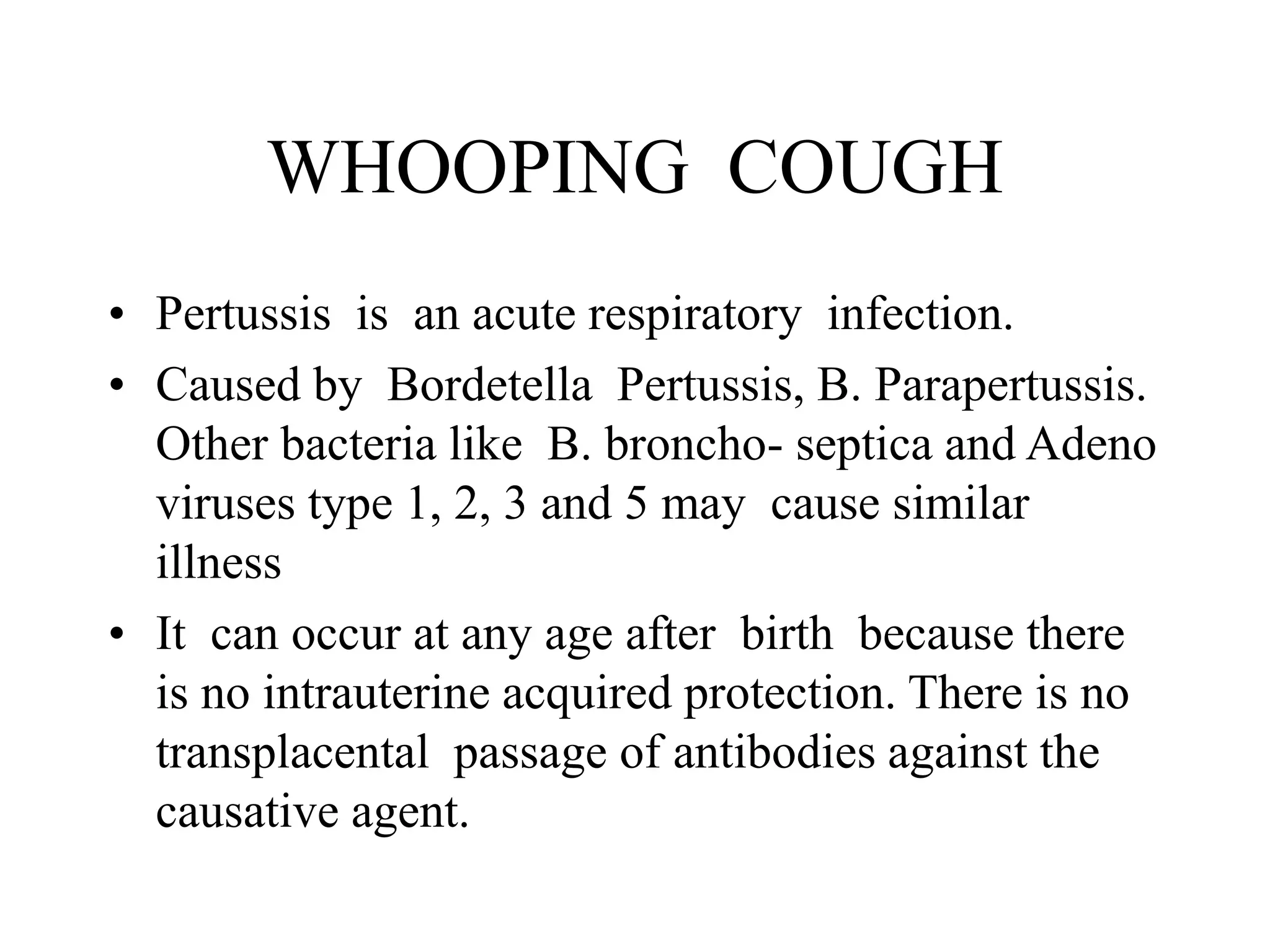
WHOOPING COUGH
• Pertussis is an acute respiratory infection.
• Caused by Bordetella Pertussis, B. Parapertussis.
Other bacteria like B. broncho- septica and Adeno
viruses type 1, 2, 3 and 5 may cause similar
illness
• It can occur at any age after birth because there
is no intrauterine acquired protection. There is no
transplacental passage of antibodies against the
causative agent.
• Incubation period is 7-14 days.
• Many patients are under 6 years old.
• Overcrowding in communities in the
tropics exposes young infants early to a
high infection pressure.
• This is more so in poorly ventilated homes
since transmission is by droplet infection
• Highest infectivity is in the early stage of
the disease. Attack rate is 80– 100 percent.
• Severity of infection decreases with age.
• Highest mortality is among infants worse
in females.
PATHOGENESIS
• Following inhalation of infected droplets.
• Bacteria attach to ciliated epithelial cells of
the respiratory tract and multiply on the
lining.
• Multiplication occurs from nasopharynx to
the alveoli with severity in the bronchioles.
• This leads to inflammation of the mucosal
lining.
• Accompanied by much mucus, congestion and
infiltration of mucosal lining with lymphocytes.
• This narrows the lumen of the bronchioles
resulting in atelectasis and emphysema in areas of
incomplete obstruction.
• Inflammation in the bronchioles weaken their
walls leading to bronchiectasis at later date.
• Organism secret an endotoxin at site of their
multiplication thought to cause the paroxysmal
cough.
CLINICAL PRESENTATION
• Divided into 3 stages:
• Catarrhal, paroxysmal and convalescent
each lasting approximately 2 weeks.
• Catarrhal stage last about 2- 3 weeks.
Has an insidious onset, with non specific
features like rhinorrhea, low grade fever,
sneezing, lacrimation and conjunctival
suffusion.
• PAROXYSMAL STAGE
Last 2 – 4 weeks.
Characterised by a repetitive series of forceful
coughs in a single expiration.
cough is associated with choking and vomiting.
Production of sticky stringy sputum.
There is a thin nasal discharge.
Whoop occurs at this stage.
• Whoop is a massive inspiratory effort as air is
inhaled forcefully through the narrowed bronchial
tree.
• Does not occur up to age of 2 years.
• Instead replaced by period of apnoea. Prolonged
may cause sudden infant death.
• Whoop usually followed by vomiting and profuse
sweating.
• Convulsions may occur due to hypoxia and
intracerebral haemorrhage.
• Poor feeding and vomiting cause loss of weight,
dehydration and eventually malnutrition.
• Intense cough and resulting increased abdominal
pressure may result in umbilical hernia, inguinal
hernia and rectal prolapse.
• Increased venous pressure lead to oedema of the
orbits and subconjuctival haemorrhage.
• Ulceration of the frenulum of the tongue and
epistaxis also occur.
•
• Older children complain of headache and chest
pain due to intensity of coughing.
• CONVALESCENT
• Both severity and frequency of paroxyms
decrease.
• But cough and whoop may persist for several
months.
• Appetite returns, vomiting ceases and the child
gains weight.
COMPLICATIONS
• Bronchopneumonia is the commonest
• Pulmonary collapse and bronchial wall
destruction lead to bronchiectasis which maybe
permanent.
• Malnutrition due to prolonged paroxysmal cough
which interferes with food intake.
• Develops early in children whose diets are
marginal at the start of illness.
• Convulsions and hemiplegia
•
• Surgical emphysema
• Reactivation of quiescent primary TB
• INVESTIGATIONS
• Diagnosis confirmed by positive cultures of
Bordetella pertussis on Bordet-Gengou
medium cough plate
• Fluorescent antibodies in stained
postnasopharyngeal specimen.
TREATMENT
Antibiotics started early may shorten course
of the disease and reduce the period of
communicability.
Antibiotics started late do not alter the course
of the disease.
Erythromycin 20- 40mg /kg per day.
Ampicillin 100/kg per day
Chloraphenical 50- 100 mg / kg per day
• Supportive treatment reduces morbidity and
mortality e.g skilled nursing care and feeding.
• Monitor apnoeic spells which need immediate
clearance of the airway and manual respiration.
• Mild sedation with phenergan or phenobarbitone
rests the child and reduces paroxysmal cough.
• This allows feeding.
• Gentle sunctioning of mucus and the stringy
viscid saliva removed by cotton wool.
•
• Oxygen
• Intravenous fluids if vomiting severe and
there is dehydration.
• PREVENTION
• Vaccination. DPT
• Adverse reactions may occur such as fever,
encephalitis and convulsions.
• Once this happens no further vaccinations
with this vaccine as it may lead to
permanent neurological disorder.
•
• Thank you

WHOOPING COUGH.ppt

  • 1.
    WHOOPING COUGH • Pertussisis an acute respiratory infection. • Caused by Bordetella Pertussis, B. Parapertussis. Other bacteria like B. broncho- septica and Adeno viruses type 1, 2, 3 and 5 may cause similar illness • It can occur at any age after birth because there is no intrauterine acquired protection. There is no transplacental passage of antibodies against the causative agent.
  • 2.
    • Incubation periodis 7-14 days. • Many patients are under 6 years old. • Overcrowding in communities in the tropics exposes young infants early to a high infection pressure. • This is more so in poorly ventilated homes since transmission is by droplet infection
  • 3.
    • Highest infectivityis in the early stage of the disease. Attack rate is 80– 100 percent. • Severity of infection decreases with age. • Highest mortality is among infants worse in females.
  • 4.
    PATHOGENESIS • Following inhalationof infected droplets. • Bacteria attach to ciliated epithelial cells of the respiratory tract and multiply on the lining. • Multiplication occurs from nasopharynx to the alveoli with severity in the bronchioles. • This leads to inflammation of the mucosal lining.
  • 5.
    • Accompanied bymuch mucus, congestion and infiltration of mucosal lining with lymphocytes. • This narrows the lumen of the bronchioles resulting in atelectasis and emphysema in areas of incomplete obstruction. • Inflammation in the bronchioles weaken their walls leading to bronchiectasis at later date. • Organism secret an endotoxin at site of their multiplication thought to cause the paroxysmal cough.
  • 6.
    CLINICAL PRESENTATION • Dividedinto 3 stages: • Catarrhal, paroxysmal and convalescent each lasting approximately 2 weeks. • Catarrhal stage last about 2- 3 weeks. Has an insidious onset, with non specific features like rhinorrhea, low grade fever, sneezing, lacrimation and conjunctival suffusion.
  • 7.
    • PAROXYSMAL STAGE Last2 – 4 weeks. Characterised by a repetitive series of forceful coughs in a single expiration. cough is associated with choking and vomiting. Production of sticky stringy sputum. There is a thin nasal discharge. Whoop occurs at this stage.
  • 8.
    • Whoop isa massive inspiratory effort as air is inhaled forcefully through the narrowed bronchial tree. • Does not occur up to age of 2 years. • Instead replaced by period of apnoea. Prolonged may cause sudden infant death. • Whoop usually followed by vomiting and profuse sweating. • Convulsions may occur due to hypoxia and intracerebral haemorrhage.
  • 9.
    • Poor feedingand vomiting cause loss of weight, dehydration and eventually malnutrition. • Intense cough and resulting increased abdominal pressure may result in umbilical hernia, inguinal hernia and rectal prolapse. • Increased venous pressure lead to oedema of the orbits and subconjuctival haemorrhage. • Ulceration of the frenulum of the tongue and epistaxis also occur. •
  • 10.
    • Older childrencomplain of headache and chest pain due to intensity of coughing. • CONVALESCENT • Both severity and frequency of paroxyms decrease. • But cough and whoop may persist for several months. • Appetite returns, vomiting ceases and the child gains weight.
  • 11.
    COMPLICATIONS • Bronchopneumonia isthe commonest • Pulmonary collapse and bronchial wall destruction lead to bronchiectasis which maybe permanent. • Malnutrition due to prolonged paroxysmal cough which interferes with food intake. • Develops early in children whose diets are marginal at the start of illness. • Convulsions and hemiplegia •
  • 12.
    • Surgical emphysema •Reactivation of quiescent primary TB • INVESTIGATIONS • Diagnosis confirmed by positive cultures of Bordetella pertussis on Bordet-Gengou medium cough plate
  • 13.
    • Fluorescent antibodiesin stained postnasopharyngeal specimen.
  • 14.
    TREATMENT Antibiotics started earlymay shorten course of the disease and reduce the period of communicability. Antibiotics started late do not alter the course of the disease. Erythromycin 20- 40mg /kg per day. Ampicillin 100/kg per day Chloraphenical 50- 100 mg / kg per day
  • 16.
    • Supportive treatmentreduces morbidity and mortality e.g skilled nursing care and feeding. • Monitor apnoeic spells which need immediate clearance of the airway and manual respiration. • Mild sedation with phenergan or phenobarbitone rests the child and reduces paroxysmal cough. • This allows feeding. • Gentle sunctioning of mucus and the stringy viscid saliva removed by cotton wool. •
  • 17.
    • Oxygen • Intravenousfluids if vomiting severe and there is dehydration. • PREVENTION • Vaccination. DPT • Adverse reactions may occur such as fever, encephalitis and convulsions.
  • 18.
    • Once thishappens no further vaccinations with this vaccine as it may lead to permanent neurological disorder. •
  • 19.