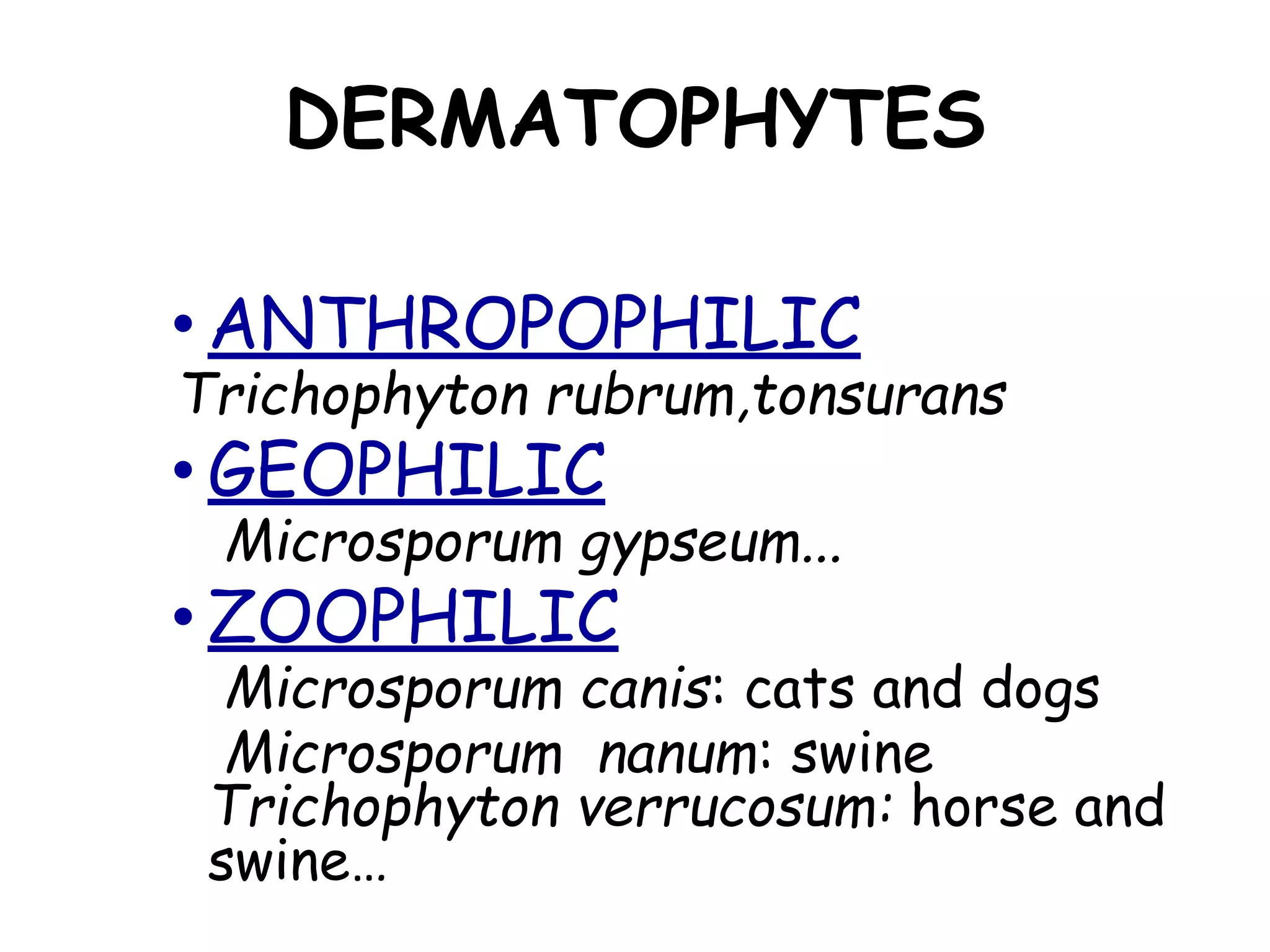
SUPERFICIAL
MYCOSES
SUPERFICIAL MYCOSES
●Dermatophytosis
●Pityriasis versicolor
●Keratomycosis
●Tinea nigra
●Black piedra
●White piedra
●candidiasis
DERMATOPHYTOSIS
(=Tinea = Ringworm)
• Infection of the skin, hair or nails c
keratinophilic fungi, called
dermatophytes
¨ Microsporum Hair, skin
¨ Epidermophyton Skin, nail
¨ Trichophyton Hair, skin, nail
DERMATOPHYTES
• ANTHROPOPHILIC
Trichophyton rubrum,tonsurans
• GEOPHILIC
Microsporum gypseum...
• ZOOPHILIC
Microsporum canis: cats and dogs
Microsporum nanum: swine
Trichophyton verrucosum: horse and
swine…
Factors:
• Contact and trauma
• Moisture
• Crowded living conditions
• Cellular immunodeficiency →
(chronic inf.)
DERMATOPHYTOSIS
Clinical Classification
• Infection is named according to
the anatomic location involved:
a. Tinea barbae e. Tinea pedis
(Athlete’s foot)
b. Tinea corporis f. Tinea manuum
c. Tinea capitis g. Tinea unguium
d. Tinea cruris
(Jock itch)
Clinical features –Tinea capitis
• Cardinal features of partial hair loss
with some degree of inflammation
It ranges from:
1. Non inflammatory patches of alopecia
2. Alopecia with black dots
3. Favus type-yellow cup shaped crust
‘SCUTULA’ which later leads to patchy
hair loss with cicatricial alopecia
andatrophy
• 4.Kerion: inflammation and alopecia
resembling bacterial furunculosis
Painful boggy mass discharging pus with
crusting and matting of adjacent hair
Lymphadenopathy is (+)nt
T.verrucosum
T.mentagrophytes var mentagrophytes
5. Seborrhic dermatitis like
Tinea Barbae
• Ringworms of beard & moustache area
with invasion of coarse hair
Tinea corporis/ T.Circinata
• Ringworm of glabourous skin
• Etiology
- M/C cause is T.rubrum
- T.mentagrophytes,M.canis, T.tonsurans
are also common
Clinical features-T.Corporis
• Annular lesion,sharply marginated with
raised edge & scales across the entire
erythematous border
Tinea cruris
- Warm humid condition & sharing of
towels & sport clothing are imp. factors
Clinical features-T.Cruris
• Itching is predominant
• Erythematous plaques curved with sharp
margins with scaling from groin down
the thigh
Tinea pedis
- Occlusive footwear,communal baths,
showers or swimming pools increase
chance of infection.
Clinical features-T.Pedis
1. Interdigital
2. Squamous hyperkeratotic variety
3. Vesicobullous type
Tinea manuum
• Ringworm of palmer skin
Clinical features
• u/l diffuse hyperkeratosis of palm &
finger with accentuation of flexural
creases
TINEA UNGUIUM
• Invasion of nail plate by dermatophytes
Clinical features
• 1.DLSO-
- strek/patch of yellow/white
discolouration at the free edge
• 2. SWO-
• 3. PROXIMAL SUBUNGUAL
ONYCHOMYCOSIS
• 4. Endonyx type
Tinea incognito
- Steroid modified tinea
Dermatophytide reactions
• Non infective cutaneus eruption
• Essential criteria for diagnosis
1 Proven dermatophytic infection
2 Distant eruption free of ringworm
3 Spontaneous disappearance when
ringworm infection settles
4 Morphology should match one of
recognise type
DERMATOPHYTOSIS
Diagnosis
I. Clinical
Appearance
Wood lamp (UV, 365 nm)
II. Lab
A. Collection of sample
B. Direct microscopic examination
(10-30% KOH)
Hair should be examined immediately
• Skin scrapings-warm gently
DERMATOPHYTOSIS
Diagnosis
C. Culture
Sabouraud dextrose agar
chloramphenicol(.005%),
cycloheximide(.04%) are added
Moulds-26-28°c for 3 to 4 wks
Candida-37°c for 1 wk
3-4% malt extract agar
Treatment of ringworm
• antifungals
Pityrosporum infection of skin
● PITYRIASIS VERSICOLOR
● Etio: Malassezia furfur (Pityrosporum
orbiculare) (Lipophilic yeast)
● Clinical findings: Hyperpigmented or
depigmented maculae on chest, back,
arms, abdomen
● Fine branny scales are characterstic
• P.folliculitis
• Site-back,chest
• Perifollicular,erythematous 2-3mm
papule or pustules
• Pruritus present
• DM,steroid,antibiotic use predisposes
• D.D. bacterial folliculitis,acne
Diagnosis
• micro: Short hyphae, yeast cells
Sphagetti and meat ballappaearance
• Culture: Yeast (suppl.: olive oil)
• Treatment antifungals
TINEA NIGRA
• Superficial chronic infection of Stratum
corneum
• Etio: Hortae werneckii (pigmented)
• Clinical findings: asymptomatic nonscaly
sharply defined Brownish maculae on
palms, fingers, face
BLACK PIEDRA
• Fungal infection of the scalp hair
• Etio: Piedraia hortae
• Frequent in tropical areas
• Clinical findings: Discrete,
hard,firmly adherent, dark brown to
black nodules on the hair
WHITE PIEDRA
• Fungal infection of facial, axillary
or genital hair
• Etio: Trichosporon beigelli
,T.ovoides,T.inkin(pubic)
• Frequent in tropical and
temperate zones
CANDIDIASIS
• CANDIDA ALBICANS
Oral cavity- whitish membrane that can be scraped
off
pseudomembrane
Angular cheilitis
Atrophic
hyperplastic
Flexures- intertrigo
Nails- paronychia
• Microscopy- budding yeast cellsin 10% koh
• Culture
• Treatment- fluconazole
• Glabrata and krussie resistant to antifungals

Superficial mycoses