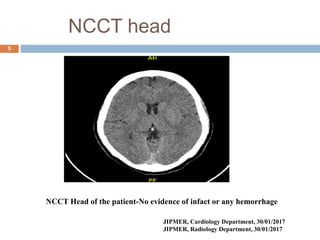
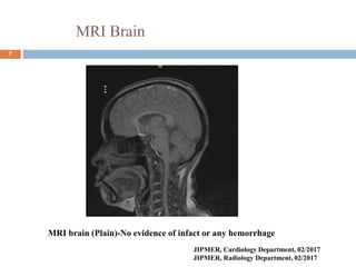
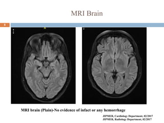
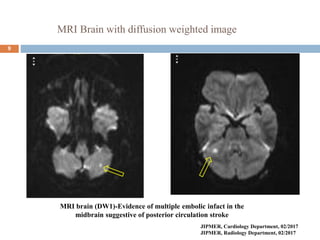
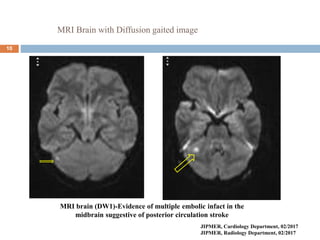
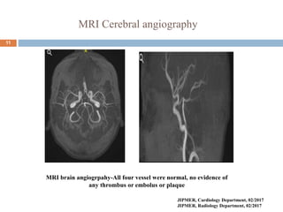
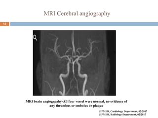
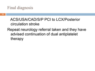
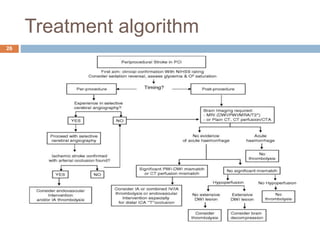
In this ppt i am going to describe about one patient who develop acute stroke after PCI in our hospital. Also i am going to discuss how to diagnose, manage and treat such patient, risk factor associated with stroke after PCI.
Primary PCI involves performing urgent angioplasty and potentially stenting of the culprit artery in STEMI patients, with the goal of reopening the blocked vessel within 90 minutes of first medical contact. It is the preferred reperfusion strategy when it can be performed promptly by an experienced team. Factors such as patient age, time to treatment, comorbidities, and initial flow in the artery help determine whether primary PCI or thrombolysis is most appropriate. Optimal anticoagulation and antiplatelet regimens along with adjunctive therapies like manual thrombectomy can improve outcomes of primary PCI.
No reflow and slow flow phenomenon during pcirahul arora
This document discusses strategies and prevention of slow flow and no-reflow phenomenon during percutaneous coronary intervention (PCI). It defines no-reflow as inadequate myocardial perfusion through a coronary artery without mechanical obstruction. No-reflow occurs in 8-11% of primary PCIs and is associated with worse clinical outcomes. The pathophysiology involves distal embolization, ischemic injury, reperfusion injury, and individual patient susceptibility. Preventing no-reflow requires reducing thrombus burden, ischemia time, reperfusion injury through anti-inflammatory drugs, and addressing risk factors like diabetes.
Cardiac arrhythmias occur frequently in ICU patients, with the most common being sinus tachycardia. Arrhythmias are often seen in patients with structural heart disease and can be exacerbated by critical illness. Management involves treating any imbalances that may be triggering the arrhythmia as well as directed medical therapy. Arrhythmias in the ICU represent a major source of morbidity and increased length of stay.
A 52-year-old male presented with chest pain. His ECG showed evolving inferior wall myocardial infarction. ST depression is more frequently seen in lead aVL than other leads for inferior MI. A 51-year-old female presented with prior chest pain and is now pain-free. Her ECG shows Wellens' syndrome type I pattern and she should be monitored closely in the ICCU. Fragmented QRS complexes can indicate ischemia or scar tissue and are associated with worse cardiac outcomes.
No-reflow occurs when there is a lack of reperfusion to the myocardium after successful coronary recanalization and is defined as inadequate perfusion without angiographic evidence of vessel obstruction. It occurs in 0.6-3.2% of PCI cases and is associated with increased risk of LV dysfunction, remodeling, arrhythmias, heart failure and cardiac rupture. Diagnosis is typically done using myocardial contrast echocardiography or cardiac MRI. Treatment focuses on improving perfusion and includes vasodilators like adenosine, antithrombotics, mechanical strategies like thrombectomy, and preventing no-reflow through measures like pre-conditioning.
Dr. Abraham discusses sinus node dysfunction and atrioventricular block. Key points include:
- The sinus node is usually located in the right atrium and receives blood supply from the right coronary artery or left circumflex artery.
- Symptoms of sinus node dysfunction range from asymptomatic ECG changes to tachycardia, bradycardia, and tachy-brady syndrome.
- Treatment options include pharmacotherapy with drugs like atropine or theophylline for short term use, and pacemaker implantation for long term treatment of sinus node disease.
- The atrioventricular node receives innervation from the arteries of Koch and shows minimal autonomic innervation. AV block can be first,
This document discusses localization of accessory pathways using electrocardiography. It describes that accessory pathways can be located in eight anatomical positions along the tricuspid and mitral valve annuli. Several algorithms are proposed to determine the location based on delta wave polarity and amplitude in various leads. The most accurate is the Arruda approach, which uses step-wise analysis of delta wave characteristics in leads I, II, aVL, aVF and V1 to identify the specific accessory pathway location with 90% sensitivity and 99% specificity. Characteristic ECG patterns are presented that help localize right anteroseptal, right posteroseptal, left lateral and right free wall accessory pathways.
This document contains 16 medical cases involving electrocardiogram (ECG) readings and interpretations. Each case provides an ECG image and description of a patient's symptoms or medical history, and asks the reader to identify the diagnosis or next step. The document also includes explanations of various ECG patterns and conditions, such as R on T phenomenon, hyperkalemia, Brugada syndrome, lead reversals, and long QT interval with T-wave alternans. The goal is to teach readers how to properly analyze ECGs and apply that analysis to diagnosing cardiac conditions.
The document provides an overview of electrocardiography (ECG), including its history, importance, physiology, leads, waves, intervals, and abnormalities. Key points covered include the names and functions of the P, QRS, and T waves, as well as common abnormalities like ST segment elevation/depression, T wave inversion, and arrhythmias. The summary analyzes ECGs to recognize conditions like myocardial infarction and ventricular hypertrophy.
Percutaneous Revascularization for Ischemic Left Ventricular Dysfunction REV...hospital
This study compared percutaneous coronary intervention (PCI) plus optimal medical therapy to optimal medical therapy alone in patients with severe left ventricular systolic dysfunction. Patients were randomly assigned to receive either PCI plus medical therapy or medical therapy alone. The primary outcome was death from any cause or hospitalization for heart failure. At 12 months, PCI did not result in a lower rate of the primary outcome compared to medical therapy alone. Extensive medical therapy was optimized for heart failure in both groups.
Stress echocardiography combines echocardiography with physical, pharmacological, or electrical stress to effectively evaluate for myocardial ischemia. It is used to screen for coronary artery disease and identify affected coronary territories. Stress echocardiography can also differentiate viable myocardium from scarred tissue and provides important prognostic information after myocardial infarction and before noncardiac surgery. Dobutamine stress echocardiography is widely used to assess viable myocardium while exercise stress echocardiography is preferred when possible due to its safety. Stress echocardiography techniques are safe and relatively inexpensive options for evaluating myocardial ischemia and viability.
This document describes equipment, catheters, and basic intervals used in electrophysiology (EP) studies. It discusses radiographic tables, EP equipment like cardiac stimulators and mapping/ablation catheters. Patient preparation includes fasting, IV access, monitoring equipment. EP catheters come in different sizes and have electrodes for recording electrical activity. Basic intervals measured include P wave to atrial interval, atrial-His bundle interval, His-ventricular interval, and sinus node recovery time. Drive train stimulation with single, double, or triple extra stimuli is used. The document continues with further discussions of EP protocols, arrhythmias, ablation, and pre-excitation pathways.
Spontaneous coronary artery dissection (SCAD) is a rare cause of acute myocardial infarction characterized by a non-traumatic separation of the coronary arterial wall. It most commonly affects younger women and the left anterior descending artery. SCAD results from an intimal tear or bleeding of vasa vasorum that leads to the formation of a false lumen filled with blood. This can cause the artery to narrow and restrict blood flow. SCAD is increasingly recognized and can be caused by conditions affecting connective tissue or hormonal factors during pregnancy. Angiography is used to diagnose SCAD but findings may be subtle, with long diffuse narrowing being most common. Management involves conservative treatment but revascularization may be needed for ongoing ischemia
This document discusses heart failure, including its increasing prevalence globally, definitions, classifications, management, and new strategies. Some key points:
- Heart failure prevalence is increasing worldwide and mortality remains high, around 50% within 5 years of diagnosis.
- The universal definition characterizes heart failure as a clinical syndrome caused by structural or functional cardiac abnormalities, accompanied by typical symptoms and signs.
- Management focuses on guideline-directed medical therapies (GDMT) including ACE inhibitors, ARBs, beta-blockers, and MRAs, though utilization remains suboptimal.
- The PARADIGM-HF trial showed the ARNI drug sacubitril/valsartan reduced cardiovascular death and heart failure
An aortic dissection is a tear within the layers of the aortic wall. It can cause severe chest or back pain and risks damage to organs if blood flow is blocked. Prompt diagnosis through imaging tests and treatment are critical to survival. While type A dissections involving the ascending aorta require emergency surgery, type B dissections of the descending aorta can often be treated through intensive blood pressure control and medication to prevent expansion of the tear. Risk factors include hypertension, genetics, trauma, and certain medical conditions.
This document provides an overview of echocardiographic evaluation of restrictive cardiomyopathy. Key points include:
- Restrictive cardiomyopathy is characterized by a nondilated left ventricle with abnormal diastolic function and typically normal systolic function.
- Causes include infiltrative diseases like amyloidosis and storage diseases. Echocardiography can help diagnose but it is more difficult than other cardiomyopathies.
- Findings include low diastolic volume, normal ejection fraction, diastolic dysfunction with rapid early mitral inflow. Echocardiography helps differentiate restrictive cardiomyopathy from constrictive pericarditis.
Spinal cord protection in aortic surgeriesthanigai arasu
Spinal cord protection is important during aortic surgeries to prevent neurological deficits. The risk is highest with open thoracoabdominal aortic aneurysm (TAAA) repair. Techniques to protect the spinal cord include minimizing ischemia time, increasing cord tolerance through hypothermia, augmenting perfusion, and monitoring for ischemia. Early detection of ischemia allows interventions like reattachment of segmental arteries or modifying perfusion to salvage the cord. While endovascular repair reduces risk compared to open surgery, open repair requires strategies like distal aortic perfusion, cerebrospinal fluid drainage and evoked potential monitoring to optimize spinal cord protection.
A 45-year-old female presented with difficulty breathing, palpitations, and sweating for 4 hours. An ECG showed Wolff-Parkinson-White (WPW) syndrome, characterized by a short PR interval, delta wave, and widened QRS complex. WPW is a congenital condition involving an accessory pathway that allows supraventricular impulses to bypass the AV node and activate the ventricles early. Treatment options include antiarrhythmic drugs or radiofrequency ablation to destroy the accessory pathway.
This document discusses ST-elevation myocardial infarction (STEMI) equivalents that can be identified on electrocardiograms (ECGs). It describes atypical ECG patterns including ST depression or T-wave changes in leads indicating posterior or lateral wall ischemia. Other STEMI equivalents include isolated ST depression in lead AVL, hyperacute T-waves, Wellens' syndrome, ST elevation in lead AVR, and presumed new left bundle branch block with Sgarbossa criteria. The document stresses the importance of recognizing these STEMI-equivalent patterns to identify patients who could benefit from primary percutaneous coronary intervention.
1) Guide catheter selection depends on factors like patient anatomy, access site, and complexity of the procedure.
2) Judkins and Amplatz catheters are commonly used for transfemoral cases while downsized versions and specialized catheters are used for transradial cases.
3) Characteristics like size, shape, curve, and support profile must be considered to provide coaxial engagement and backup support for device delivery.
This document discusses coronary guidewires used in percutaneous coronary intervention (PCI). It describes the components, classifications, and appropriate uses of guidewires for different clinical scenarios. Guidewires are classified based on tip flexibility, device support, coating, and tip load. Commonly used guidewires include Balance Middleweight Universal, Choice Floppy, and BMW. Guidewire selection depends on vessel anatomy, lesion morphology, devices used, and operator experience. Special guidewires are discussed for procedures like left main PCI, bifurcation PCI, dissections, calcified lesions, and chronic total occlusions.
This document discusses different types of bradyarrhythmias which are heart rates less than 60 beats per minute. The types include sinus bradycardia, sick sinus syndrome, and AV blocks of varying degrees. Causes include drugs, ischemia, structural issues, and electrolyte imbalances. Treatment depends on whether the bradycardia is stable or unstable. For stable patients, no treatment may be needed but unstable patients require treatment of the underlying cause if known and use of drugs like atropine, epinephrine, or isoproterenol to increase heart rate. Pacing may also be used through temporary or permanent pacemakers.
This document provides information on various types of supraventricular tachyarrhythmias including AV nodal reentrant tachycardia (AVNRT), orthodromic reciprocating tachycardia (ORT), atrial tachycardia, junctional tachycardias, Wolff-Parkinson-White (WPW) syndrome, and atrial fibrillation. It discusses the mechanisms, ECG patterns, symptoms, diagnostic approaches, and management options for these arrhythmias in 1-3 sentences per type of arrhythmia.
The document discusses the evaluation and differential diagnosis of acute dyspnea in adult patients presenting to the emergency department. It outlines the importance of obtaining a thorough history and physical exam to identify potential life-threatening causes of dyspnea such as heart failure, pulmonary embolism, pneumonia, and asthma. Common signs and symptoms associated with different conditions are described. The emergency clinician must work through a wide range of diagnoses while providing initial treatment for what may be a serious underlying illness.
This document provides an overview of the approach to evaluating and diagnosing wide complex tachycardias. It begins with definitions of terms like wide complex tachycardia, ventricular tachycardia, and supraventricular tachycardia. It then discusses the importance of making an accurate diagnosis to avoid inappropriate treatment. Various ECG criteria are presented to help distinguish ventricular from supraventricular rhythms based on features like AV dissociation, QRS morphology, axis, and precordial patterns. Specific criteria for right bundle branch block and left bundle branch block morphologies are also outlined. The document emphasizes taking a stepwise approach and considering clinical history in narrowing the differential diagnosis of wide complex tachycardias.
This document discusses the implications of 3D mapping in electrophysiology procedures. It provides an overview of common arrhythmias treated with catheter ablation such as WPW syndrome, AVNRT, atrial flutter, and atrial fibrillation. It describes the typical sequence of an EP study and ablation procedure. It also discusses classification of tachycardias as focal or macroreentrant, and different reentry patterns. The document highlights the development of 3D mapping technologies including contact and non-contact mapping systems, and their ability to create 3D geometry and electroanatomic maps with integration of CT/MRI images. It reviews studies validating the reduction of fluoroscopy time with 3D mapping approaches.
4. stroke- investigations and managementmariam hamzah
The document summarizes the investigations and management of stroke. Key points include:
1. Imaging such as CT or MRI is used to distinguish between hemorrhagic and ischemic stroke and identify underlying causes. CT is more widely available while MRI is more sensitive.
2. Risk factors, cardiac investigations, and vascular imaging are also conducted to determine the cause of ischemic stroke.
3. Management of ischemic stroke involves supportive care, thrombolysis within 3 hours, aspirin to prevent recurrence, and carotid surgery for severe stenosis to reduce risk of further stroke.
4. For hemorrhagic stroke, reversal of coagulopathy and surgical evacuation may be considered to control bleeding and intracranial pressure
Combined carotid and coronary disease the strategy should beuvcd
1. Combined carotid and coronary artery disease presents challenges in determining the optimal treatment strategy. Performing carotid endarterectomy and coronary artery bypass grafting simultaneously or in stages both carry risks.
2. Factors such as the severity of stenosis in the carotid and coronary arteries, and a patient's surgical risk profile must be considered. High grade stenosis in both territories typically warrants staged procedures to avoid complications.
3. Preventing embolic sources, maintaining adequate cerebral perfusion and temperature, and using monitoring techniques can help reduce risks of central nervous system injuries during combined or staged carotid and cardiac surgeries. Close evaluation of individual patient characteristics is important for surgical planning.
This document provides information on acute stroke, including its epidemiology, definition, risk factors, clinical presentation, investigations, imaging, and management. Some key points:
- Stroke is a leading cause of death worldwide and in South Africa. Incidence rates in SA are estimated to be 244 per 100,000 people.
- Risk factors for ischemic stroke include hypertension, tobacco use, diabetes, high cholesterol, physical inactivity, and others.
- Clinical presentation depends on location of stroke in the brain. Imaging such as CT scan is important to distinguish ischemic from hemorrhagic stroke.
- Management involves supportive care as well as specific treatments depending on stroke type, such as intravenous thrombolysis for ischemic strokes within
This document provides an overview of radiological imaging in the management of stroke. It discusses:
1) Various imaging modalities used including unenhanced CT, CT angiography, MRI, and their benefits. Diffusion weighted MRI can detect acute ischemia within 30 minutes.
2) Examples of imaging findings for different stroke types like ischemic and hemorrhagic strokes. Ischemic strokes appear as bright lesions on DWI MRI.
3) Surgical interventions for acute stroke management include decompressive hemicraniectomy to reduce intracranial pressure for large hemispheric infarcts, and external ventricular drainage for intraventricular hemorrhage and hydrocephalus.
This document provides an overview of stroke, including epidemiology, etiology, pathophysiology, clinical features, diagnosis, treatment and evaluation. It discusses the differences between ischemic and hemorrhagic stroke, risk factors, treatment goals, and management strategies for both acute and long-term prevention of recurrent stroke. Pharmacologic therapies for ischemic stroke, including alteplase and aspirin, are described. Non-pharmacologic interventions like endovascular procedures and surgery are also summarized.
1. A transient ischemic attack (TIA) is a brief episode of neurological dysfunction caused by focal brain or retinal ischemia, with symptoms typically lasting less than one hour without evidence of acute infarction.
2. The risk of stroke is highest in the first few days after a TIA, with about a 10% risk of stroke in the first week and 15% risk within the first 90 days.
3. Evaluation of patients with suspected TIA involves detailed history, neurological exam, prognostic testing like the ABCD2 score, and investigations including blood tests, brain and vascular imaging to identify the cause and risk of future stroke.
This document provides an overview of stroke neuroimaging essentials. It begins with an introduction to stroke basics, including definitions of ischemic and hemorrhagic strokes. It then covers typical stroke presentations based on the affected territory. The document outlines the imaging approach to acute stroke, including the role of non-contrast CT, CTA, and MRA. It reviews common early signs on non-contrast CT such as the hyperdense vessel sign. Later signs like hypoattenuation and mass effect are also discussed. The document concludes with an example case walking through the imaging and management of an acute stroke patient.
Ischaemic stroke is caused by blockage of arteries in the brain and accounts for 87% of all strokes. The main types are thrombosis, embolism, and hypoperfusion. Risk factors include atherosclerosis, small vessel disease, and cardiogenic embolism from conditions like atrial fibrillation. Symptoms depend on the affected brain region and may include weakness, sensory loss, speech problems, and visual issues. Treatment involves stabilizing vital functions, managing blood pressure and glucose, and administering thrombolysis within 4.5 hours or revascularization procedures for selected patients to restore blood flow. Secondary prevention focuses on controlling risk factors and long-term anticoagulation or antiplatelet therapy.
STROKE is also known as CVA. (cerebrovascular accident). it is a medical emergency. damage to the brain from interruption of its blood supply .early action can reduce brain damage and other complication.
signs and symptoms slur words or difficulty understanding speech.
This document discusses the challenges in nursing care for patients experiencing a cerebrovascular accident (CVA) or stroke. It begins by defining a CVA as a sudden loss of brain function caused by disrupted blood flow to the brain. The document then covers the types, risk factors, clinical manifestations, investigations, and management of strokes. It emphasizes the nursing priorities of initial treatment to prevent further deterioration, ongoing risk assessment, and interventions to address impaired mobility, vital signs, nutrition, and more. The overall goal of nursing management is to control symptoms, prevent complications, and optimize recovery through a coordinated, multidisciplinary approach.
The document discusses the radiological pathology of various cerebral conditions. It focuses on cerebral infarction, providing details on the pathophysiology and timeline of ischemic changes. Key CT findings of acute infarction include the hyperdense middle cerebral artery sign indicating vessel occlusion, and subtle early parenchymal edema visible as hypoattenuation. The significance of accurate early detection by CT for determining treatment eligibility for thrombolysis is emphasized.
Concerns and challenges during anesthetic management of aneurysmalChamika Huruggamuwa
Anesthetic management of patients with aneurysmal subarachnoid hemorrhage is challenging due to the emergency presentation, complex pathology, varied intracranial and systemic manifestations, and special management requirements. Successful outcomes rely on understanding the pathophysiology, associated complications, preoperative optimization, definitive therapy choice, vigilant monitoring, and optimal postoperative care. Key concerns include effects of the ruptured aneurysm, maintaining a relaxed brain during surgery, monitoring for ischemia during temporary vessel occlusion, and detecting postoperative complications.
From vertigo to coma basilar artery occlusion بالاتر از سیاهی رنگی نیستramtinyoung
This document discusses standards of care for acute management of posterior circulation stroke patients. It summarizes that the patient presented with vertigo, blurred vision and other symptoms from an occlusion of the basilar artery, and received IV thrombolysis followed by a drug to promote recanalization, with improvement in symptoms. It also reviews general treatment approaches for posterior circulation strokes, including antiplatelet therapy, anticoagulation, management of blood pressure, and cautions around hemorrhagic transformation.
This document discusses cerebral aneurysms and subarachnoid hemorrhage. It provides details on:
- The incidence, risk factors, locations, and causes of cerebral aneurysms
- Grading scales used to assess the severity of subarachnoid hemorrhage
- Complications associated with subarachnoid hemorrhage such as vasospasm, rebleeding, and elevated intracranial pressure
- Management strategies for unruptured and ruptured aneurysms including surgical clipping and endovascular coiling to prevent rebleeding
This document discusses cerebral aneurysms and subarachnoid hemorrhage. It notes that ruptured cerebral aneurysms account for 75% of subarachnoid hemorrhages, which affect 27,000 Americans per year. The majority of cerebral aneurysms occur at specific locations on major cerebral vessels. Ruptured aneurysms have high rates of morbidity and mortality, while unruptured aneurysms have lower risks if left untreated. The document discusses risks factors, classifications, complications including vasospasm, and approaches to treatment and anesthesia management for patients with cerebral aneurysms and subarachnoid hemorrhage.
This document provides an overview of carotid artery stenosis. It discusses the anatomy of the carotid arteries and how stenosis can increase the risk of stroke by reducing blood flow to the brain. Symptoms of stenosis range from transient ischemic attacks to full strokes, depending on the location and severity of the blockage. Imaging plays a key role in detecting and evaluating carotid artery stenosis. Treatment may involve medications, lifestyle changes, or carotid endarterectomy surgery to remove plaque buildup.
"Revolutionizing Stroke Care: Endovascular Therapy and Neuro Intervention in Acute Ischemic Stroke with Dr. Ganesh"
🌟 Greetings, everyone! Dr. Ganesh here, and today, we're exploring a groundbreaking topic that's transforming the landscape of stroke care: Endovascular Therapy and Neuro Intervention in Acute Ischemic Stroke (AIS). Whether you're a healthcare professional, a patient, or simply intrigued by medical advancements, this discussion is tailored for you.
Thrombolysis and thrombectomy for acute ischaemic strokeHan Naung Tun
Reperfusion by intravenous thrombolysis or endovascular
mechanical thrombectomy improves functional outcomes
after stroke, but benefit for both treatment modalities is highly
time-dependent. Maximum benefit requires minimisation
of onset-to-treatment times. The safety and efficacy of IV
rtPA is established across a broad range of clinical scenarios.
Endovascular treatment now offers greatly improved outcome
among patients with poor response to IV rtPA but efficacy
has been established only in the context of highly organised
neurovascular interventional services.
This document provides information about stroke, including intracerebral hemorrhage (ICH). It defines stroke and notes that ischemic stroke accounts for 80% of cases. Globally, 15 million people suffer strokes annually, making it a leading cause of death and disability. The document discusses risk factors, pathophysiology, clinical presentation, investigations, management, and rehabilitation for stroke. It provides details on diagnosing and treating ICH, including protocols for stabilizing patients, controlling blood pressure and coagulopathy, and indications for surgery.
Stroke is a major cause of death and disability globally. Diagnosis depends on clinical features and brain imaging to differentiate between ischemic stroke and intracerebral hemorrhage. Management of ischemic stroke has advanced with therapies like intravenous thrombolysis and endovascular thrombectomy shown to reduce disability if applied rapidly. Both hemorrhagic and ischemic strokes require identifying risk factors and mechanisms to target prevention interventions, while lifestyle changes are common to reducing risk for all stroke subtypes.
Similar to Stroke a rare complication in Post PCI patient (20)
Praveen's Indian cardiology board review and self-assessmentPRAVEEN GUPTA
This document announces the upcoming release of a book titled "Praveen's Indian Cardiology Board Review And Self-assessment DM cardiology entrance exam multiple choice questions book". The book contains solved papers from AIIMS and JIPMER entrance exams, 398 multiple choice questions, high-resolution images, and detailed explanations with references from medical textbooks and journals. It is authored by Praveen Gupta, an Assistant Professor of Cardiology at VMMC & Safdarjung Hospital in New Delhi.
Evaluation and management of Pacemaker malfunctionPRAVEEN GUPTA
The document discusses the evaluation and management of pacemaker malfunctions. It describes how to differentiate between various types of single chamber pacemaker malfunctions including pacing stimuli present with failure to capture, pacing stimuli present with failure to sense, and pacing stimuli absent. Common causes of these malfunctions are then outlined such as lead dislodgment, insulation defects, threshold increases, and undersensing. The document stresses the importance of obtaining baseline pacemaker data during initial programming and follow-up to properly diagnose malfunctions.
Air pollution and cardiovascular diseasePRAVEEN GUPTA
The document discusses the relationship between air pollution and cardiovascular disease. It notes that levels of air pollutants have increased since the Industrial Revolution and are associated with hundreds of thousands of deaths per year from cardiovascular causes. Air pollution consists of particulate matter and gases that can be inhaled and cause oxidative stress, pulmonary and systemic inflammation, and autonomic nervous system imbalances leading to increased risk of cardiac arrhythmias, myocardial infarction, heart failure and mortality. Both outdoor and indoor air pollution exposure are linked to elevated cardiovascular disease risks.
Chronic thromboembolic pulmonary hypertension (CTEPH) is caused by chronic obstruction of the major pulmonary arteries by thrombi. It is amenable to cure by pulmonary endarterectomy (PEA). Diagnosis involves ventilation/perfusion scanning, CT pulmonary angiography, pulmonary angiography, and right heart catheterization to assess pulmonary hypertension. PEA aims to surgically remove the obstructive thrombi and can cure CTEPH if performed at an expert center, where the condition is often underdiagnosed or referred to late.
In this ppt, I am going to discuss the role of ICD in the patient with Non-ischemic cardiomyopathy. I am going to discuss all the major trials done in the patient with non-ischemic cardiomyopathy.
2015 Jones criteria for the diagnosis of rheumatic feverPRAVEEN GUPTA
In this ppt, I am going to discuss 2015 Jones criteria for the diagnosis of rheumatic fever and various modification made into it and basis for those modifications
This document summarizes a 64-Cardiology mcq book that provides a fully solved and explained AIIMS, Delhi December 2014 DM cardiology exam entrance paper. The book was released on December 22, 2017 and is available for purchase online from the Bluerose publisher website, as well as Amazon, Flipkart and Shopclues. It discusses the AIIMS, New Delhi December 2014 cardiology DM entrance exam paper with colored pictures, full explanations and references.
Dr. Praveen Gupta of JIPMER in Pondicherry, India gave a presentation on heart block classifications and characteristics. Heart block occurs when the electrical impulse conducting from the atria to the ventricles is delayed or blocked. It can occur in the AV node, His bundle or bundle branches. Heart block is classified by severity from first to third-degree. First-degree block delays conduction but all impulses pass through. Second-degree block intermittently blocks impulses via Mobitz types I and II. Third-degree or complete heart block results in no conduction between atria and ventricles. Symptoms range from none to fainting, and treatment involves drugs, pacemakers or
1) Pulseless electrical activity (PEA) occurs when organized cardiac electrical activity is present but fails to produce adequate mechanical activity and blood flow. 2) Causes of PEA include hypoxia, acidosis, decreased contractility, and increased afterload. 3) Ventricular flutter and fibrillation represent severe irregularities of the heartbeat that can quickly lead to loss of consciousness and death if not treated with defibrillation.
This document summarizes the results of a journal club presentation on using stellate ganglion block (SGB) to treat electrical storm. The following key points were discussed:
1. SGB significantly reduced the number of ventricular arrhythmia episodes and defibrillator shocks per day compared to before SGB.
2. The efficacy of SGB in reducing arrhythmias was independent of left ventricular function, presence or type of cardiomyopathy, and subtype of ventricular arrhythmia.
3. While SGB shows promise as an effective treatment for electrical storm, larger prospective randomized studies are still needed due to limitations of current retrospective studies.
This document summarizes optimal programming of implantable cardiac defibrillators. It discusses sequential steps in detection and treatment of ventricular arrhythmias by ICDs to minimize unnecessary shocks, including programming detection rates and durations, use of antitachycardia pacing, discriminating supraventricular tachycardia from ventricular tachycardia, and programming to minimize noise sensing. It also reviews dual chamber discrimination algorithms and factors affecting efficacy of antitachycardia pacing.
Cardiovascular complication of cancer chemotherapyPRAVEEN GUPTA
Cardiovascular complications are an increasing problem for cancer patients due to improved survival rates. Anthracyclines like doxorubicin are highly cardiotoxic and can cause heart failure, especially at cumulative doses over 450 mg/m2. Detection methods include cardiac troponin levels, echocardiography, and cardiac MRI. Risk factors include older age, previous cardiac disease, and concurrent radiation therapy. Prevention strategies involve careful monitoring, risk stratification, limiting cumulative doses, using less toxic agents like epirubicin, and administering cardioprotective drugs.
Diabetic cardiomyopathy is characterized by ventricular dysfunction that occurs independently of coronary artery disease or hypertension. Hyperglycemia causes structural and functional damage to cardiac myocytes through increased advanced glycation end products and oxidative stress that impair calcium handling. Other risk factors include insulin resistance, dyslipidemia, hypertension, and obesity. Over time, these factors can lead to myocardial fibrosis and decreased systolic and diastolic function. Strict control of blood sugar and associated cardiovascular risk factors is important to prevent and manage diabetic cardiomyopathy.
This document discusses heart sounds and their clinical significance. It describes the two main types of heart sounds as well as abnormal heart sounds including splitting of S1 and S2. Specific heart sounds are examined in different cardiac conditions. S1 components mitral closure (M1) and tricuspid closure (T1) are defined. Abnormal splitting of S1 and S2 are described in various cardiac diseases. Other sounds like S3, S4, ejection sounds, opening snaps are also detailed along with the factors affecting their production and interpretation.
বাংলাদেশের অর্থনৈতিক সমীক্ষা ২০২৪ [Bangladesh Economic Review 2024 Bangla.pdf] কম্পিউটার , ট্যাব ও স্মার্ট ফোন ভার্সন সহ সম্পূর্ণ বাংলা ই-বুক বা pdf বই " সুচিপত্র ...বুকমার্ক মেনু 🔖 ও হাইপার লিংক মেনু 📝👆 যুক্ত ..
আমাদের সবার জন্য খুব খুব গুরুত্বপূর্ণ একটি বই ..বিসিএস, ব্যাংক, ইউনিভার্সিটি ভর্তি ও যে কোন প্রতিযোগিতা মূলক পরীক্ষার জন্য এর খুব ইম্পরট্যান্ট একটি বিষয় ...তাছাড়া বাংলাদেশের সাম্প্রতিক যে কোন ডাটা বা তথ্য এই বইতে পাবেন ...
তাই একজন নাগরিক হিসাবে এই তথ্য গুলো আপনার জানা প্রয়োজন ...।
বিসিএস ও ব্যাংক এর লিখিত পরীক্ষা ...+এছাড়া মাধ্যমিক ও উচ্চমাধ্যমিকের স্টুডেন্টদের জন্য অনেক কাজে আসবে ...
How to Setup Warehouse & Location in Odoo 17 InventoryCeline George
In this slide, we'll explore how to set up warehouses and locations in Odoo 17 Inventory. This will help us manage our stock effectively, track inventory levels, and streamline warehouse operations.
This presentation includes basic of PCOS their pathology and treatment and also Ayurveda correlation of PCOS and Ayurvedic line of treatment mentioned in classics.
How to Build a Module in Odoo 17 Using the Scaffold MethodCeline George
Odoo provides an option for creating a module by using a single line command. By using this command the user can make a whole structure of a module. It is very easy for a beginner to make a module. There is no need to make each file manually. This slide will show how to create a module using the scaffold method.
Strategies for Effective Upskilling is a presentation by Chinwendu Peace in a Your Skill Boost Masterclass organisation by the Excellence Foundation for South Sudan on 08th and 09th June 2024 from 1 PM to 3 PM on each day.
ISO/IEC 27001, ISO/IEC 42001, and GDPR: Best Practices for Implementation and...PECB
Denis is a dynamic and results-driven Chief Information Officer (CIO) with a distinguished career spanning information systems analysis and technical project management. With a proven track record of spearheading the design and delivery of cutting-edge Information Management solutions, he has consistently elevated business operations, streamlined reporting functions, and maximized process efficiency.
Certified as an ISO/IEC 27001: Information Security Management Systems (ISMS) Lead Implementer, Data Protection Officer, and Cyber Risks Analyst, Denis brings a heightened focus on data security, privacy, and cyber resilience to every endeavor.
His expertise extends across a diverse spectrum of reporting, database, and web development applications, underpinned by an exceptional grasp of data storage and virtualization technologies. His proficiency in application testing, database administration, and data cleansing ensures seamless execution of complex projects.
What sets Denis apart is his comprehensive understanding of Business and Systems Analysis technologies, honed through involvement in all phases of the Software Development Lifecycle (SDLC). From meticulous requirements gathering to precise analysis, innovative design, rigorous development, thorough testing, and successful implementation, he has consistently delivered exceptional results.
Throughout his career, he has taken on multifaceted roles, from leading technical project management teams to owning solutions that drive operational excellence. His conscientious and proactive approach is unwavering, whether he is working independently or collaboratively within a team. His ability to connect with colleagues on a personal level underscores his commitment to fostering a harmonious and productive workplace environment.
Date: May 29, 2024
Tags: Information Security, ISO/IEC 27001, ISO/IEC 42001, Artificial Intelligence, GDPR
-------------------------------------------------------------------------------
Find out more about ISO training and certification services
Training: ISO/IEC 27001 Information Security Management System - EN | PECB
ISO/IEC 42001 Artificial Intelligence Management System - EN | PECB
General Data Protection Regulation (GDPR) - Training Courses - EN | PECB
Webinars: https://pecb.com/webinars
Article: https://pecb.com/article
-------------------------------------------------------------------------------
For more information about PECB:
Website: https://pecb.com/
LinkedIn: https://www.linkedin.com/company/pecb/
Facebook: https://www.facebook.com/PECBInternational/
Slideshare: http://www.slideshare.net/PECBCERTIFICATION
Main Java[All of the Base Concepts}.docxadhitya5119
This is part 1 of my Java Learning Journey. This Contains Custom methods, classes, constructors, packages, multithreading , try- catch block, finally block and more.
2. History
42 year old female
known case of Diabetes mellitus since 3 years on OHA
Acute onset retrosternal chest pain since 1 day
ECG-NSR@60 beats/min, T wave inversion in lead 2,3,avF,V4-V6
ECHO-Normal, LVEFF-60%
ACS/Unstable angina
2
3. History
CAG-SVD of LCX (80-90% stenosis)
Transradial PCI to LCX with DES (Successful)
3
4. Course after procedure
Immediately post PCI patient was complaiting of
Headache
Giddiness
Diplopia
Vomiting
4
5. NCCT head
5
NCCT Head of the patient-No evidence of infact or any hemorrhage
JIPMER, Cardiology Department, 30/01/2017
JIPMER, Radiology Department, 30/01/2017
6. Course during stay
Neurology opinion taken
During examination, right eye has CN III palsy in the form of
adduction and elevation limitation
Rest of the motor and sensory system were normal
??Posterior circulation stroke
Advised MRI brain with MRI angiography
6
7. MRI Brain
7
JIPMER, Cardiology Department, 02/2017
JIPMER, Radiology Department, 02/2017
MRI brain (Plain)-No evidence of infact or any hemorrhage
8. MRI Brain
8
MRI brain (Plain)-No evidence of infact or any hemorrhage
JIPMER, Cardiology Department, 02/2017
JIPMER, Radiology Department, 02/2017
9. MRI Brain with diffusion weighted image
9
MRI brain (DW1)-Evidence of multiple embolic infact in the
midbrain suggestive of posterior circulation stroke
JIPMER, Cardiology Department, 02/2017
JIPMER, Radiology Department, 02/2017
10. MRI Brain with Diffusion gaited image
10
MRI brain (DW1)-Evidence of multiple embolic infact in the
midbrain suggestive of posterior circulation stroke
JIPMER, Cardiology Department, 02/2017
JIPMER, Radiology Department, 02/2017
11. MRI Cerebral angiography
11
MRI brain angiogrpahy-All four vessel were normal, no evidence of
any thrombus or embolus or plaque
JIPMER, Cardiology Department, 02/2017
JIPMER, Radiology Department, 02/2017
12. MRI Cerebral angiography
12
MRI brain angiogrpahy-All four vessel were normal, no evidence of
any thrombus or embolus or plaque
JIPMER, Cardiology Department, 02/2017
JIPMER, Radiology Department, 02/2017
13. Final diagnosis
ACS/USA/CAD/S/P PCI to LCX/Posterior
circulation stroke
Repeat neurology referral taken and they have
advised continuation of dual antiplatelet
therapy
13
14. Introduction
Stroke third-leading cause of death and the leading cause of disability
Risk factors are similar to those for coronary disease
Patients undergoing cardiac interventions for coronary disease have a
periprocedural risk for stroke
Stroke during and after diagnostic cardiac catheterization from 0.11% to 0.4%
Stroke during or after PCI 0.18-0.44%
Incidence of cerebral hemorrhage specifically after PCI is 0.2-0.3%
Asymptomatic cerebral infarction after cardiac catheterization has incidence of
15%
14
15. JIPMER Hospital data
15
Total procedure Approximately
No of patient
with stroke
PCI in 2014 409 0.73-1.63/year
PCI in 2015 673 0.74-2.69/year
PCI in 2016 540 0.59-2.16/year
Coroanry
Angio/Year
3000 4- 12/year
Total
hemorrhageic
stroke(over3 yr)
1622(Total PCI
done)
3.2-4.86/year
The no of stroke/hemorrhage was calculated by multiplying the
incidence of stroke/hemorrhage with no of procedure done per year
16. Introduction
Patients who experience a stroke have an increased length of hospital stay and
moderate to severe disability post-discharge
In-hospital mortality from 25-44%
Rapid recognition of a stroke and immediate intervention improve outcomes
Identifying patients at risk, and understanding symptoms and treatment is vital
Cath lab team must be aware of hospital protocol should a PCI patient suffer a
stroke during or after their procedure
16
17. Risk factor for stroke after PCI
Advanced age
Female gender
History of stroke
Renal failure
Diabetes mellitus
Arterial hypertension
Peripheral vascular disease
Dyslipidemia
Tobacco use
Atrial fibrillation
Previous myocardial infarction
Congestive heart failure
Left-sided valvular disease
Poor left ventricular systolic function
Prior coronary artery bypass graft
No or irregular use of needed antiplatelet
medications
PCI done under emergent conditions and
the use of an intra-aortic balloon pump
17
18. Stroke symptoms
Stroke symptoms vary with the location of infarct or hemorrhage
General population, 80-90% of embolic stroke affects anterior cerebral circulation
Cardiac catheterization population >50% emboli affect vertebrobasilar circulation.
20% of the cerebral blood flow traverses the posterior circulation and even very
small emboli in the can cause significant neuro deficits
Symptoms of vertebrobasilar circulation disruption include facial paresthesias,
dysphagia, dysarthria, hoarseness, hemisensory extremity symptoms, motor
weakness, diplopia, and sudden sensorineural hearing loss
18
19. Stroke symptoms
Common neurological deficits noted in general during stroke in the cath
lab are motor weakness, aphasia, change in mental status and visual
disturbances, with the most common being motor or speech deficits
Stroke symptoms can be camouflaged by or mimic the effects of sedation,
making difficult to identify stroke
Seizures, hypoglycemia, and migraine can mimic stroke symptoms
19
20. Sources of infarcts
Arise from various embolic sources
The composition of the emboli also varies, from air to soft clot to calcified
atheroma, or multiple compositions such as atheroma with a fibrin clot around it
Air emboli result from microbubbles injected with contrast or saline
PCI use of a larger guide catheter, and more and stiffer-caliber catheters than
diagnostic catheterizations
This raises the risk of trauma to the aorta and the dislodgement of aortic atheroma
during catheter manipulation
20
21. Sources of infarcts
Thrombus formed within the catheter or catheter tip during the procedure can also become a
source of emboli.
The transradial approach to catheterization is thought to lead to a higher number of solid
emboli due to mechanical forces near the apertures of the right vertebral and common carotid
arteries; plaques in those areas risk becoming dislodged and embolizing to the brain
Transcranial Doppler (TCD) studies have shown multiple cerebral microemboli released
during cardiac catheterization
21
22. Sources of infarcts
Recognition of the source and type of infarct will aid in determining which type of
immediate intervention will be most beneficial for the patient and in formulating
overall acute care treatment and secondary stroke prevention plans
Brain has minimal oxygen reserves, cannot withstand ischemic situation
Interventions are instituted as soon as possible after stroke symptoms evident
In the case of the CCL, treatment should be initiated within 60 minutes of
symptom discovery
22
23. Response to stroke
Once symptoms evident, the stroke team responders should be notified
Assessment includes vital signs and basic neuro exam at least every 15 minutes,
and performing the National Institutes of Health Stroke Scale (NIHSS)
The symptoms must be confirmed as the result of stroke, rather than other possible
neurological events, such as seizures or brain tumor
Confirmation via CT or MRI
CT most readily available
23
24. Response to stroke
Procedure catheter can remain in place for the CT if there is a potential to use it for
an intra-arterial lytic intervention
If the sheath in place, a cerebral angiogram can be performed
An angiogram will better determine thrombus morphology, the location and degree
of the occlusion, and the status of collateral circulation, when compared to CT
Sheath provide access for mechanical retrieval of the occluding material
Selective intra-arterial treatment may be preferred if the patient has recently
received antiplatelets and anticoagulants which would increase the risk of bleeding.
24
25. Response to stroke
If CT suggests infarct, t-PA inclusion/exclusion criteria list should be reviewed
If meets criteria for intravenous t-PA, the drug should be started immediately
t-PA dosage is weight-based at 0.9 mg/kg to a maximum of 90 mg
It is given in two stages: 10% of the total dose is given through a dedicated IV line
over one minute, with the remaining 90% of the dose given over 60 minutes via IV
infusion pump
25
27. Response to stroke
Vital signs and neuro exams are performed every 15 minutes for two hours, every
half hour for six hours, then every hour for the next 16 hours.
The patient should be admitted to an intensive care unit for close monitoring for
neurological changes and complications due to the t-PA
Intra-arterial t-PA will be administered at a lesser dose
If the patient is not a candidate for t-PA, mechanical extravasation of the embolus
or multimodal endovascular therapy may be considered
Two critical complications that can occur with t-PA are intracranial or systemic
bleeding, and angioedema, both of which require immediate intervention
27
28. Response to stroke
During or shortly after cardiac catheterization, retroperitoneal bleeding and groin
hematoma can also occur. If the sheath is in place during lysis, leaving it there for
several hours after t-PA infusion helps to minimize the risk of bleeding
The risk of retroperitoneal blood loss from compressible access site is lower with
intra-arterial than with intravenous t-PA
If the stroke is due to an intracranial hemorrhage, anticoagulation should be
reversed and a neurosurgeon consulted to determine if any surgical intervention is
indicated
If cerebral embolism is due to air, 100% oxygen should be administered by face
mask and the patient considered for hyperbaric oxygen therapy
28
30. Conclusion
Stroke an uncommon but devastating complication of cardiac catheterization
Pre-procedure identification of the high-risk patient
Having patient well hydrated prior to the procedurre
Using catheter techniques to minimize trauma
Judicious use of ventriculography
Initiating immediate patient assessment and intervention in case stroke event
30
31. Reference
31
Thanks to department of Cardiology(JIPMER) for providing patient details and
Department of Radiology(JIPMER) for providing NCCT head/MRI brain images of
the patient
Stroke and PCI: Best Practice in the Cardiac Cath Lab, Jan Yanko, Consultant ,Corazon,
Inc., Pittsburgh, Pennsylvania, Volume 20 - Issue 7 - July 2012,Cath Lab digest
Hamon M, Baron JC, Viader F, Hamon M. Periprocedural stroke and cardiac
catheterization. Circulation. 2008 Aug 5;118(6):678-83
Naik BI, Keeley EC, Gress DR, Zuo Z. Case scenario: A patient on dual antiplatelet
therapy with an intracranial hemorrhage after percutaneous coronary intervention. The
Journal of the American Society of Anesthesiologists. 2014 Sep 1;121(3):644-53.