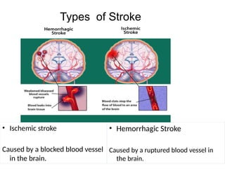
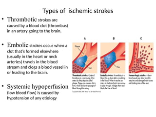
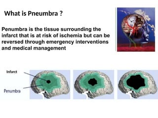
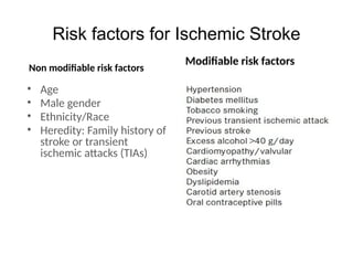
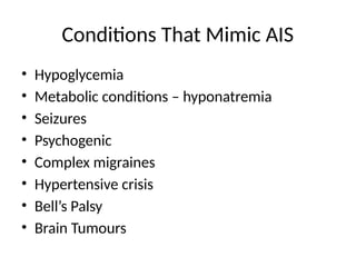
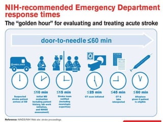
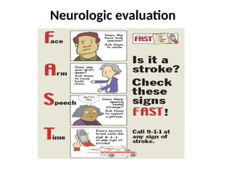
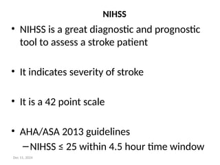
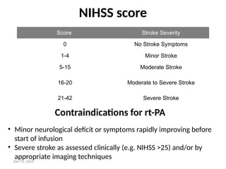
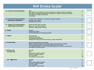
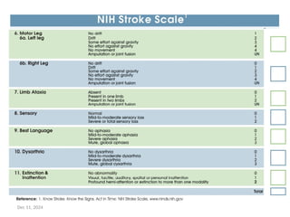
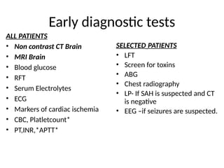
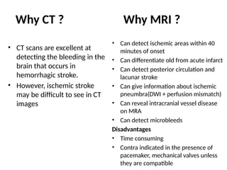
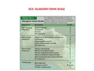
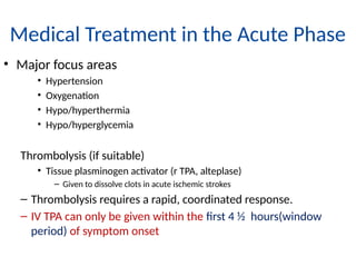
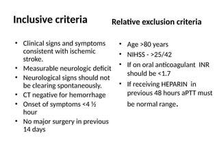
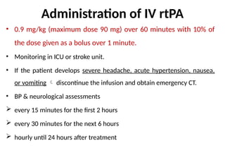
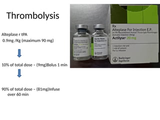
Acute Ischemic stroke
Dr.S.Maheshwari.,M.D
Assistant Professor
General Medicine
Anatomy - Circulation of the Brain
DEFINITION OF STROKE
Stroke is defined by the World Health Organization
as
‘A clinical syndrome consisting of rapidly
developing clinical signs of focal (or global in case
of coma) disturbance of cerebral function lasting
more than 24 hours or leading to death with no
apparent cause other than a vascular origin.’
Transient Ischemic Attack (TIA)
• A Transient ischemic attack (TIA) is a type of ischemic stroke—sometimes
referred to as a “warning stroke”
• Transient ischemic attack (TIA) is a brief episode of neurologic dysfunction
resulting from focal temporary cerebral ischemia with symptoms lasting
<24 hours but not associated with cerebral infarction.
• It usually causes no permanent injury to the brain.
• All neurologic signs and symptoms resolve within 24 h without evidence
of brain infarction on brain imaging.
• TIA symptoms occur rapidly and last a relatively short time.
• The majority last < 60 minutes
Time is brain
• Every minute matters
• Time is Brain…why?
The typical patient loses 1.9 million neurons each minute
in which stroke is untreated
• Normal cerebral blood flow (CBF)
– 50-to 60 ml/100g/ min
• when the CBF is
– < 20 ml/100g/min diminished electrical activity
– < 10ml/100g/min results in irreversible neuronal injury
Types of Stroke
• Ischemic stroke
Caused by a blocked blood vessel
in the brain.
• Hemorrhagic Stroke
Caused by a ruptured blood vessel in
the brain.
Types of ischemic strokes
• Thrombotic strokes are
caused by a blood clot (thrombus)
in an artery going to the brain.
• Embolic strokes occur when a
clot that’s formed elsewhere
(usually in the heart or neck
arteries) travels in the blood
stream and clogs a blood vessel in
or leading to the brain.
• Systemic hypoperfusion
(low blood flow) is caused by
hypotension of any etiology
Penumbra is the tissue surrounding the
infarct that is at risk of ischemia but can be
reversed through emergency interventions
and medical management
What is Pneumbra ?
Infarct
• Age
• Male gender
• Ethnicity/Race
• Heredity: Family history of
stroke or transient
ischemic attacks (TIAs)
Non modifiable risk factors
Modifiable risk factors
Risk factors for Ischemic Stroke
Conditions That Mimic AIS
• Hypoglycemia
• Metabolic conditions – hyponatremia
• Seizures
• Psychogenic
• Complex migraines
• Hypertensive crisis
• Bell’s Palsy
• Brain Tumours
Neurologic evaluation
NIHSS
• NIHSS is a great diagnostic and prognostic
tool to assess a stroke patient
• It indicates severity of stroke
• It is a 42 point scale
• AHA/ASA 2013 guidelines
–NIHSS ≤ 25 within 4.5 hour time window
Dec 11, 2024
NIHSS score
Score Stroke Severity
0 No Stroke Symptoms
1-4 Minor Stroke
5-15 Moderate Stroke
16-20 Moderate to Severe Stroke
21-42 Severe Stroke
Contraindications for rt-PA
• Minor neurological deficit or symptoms rapidly improving before
start of infusion
• Severe stroke as assessed clinically (e.g. NIHSS >25) and/or by
appropriate imaging techniques
Dec 11, 2024
It should be recorded at -
–0 hrs
–2 hrs post thrombolysis
–24 hrs after the onset of symptoms
–7 – 10 days
–3 months
Dec 11, 2024
Dec 11, 2024
Dec 11, 2024
Early diagnostic tests
ALL PATIENTS
• Non contrast CT Brain
• MRI Brain
• Blood glucose
• RFT
• Serum Electrolytes
• ECG
• Markers of cardiac ischemia
• CBC, Platletcount*
• PT,INR,*APTT*
SELECTED PATIENTS
• LFT
• Screen for toxins
• ABG
• Chest radiography
• LP- If SAH is suspected and CT
is negative
• EEG –if seizures are suspected.
• CT scans are excellent at
detecting the bleeding in the
brain that occurs in
hemorrhagic stroke.
• However, ischemic stroke
may be difficult to see in CT
images
• Can detect ischemic areas within 40
minutes of onset
• Can differentiate old from acute infarct
• Can detect posterior circulation and
lacunar stroke
• Can give information about ischemic
pneumbra(DWI + perfusion mismatch)
• Can reveal intracranial vessel disease
on MRA
• Can detect microbleeds
Disadvantages
• Time consuming
• Contra indicated in the presence of
pacemaker, mechanical valves unless
they are compatible
Why CT ? Why MRI ?
GCS- GLASGOW COMA SCALE
Medical Treatment in the Acute Phase
• Major focus areas
• Hypertension
• Oxygenation
• Hypo/hyperthermia
• Hypo/hyperglycemia
Thrombolysis (if suitable)
• Tissue plasminogen activator (r TPA, alteplase)
– Given to dissolve clots in acute ischemic strokes
– Thrombolysis requires a rapid, coordinated response.
– IV TPA can only be given within the first 4 ½ hours(window
period) of symptom onset
• Clinical signs and symptoms
consistent with ischemic
stroke.
• Measurable neurologic deficit
• Neurological signs should not
be clearing spontaneously.
• CT negative for hemorrhage
• Onset of symptoms <4 ½
hour
• No major surgery in previous
14 days
• Age >80 years
• NIHSS - >25/42
• If on oral anticoagulant INR
should be <1.7
• If receiving HEPARIN in
previous 48 hours aPTT must
be normal range.
Inclusive criteria Relative exclusion criteria
Administration of IV rtPA
• 0.9 mg/kg (maximum dose 90 mg) over 60 minutes with 10% of
the dose given as a bolus over 1 minute.
• Monitoring in ICU or stroke unit.
• If the patient develops severe headache, acute hypertension, nausea,
or vomiting  discontinue the infusion and obtain emergency CT.
• BP & neurological assessments
 every 15 minutes for the first 2 hours
 every 30 minutes for the next 6 hours
 hourly until 24 hours after treatment
Thrombolysis
Alteplase r tPA
0.9mg /Kg (maximum 90 mg)
10% of total dose – (9mg)Bolus 1 min
90% of total dose – (81mg)Infuse
over 60 min
Alteplace
reconstitution
rTPA Alteplase
• Do not mix rTPA with any other medications.
• Secure two iv lines with wide bore needle
• Do not use IV tubing with infusion filters.
• All patients must be on a cardiac monitors
• When infusion is complete, flush with Normal saline
• t-PA must be used within 8 hours of mixing when
stored at room temperature or within 24 hours if
refrigerated
Post thrombolytic care
• Bp every 15min x2 hours,
every 30min x 6 hours, then
hourly for 24 hours
• Treat Bp > 180/105
• Avoid invasive procedures for 24
hours i.e central line, arterial line &
Ryles tube
• Avoid urinary catheterization
during and for ½ hrs post Tpa
• 3% Nacl to reduce ICP
• No platelets, anticoagulants for 24
hrs
Complications
• Intra -cerebral haemorrhage
• Major Haemorrhage (eg GI)
• Bleeding-minor bleeding is
common (IV site)
• Anaphylaxis
• Angioedema
Angioedema
Stroke team
Stroke
Team
Emergency
Radiology
Nurse
Neurologist
Neuro
Intervention/
Surgery
Physician
Administrator
Dec 11, 2024
Dec 11, 2024
Dec 11, 2024
Blood Pressure Management
>
Dec 11, 2024
Dec 11, 2024
Supportive management
Head Position
• Head in a neutral position & elevate to 30 – 40 degrees in
the acute phase of stroke. It helps to prevent;
• Increasing ICP
• Aspiration (eg, those with dysphagia and/or decrease level of
consciousness)
• Cardiopulmonary decompensation or oxygen desaturation
Airway and Breathing
1. Maintain a patent airway by proper positioning of the patient.
2. Position the patient on lateral position with chin extended.
3. This prevents the tongue from obstructing the airway
4. Administer oxygen as required.
Physiological Monitoring
• Hypoxia
– Monitor Respirations & Spo2 <92% is associated with neurological
– deterioration
• Temperature
– >100 F(38C) must be treated. It lead to further worsening of infarct.
• Arrhythmias
– Continuous ECG to identify and treat AF as earlier
• Blood Sugar
– Hyperglycaemia is associated with poor clinical outcome
• Hydration
• Monitor urine output
Cont…..
• Blood pressure
• Non thrombolysed patients
– Dont treat BP unless
–
– BP is >220/120mmHg with 2 consecutive readings
• Thrombolysed patients
– BP is treated if
– BP is >185/110mmHg with 2 consecutive readings
• Abrupt fall in BP may affect cerebral perfusion pressure
Surgical intervention
Mechanical thrombectomy
Intra arterial fibrinolysis
Carotid endarterectomy
Carotid angioplasty and stenting
Decompressive hemicraniectomy is being increasingly performed
if patient having;
– pupilary asymmetry
– impending herniation
– decerebrate , decorticate posturing
– bleeding into the infract
– increase in mass effect & midline shift with raised ICP in imaging of
brain
Complications of Stroke
• Cerebral edema and herniation
(within 96 hr)
• Expansion of the
infarct/recurrent infarction
• Hemorrhage
• Seizure
• Aspiration pnemonitis
• Gastrointestinal ulcers and/or
bleeding
• Deep vein thrombosis and
pulmonary embolism
• Myocardial infarction
• Recurrent stroke
• Seizure
• Aspiration pnemonitis
• Deep vein thrombosis and
pulmonary embolism
• Decubitus ulcer
• Persistent language
dysfunction
• Persistent loss of mobility
• Spasticity
Early Complications of Stroke
(within 7 days) Late Complications (>7 days later)
Nursing management
cont……
Musculoskeletal system
• Full ROM exercises – to prevent joint contractures
• Paralysed hand/ leg – positioned higher with the use of pillows to prevent
edema
• Careful positioning and moving of affected arm
Skin care
• Skin care should be provided each time the patient is turned.
• Examine the skin for areas of irritation or breakdown.
• Gently massage the skin to stimulate circulation.
• Precautions must be taken to prevent the development of pressure sores.
Eye care
• Check the eyes frequently for signs of irritation or infection.
• Apply artificial tears/gel to prevent damage to the cornea as impaired normal
blink reflex .
45
Nutritional Needs
• Unconscious patient is fed by Ryles tube
• Always observe the patient carefully while feeding
• Maintain fowler's or semi-fowlers position to prevent
aspiration
• Provide oral hygiene twice per shift, Include the tongue, all
tooth surfaces, and all soft tissue areas.
• If oral feed started, food placed on unaffected side of
mouth
Bowel care
• The bowel should be evacuated regularly to prevent
stool impaction. Some time may require manual
evacuation
Bladder care
• Adequate fluids should be given to prevent dehydration
• Follow catheter bundle care to prevent infection in
catheterized patients
• Catheter and tubing is free of kinks and well secured
• Drainage bag is below level of the bladder and does not
touch the floor
• Maintain continuous close drainage
• Maintain unobstructed urine flow
• Drainage bag was emptied regularly into a clean
container
• Provide catheter care in each shift
• Assessment of necessity of catheter
Prevention of Deep Vein Thrombosis
•Graduated compressive stockings
•Intermittent pneumatic compressive devic
Anticoagulant therapy
(Heparin /LMWH)
Early mobilization
Patient Education /Rehabilitation
• Healthy diet — Components of a healthy diet include intakes of Fruits
and vegetables & Fiber, including cereals & Foods with a low glycemic
load & Omega-3 fatty acids (from fish, plant sources, or supplements)
• Smoking avoidance and cessation
• Hypertension control- The goal blood pressure is usually <140 systolic
as well as <90 mmHg diastolic
• Dyslipidemia control
• Physical activity - Even modest amounts of regular physical activity
such as brisk walking for 20 minutes daily
• Weight loss
• Lifestyle changes
• Regular medication & follow up
• To contact if any complication arises
THANK YOU

Stroke causes investigations and management.pptx

  • 1.
  • 2.
  • 3.
    DEFINITION OF STROKE Strokeis defined by the World Health Organization as ‘A clinical syndrome consisting of rapidly developing clinical signs of focal (or global in case of coma) disturbance of cerebral function lasting more than 24 hours or leading to death with no apparent cause other than a vascular origin.’
  • 4.
    Transient Ischemic Attack(TIA) • A Transient ischemic attack (TIA) is a type of ischemic stroke—sometimes referred to as a “warning stroke” • Transient ischemic attack (TIA) is a brief episode of neurologic dysfunction resulting from focal temporary cerebral ischemia with symptoms lasting <24 hours but not associated with cerebral infarction. • It usually causes no permanent injury to the brain. • All neurologic signs and symptoms resolve within 24 h without evidence of brain infarction on brain imaging. • TIA symptoms occur rapidly and last a relatively short time. • The majority last < 60 minutes
  • 5.
    Time is brain •Every minute matters • Time is Brain…why? The typical patient loses 1.9 million neurons each minute in which stroke is untreated • Normal cerebral blood flow (CBF) – 50-to 60 ml/100g/ min • when the CBF is – < 20 ml/100g/min diminished electrical activity – < 10ml/100g/min results in irreversible neuronal injury
  • 6.
    Types of Stroke •Ischemic stroke Caused by a blocked blood vessel in the brain. • Hemorrhagic Stroke Caused by a ruptured blood vessel in the brain.
  • 7.
    Types of ischemicstrokes • Thrombotic strokes are caused by a blood clot (thrombus) in an artery going to the brain. • Embolic strokes occur when a clot that’s formed elsewhere (usually in the heart or neck arteries) travels in the blood stream and clogs a blood vessel in or leading to the brain. • Systemic hypoperfusion (low blood flow) is caused by hypotension of any etiology
  • 8.
    Penumbra is thetissue surrounding the infarct that is at risk of ischemia but can be reversed through emergency interventions and medical management What is Pneumbra ? Infarct
  • 9.
    • Age • Malegender • Ethnicity/Race • Heredity: Family history of stroke or transient ischemic attacks (TIAs) Non modifiable risk factors Modifiable risk factors Risk factors for Ischemic Stroke
  • 10.
    Conditions That MimicAIS • Hypoglycemia • Metabolic conditions – hyponatremia • Seizures • Psychogenic • Complex migraines • Hypertensive crisis • Bell’s Palsy • Brain Tumours
  • 12.
  • 13.
    NIHSS • NIHSS isa great diagnostic and prognostic tool to assess a stroke patient • It indicates severity of stroke • It is a 42 point scale • AHA/ASA 2013 guidelines –NIHSS ≤ 25 within 4.5 hour time window Dec 11, 2024
  • 14.
    NIHSS score Score StrokeSeverity 0 No Stroke Symptoms 1-4 Minor Stroke 5-15 Moderate Stroke 16-20 Moderate to Severe Stroke 21-42 Severe Stroke Contraindications for rt-PA • Minor neurological deficit or symptoms rapidly improving before start of infusion • Severe stroke as assessed clinically (e.g. NIHSS >25) and/or by appropriate imaging techniques Dec 11, 2024
  • 15.
    It should berecorded at - –0 hrs –2 hrs post thrombolysis –24 hrs after the onset of symptoms –7 – 10 days –3 months Dec 11, 2024
  • 16.
  • 17.
  • 18.
    Early diagnostic tests ALLPATIENTS • Non contrast CT Brain • MRI Brain • Blood glucose • RFT • Serum Electrolytes • ECG • Markers of cardiac ischemia • CBC, Platletcount* • PT,INR,*APTT* SELECTED PATIENTS • LFT • Screen for toxins • ABG • Chest radiography • LP- If SAH is suspected and CT is negative • EEG –if seizures are suspected.
  • 19.
    • CT scansare excellent at detecting the bleeding in the brain that occurs in hemorrhagic stroke. • However, ischemic stroke may be difficult to see in CT images • Can detect ischemic areas within 40 minutes of onset • Can differentiate old from acute infarct • Can detect posterior circulation and lacunar stroke • Can give information about ischemic pneumbra(DWI + perfusion mismatch) • Can reveal intracranial vessel disease on MRA • Can detect microbleeds Disadvantages • Time consuming • Contra indicated in the presence of pacemaker, mechanical valves unless they are compatible Why CT ? Why MRI ?
  • 20.
  • 21.
    Medical Treatment inthe Acute Phase • Major focus areas • Hypertension • Oxygenation • Hypo/hyperthermia • Hypo/hyperglycemia Thrombolysis (if suitable) • Tissue plasminogen activator (r TPA, alteplase) – Given to dissolve clots in acute ischemic strokes – Thrombolysis requires a rapid, coordinated response. – IV TPA can only be given within the first 4 ½ hours(window period) of symptom onset
  • 22.
    • Clinical signsand symptoms consistent with ischemic stroke. • Measurable neurologic deficit • Neurological signs should not be clearing spontaneously. • CT negative for hemorrhage • Onset of symptoms <4 ½ hour • No major surgery in previous 14 days • Age >80 years • NIHSS - >25/42 • If on oral anticoagulant INR should be <1.7 • If receiving HEPARIN in previous 48 hours aPTT must be normal range. Inclusive criteria Relative exclusion criteria
  • 23.
    Administration of IVrtPA • 0.9 mg/kg (maximum dose 90 mg) over 60 minutes with 10% of the dose given as a bolus over 1 minute. • Monitoring in ICU or stroke unit. • If the patient develops severe headache, acute hypertension, nausea, or vomiting  discontinue the infusion and obtain emergency CT. • BP & neurological assessments  every 15 minutes for the first 2 hours  every 30 minutes for the next 6 hours  hourly until 24 hours after treatment
  • 24.
    Thrombolysis Alteplase r tPA 0.9mg/Kg (maximum 90 mg) 10% of total dose – (9mg)Bolus 1 min 90% of total dose – (81mg)Infuse over 60 min
  • 25.
  • 26.
    rTPA Alteplase • Donot mix rTPA with any other medications. • Secure two iv lines with wide bore needle • Do not use IV tubing with infusion filters. • All patients must be on a cardiac monitors • When infusion is complete, flush with Normal saline • t-PA must be used within 8 hours of mixing when stored at room temperature or within 24 hours if refrigerated
  • 27.
    Post thrombolytic care •Bp every 15min x2 hours, every 30min x 6 hours, then hourly for 24 hours • Treat Bp > 180/105 • Avoid invasive procedures for 24 hours i.e central line, arterial line & Ryles tube • Avoid urinary catheterization during and for ½ hrs post Tpa • 3% Nacl to reduce ICP • No platelets, anticoagulants for 24 hrs Complications • Intra -cerebral haemorrhage • Major Haemorrhage (eg GI) • Bleeding-minor bleeding is common (IV site) • Anaphylaxis • Angioedema Angioedema
  • 28.
  • 29.
  • 30.
  • 31.
  • 32.
  • 33.
  • 34.
    Supportive management Head Position •Head in a neutral position & elevate to 30 – 40 degrees in the acute phase of stroke. It helps to prevent; • Increasing ICP • Aspiration (eg, those with dysphagia and/or decrease level of consciousness) • Cardiopulmonary decompensation or oxygen desaturation
  • 35.
    Airway and Breathing 1.Maintain a patent airway by proper positioning of the patient. 2. Position the patient on lateral position with chin extended. 3. This prevents the tongue from obstructing the airway 4. Administer oxygen as required.
  • 36.
    Physiological Monitoring • Hypoxia –Monitor Respirations & Spo2 <92% is associated with neurological – deterioration • Temperature – >100 F(38C) must be treated. It lead to further worsening of infarct. • Arrhythmias – Continuous ECG to identify and treat AF as earlier • Blood Sugar – Hyperglycaemia is associated with poor clinical outcome • Hydration • Monitor urine output
  • 37.
    Cont….. • Blood pressure •Non thrombolysed patients – Dont treat BP unless – – BP is >220/120mmHg with 2 consecutive readings • Thrombolysed patients – BP is treated if – BP is >185/110mmHg with 2 consecutive readings • Abrupt fall in BP may affect cerebral perfusion pressure
  • 38.
    Surgical intervention Mechanical thrombectomy Intraarterial fibrinolysis Carotid endarterectomy Carotid angioplasty and stenting Decompressive hemicraniectomy is being increasingly performed if patient having; – pupilary asymmetry – impending herniation – decerebrate , decorticate posturing – bleeding into the infract – increase in mass effect & midline shift with raised ICP in imaging of brain
  • 39.
    Complications of Stroke •Cerebral edema and herniation (within 96 hr) • Expansion of the infarct/recurrent infarction • Hemorrhage • Seizure • Aspiration pnemonitis • Gastrointestinal ulcers and/or bleeding • Deep vein thrombosis and pulmonary embolism • Myocardial infarction • Recurrent stroke • Seizure • Aspiration pnemonitis • Deep vein thrombosis and pulmonary embolism • Decubitus ulcer • Persistent language dysfunction • Persistent loss of mobility • Spasticity Early Complications of Stroke (within 7 days) Late Complications (>7 days later)
  • 40.
  • 41.
    cont…… Musculoskeletal system • FullROM exercises – to prevent joint contractures • Paralysed hand/ leg – positioned higher with the use of pillows to prevent edema • Careful positioning and moving of affected arm Skin care • Skin care should be provided each time the patient is turned. • Examine the skin for areas of irritation or breakdown. • Gently massage the skin to stimulate circulation. • Precautions must be taken to prevent the development of pressure sores. Eye care • Check the eyes frequently for signs of irritation or infection. • Apply artificial tears/gel to prevent damage to the cornea as impaired normal blink reflex .
  • 42.
  • 43.
    Nutritional Needs • Unconsciouspatient is fed by Ryles tube • Always observe the patient carefully while feeding • Maintain fowler's or semi-fowlers position to prevent aspiration • Provide oral hygiene twice per shift, Include the tongue, all tooth surfaces, and all soft tissue areas. • If oral feed started, food placed on unaffected side of mouth Bowel care • The bowel should be evacuated regularly to prevent stool impaction. Some time may require manual evacuation
  • 44.
    Bladder care • Adequatefluids should be given to prevent dehydration • Follow catheter bundle care to prevent infection in catheterized patients • Catheter and tubing is free of kinks and well secured • Drainage bag is below level of the bladder and does not touch the floor • Maintain continuous close drainage • Maintain unobstructed urine flow • Drainage bag was emptied regularly into a clean container • Provide catheter care in each shift • Assessment of necessity of catheter
  • 45.
    Prevention of DeepVein Thrombosis •Graduated compressive stockings •Intermittent pneumatic compressive devic Anticoagulant therapy (Heparin /LMWH) Early mobilization
  • 46.
    Patient Education /Rehabilitation •Healthy diet — Components of a healthy diet include intakes of Fruits and vegetables & Fiber, including cereals & Foods with a low glycemic load & Omega-3 fatty acids (from fish, plant sources, or supplements) • Smoking avoidance and cessation • Hypertension control- The goal blood pressure is usually <140 systolic as well as <90 mmHg diastolic • Dyslipidemia control • Physical activity - Even modest amounts of regular physical activity such as brisk walking for 20 minutes daily • Weight loss • Lifestyle changes • Regular medication & follow up • To contact if any complication arises
  • 47.