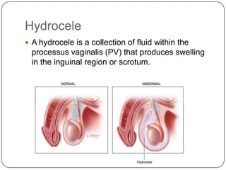
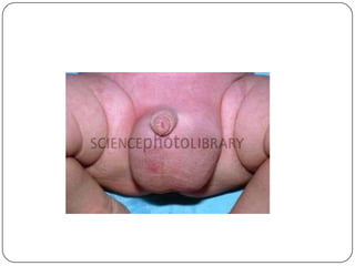
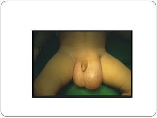
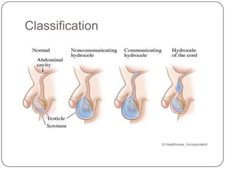
Hydrocele is a collection of fluid within the processus vaginalis that causes swelling in the scrotum. It is usually caused by a failure of the processus vaginalis to completely fuse during development. Hydroceles can be communicating or non-communicating depending on whether the processus vaginalis remains open to the abdominal cavity. The standard treatment is surgical repair to prevent the fluid from reaccumulating. Surgery involves ligating and removing the hydrocele sac to restore normal drainage from the scrotum. Complications are rare if the surgery is performed properly.
2. Hydrocele
A hydrocele is a collection of fluid within the
processus vaginalis (PV) that produces swelling
in the inguinal region or scrotum.
3.
4.
5.
6.
7. Relevant Anatomy
The developmental anatomy of the inguinal canal
is responsible for the genesis of pediatric
communicating hydroceles.
As the testis descends from the posterolateral
genitourinary ridge at the beginning of the third
trimester of fetal gestation, a saclike extension of
peritoneum descends in concert with the testis.
As descent progresses, the sac envelops the
testis and epididymis. The result is a serosal-lined
tubular communication between the abdomen
and the tunica vaginalis of the scrotum.
8. The peritoneum-derived serosal communication is
the processus vaginalis, and the serosa of the
hemiscrotum becomes the tunica vaginalis.
At term, or within the first 1-2 years of life, the
processus vaginalis of the spermatic cord
fuses, obliterating the communication between
the abdomen and the scrotum.
The processus fuses distally as far as the lower
epididymal pole and anteriorly to the upper
epididymal pole.
Failure of complete fusion may result in
communicating hydroceles, indirect inguinal
hernias, and the bell-clapper deformity of
abnormal testicular fixation in the scrotum.
9. Classification
Primary hydrocele—when there is no definitive
cause / idiopathic.
Secondary hydrocele—diseases of testis
1. TB of epididymis
2. Epididymal orchitis
3. Syphilitic orchitis
4. Testicular tumours (seminoma—5th
decade of life and onwards, teratoma—1st
and 2nd decades of life, sertoli cell
tumours, leydig cell tumours, lymphoma)
5. Orchitis arising by virus
6. Trauma
10. Difference between primary and secondary
hydrocele:
Primary Secondary
Big and tense Small and loose, lax
Testis cannot be felt Testis can be felt
No definitive history Definitive history of the disease
13. Epidemiology
Patent processus vaginalis are found in 80-90%
of term male infants at birth.
This frequency rate steadily decreases until age 2
years, when it appears to plateau at
approximately 25-40%. Indeed, autopsy series of
men have identified a frequency rate of 20% of
the processus vaginalis remaining patent until
late in life.
However, clinically apparent scrotal hydroceles
are evident in only 6% of term males beyond the
newborn period. Certain conditions, such as
breech presentation, gestational progestin
use, and low birth weight, have been associated
with an increased risk of hydroceles.
14. Etiology of Hydroceles
Congenital Hydroceles
With the descent of the testis, the parietal peritoneum forms the
processus vaginalis and the cavity of the tunica vaginalis of the
testis. The processus vaginalis normally obliterates till the fourth
month of life. Congenital hydroceles occur mostly through lack of
closure of the processus vaginalis (= communicating hydrocele).
Acquired Hydroceles
Usually, there is a balance between fluid production and outflow in
the cavity of the tunica vaginalis. The following diseases disturb this
balance: inflammation, tumors, testicular trauma, torsion of the
testis or testicular appendages, defective lymphatic drainage (after
surgery for varicoceles or inguinal hernias).
15. Pathophysiology
The pathophysiology of hydroceles requires an
imbalance of scrotal fluid production and
absorption. This imbalance can be divided further
into exogenous fluid sources or intrinsic fluid
production.
Alternatively, hydroceles can be divided into those
that represent a persistent communication with
the abdominal cavity and those that do not. Fluid
excesses are from exogenous sources (the
abdomen) in communicating hydroceles, whereas
noncommunicating hydroceles develop increased
scrotal fluid from abnormal intrinsic scrotal fluid
shifts.
16. Communicating hydroceles
With communicating hydroceles, simple Valsalva
probably accounts for the classic variation in size
during day-sleep cycles. Nonetheless, with the
incidence of patent processus so great, why children
with clinically apparent hydroceles are relatively few
remains somewhat inexplicable. Chronically
increased intra-abdominal pressure (eg, as in
chronic lung disease) or increased abdominal fluid
production (eg, children with ventriculoperitoneal
shunts) probably warrants early surgical intervention.
17. Noncommunicating hydroceles
In noncommunicating hydroceles, the
pathophysiology may occur as a result of increased
fluid production or as a consequence of impaired
absorption. A sudden onset of scrotal hydrocele in
older children has been noted after viral illnesses. In
such cases, viral-mediated serositis may account for
the net increased fluid production. Posttraumatic
hydroceles likely occur secondary to increased
serosal fluid production due to underlying
inflammation. Although rare in the United
States, filarial infestations are a classic cause of the
decreased lymphatic fluid absorption resulting in
hydroceles.
18. Symptoms
Although each child may experience symptoms
differently, the most common symptom is a fluid
mass that is usually smooth and not tender in the
scrotum.
In the case of a communicating hydrocele, the
mass fluctuates in size, getting smaller at night
while lying flat,and increasing in size during more
active periods.
24. Unlike hernias in infants, many newborn
hydroceles resolve because of spontaneous
closure of the PPV early after birth. The residual
noncommunicating hydrocele does not wax and
wane in volume, and no silk glove sign is present.
The fluid in the hydrocele is usually reabsorbed
before the infant reaches age 1 year. Because of
these facts, observation is often appropriate for
hydroceles in infants.
25. The following factors indicate
hydrocele repair:
Failure to resolve by age 2 years
Continued discomfort
Enlargement or waxing and waning in volume
Unsightly appearance
Secondary infection (very rare)
26. Specific conditions or demographics
and timing of surgery
In full-term infants with no history of
incarceration, schedule surgery as soon as
possible on an outpatient basis.
For preterm neonatal intensive care unit (NICU)
infants weighing 1800-2000 g, schedule surgery
before hospital discharge.
For formerly premature infants younger than 60
weeks’ postconceptual age, schedule surgery as
soon as possible with 24-hour postoperative
monitoring for apnea and other anesthesia-
related complications.
28. IF SAC IS SMALL THIN AND CONTAINS CLEAR
FLUID
->LORDS PLICATION –SAC IS MADE TO FORM
FIBROUS TISSUE
OR EVACUATION & EVERSION
IF SAC IS THICK IN LARGE HYDROCELE –SUBTOTAL
EXCISION
JABOULEYS OPERATION
SHARMA & JHAWER TECHNIQUE
29. Drainage
The fluid can be drained easily with a needle and
syringe. However, following this procedure, it is
common for the sac of the hydrocele to refill with
fluid within a few months. Draining every now and
then may be suitable though, if you are not fit for
surgery or if you do not want an operation.
30. Surgery for Hydroceles of the
Cord
Treatment of hydroceles of the cord starts with an
inguinal incision for exposure of the spermatic
cord. After excision of the hydrocele of the
cord, the processus vaginalis is ligated at the
internal inguinal ring.
31. Surgery for Communicating
Hydroceles
Treatment of communicating hydroceles starts
with an inguinal incision for exposure of the testis.
The processus vaginalis is isolated from the
spermatic cord, divided and ligated at the internal
inguinal ring. The distal sac is resected as far as
possible, the end of the sac can be left open.
The contralateral exploration is not a standard
therapy, but is sometimes performed. The
probability for an open contralateral processus
vaginalis in unilateral communicating hydrocele is
50%, but only about 15–22% become clinically
significant.
32. Surgery for Hydroceles of the
Testis
After scrotal incision for exposure of the scrotal
hydrocele, two surgical techniques are available.
The recurrence rate should be below 5% with
either technique, Lord's technique has probably
the lowest complication rate:
Hydrocelectomy with excision of the hydrocele
sac: Winkelmann's technique or Jaboulay's
technique
Hydrocelectomy with plication of the hydrocele
sac: Lord's technique
33. Operation Winkelmann. This surgical intervention is
one of the leaflets own shell eggs dissect the
anterior surface, turn inside out and stitch the back
of the testicle. At the same time accumulation of fluid
no longer occurs.
Operation Bergman. Part own inner layer shell eggs
are removed, the remaining part of ligated.
Postoperatively appointed antimicrobials and for
some time wearing a jockstrap.
Operation Lord. When this operation is performed
dissection egg shells, release of dropsical fluid and a
so-called corrugation of the tunica vaginalis testis
around. It goes from the surrounding tissue egg is
not released and the wound did not dislocate. This
reduces trauma to adjacent tissues and blood
vessels supplying the testicle.
34. Follow-up
At least one postoperative follow-up visit is
recommended. For small infants, chronic
recurring hydroceles, or patients with
unsuspected intraoperative findings, more
protracted follow-up evaluations may be
warranted biweekly, monthly, or every 2-3 months
to ensure complete recovery and normal
testicular size and architecture.
35. Outcome and Prognosis
Inguinal repairs of communicating hydroceles are
exceedingly successful, with a less than 1%
recurrence rate. If a unilateral approach is
completed, the small but recognized risk for a
metachronous hydrocele or inguinal hernia
developing remains, but the rate is likely less than
10%. Likewise, recurrence after tunica excision is
also uncommon.
36. COMPLICATIONS OF HYDROCELE:
1. INFECTION
2. PYOCELE,HEMATOCELE
3. INFERTILITY
4. ATROPHY OF TESTIS
5. HERNIATION OF HYDROCELE SAC (rare)
6. RUPTURE (rare)