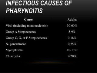
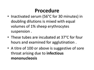
Sore
throa
t
Ajay
Sharma
1305
Sore throat
*(or throat pain) is pain or
irritation of the throat.
*A common physical symptom, it
is usually caused by
acutepharyngitis (inflammation of
the throat)
Viruses
-Adenovirus
-Influenza
-Parainfluenza virus
-Epstein-Barr
-Coronavirus
-Rhinovirus
-Enterovirus
Respiratory synctial virus
-Metapneumovirus
-Herpes simplex virus
Bacterial
Group A beta-haemolytic
strep
Group C and G
streptococci
Chlamydia pneumoniae
Diphtheria
Mycoplasma pneumonia
Neisseria gonorrhea or
chlamydia trachomatis
(related to sexual activity)
Fungi
Candida albicans
Laboratory Diagnosis
A. SPECIMEN
Throat swab from fauces (part of oropharynx
directly behind the mouth cavity )
B. COLLECTION
*Two sterile swabs should be used , one for direct
microscopy and other for culture
*Swabs should be rubbed over the tonsillar fossa ,
pseudomembrane if present and finally the post
pharyngeal wall
*These swabs should include exudate present in
the throat , should be quickly sent to lab ; if not so
then swabs should be refrigerated .
c. Direct Microscopy Gram Staining
and Albert Staining may be used for
staining the smears
1. Gram Staining
Streptococcus pyogens
Gram positive cocci may be
present in chains
c. diptheriae
seen as Gram positive, Non acid fast ,
non-motile and non-sporing bacilli
Candida albicans reveal Gram
positive budding yeast cells
2. ALBERT STAINING
• Albert staining is helpful for presumptive
diagnosis of Corynebacterium diptheriae .
• It shows green coloured , and L shaped
(Chinese letter pattern ) bacilli with bluish
black metachromatic granules .
C. diphtheriae shows green coloured V or L shaped
bacilli with bluish black metachromatic granules
D. CULTURE
• Culture media are selected according to the
organism suspected to be the causative agent
of sore throat. Following media may be used
for culture
• Blood Agar – All the organisms will grow on this
medium
• Crystal violet blood agar- It is selective medium
for Str. Pyogens
• Potassium tellurite blood agar - It is selective
media for growning C. diptheriae
• Sabouraud’s dextrose agar (SDA) – When
suspecting Candida albicans , SDA should be
included !
• These culture media are incubated at 37°C
for overnight.
• In case of potassium tellurite blood agar , it
should be incubated for 48 hours.
• After 6-8 hours a subculture should be
made from Loeffler’s serum slope onto
potassium tellurite blood agar , which is
then incubated at 37°C for 48 hours
By Colony Morphology
1. Streptococcuspyogens
Pin Point (0.5-1mm) ,
round or convex colonies
with entire margins .
Colour is greyish white ,
no specific odour
Having wide zone of β-
hemolysis
2.Coryynebacterium
diphtheriae
•On potassium tellurite
blood agar black
coloured round colonies
are seen
3. Candida
albicans
White or cream
coloured
colonies are
seen
Other Tests for confirmation
• 1. For β-haemolytic streptococci
• Bacitracin sensitivity test – For streptococcus
pyogens
• In this test Susceptibility to bacitracin is
determined by placing a bacitracin-
impregnated paper disk on a nutrient agar
plate seeded with the microbe under
investigation.
• As the microbe multiplies during incubation to
produce a lawn of confluent growth, cells are
exposed to the antibiotic diffusing into the agar
from the paper disk.
• If the bacteria are susceptible to bacitracin,
there will be a visible zone of
inhibition forming around the disk,
representing an area where the antibiotic
concentration has prevented bacterial
growth. Should the microbe be resistant,
the lawn of cells will form visible growth up
to the margin of the disk.
2) For C. diphtheriae
• Toxigenic tests
• A) Elek’s gel precipitation test
• This is an immunodiffusion described by Elek
• A rectangular strip of filter paper soaked in
diphtheria antitoxin (1000 units per ml ) is
placed on the surface of a 20% horse serum
agar plate while the medium is still fluid
• When the agar solidifies , the test strain is
streaked at right angle to the filter paper strip
• The plate is incubated at 37°C for 24 to 48
hours
The toxin produced by the
bacterial growth diffuses
in the agar and produces a
line of precipitation where
it meets the antitoxin
optimum concentration
Non toxigenic stains will
not produce any
precipitation line
B) Animal inoculation test
3) Epstein Barr Virus
• If sore throat is due to Epstein Barr Virus as seen in
Infectious Mononucleosis then Paul Bunnell test can
be done
• Principle-
* During infectious mononucleosis , heterophile
antibodies appear in the serum of the patient . These
are IgM antibodies elicited by EBV infection .
* These antibodies appear in 85-90% of patients
during acute phase of illness .
*These antibodies kill the erythrocytes of sheep
*Their titre decreases rapidly after fourth week and are
not detectable after 2-3 months .
Procedure
• Inactivated serum (56°C for 30 minutes) in
doubling dilutions is mixed with equal
volumes of 1% sheep erythrocytes
suspension .
• These tubes are incubated at 37°C for four
hours and examined for agglutination .
• A titre of 100 or above is suggestive of sore
throat arising due to infectious
mononucleosis
Sore throat
Sore throat
Sore throat
Sore throat
Sore throat

Sore throat

  • 1.
  • 2.
    Sore throat *(or throatpain) is pain or irritation of the throat. *A common physical symptom, it is usually caused by acutepharyngitis (inflammation of the throat)
  • 7.
  • 8.
    Bacterial Group A beta-haemolytic strep GroupC and G streptococci Chlamydia pneumoniae Diphtheria Mycoplasma pneumonia Neisseria gonorrhea or chlamydia trachomatis (related to sexual activity) Fungi Candida albicans
  • 16.
    Laboratory Diagnosis A. SPECIMEN Throatswab from fauces (part of oropharynx directly behind the mouth cavity ) B. COLLECTION *Two sterile swabs should be used , one for direct microscopy and other for culture *Swabs should be rubbed over the tonsillar fossa , pseudomembrane if present and finally the post pharyngeal wall *These swabs should include exudate present in the throat , should be quickly sent to lab ; if not so then swabs should be refrigerated .
  • 17.
    c. Direct MicroscopyGram Staining and Albert Staining may be used for staining the smears 1. Gram Staining Streptococcus pyogens Gram positive cocci may be present in chains
  • 18.
    c. diptheriae seen asGram positive, Non acid fast , non-motile and non-sporing bacilli
  • 19.
    Candida albicans revealGram positive budding yeast cells
  • 20.
    2. ALBERT STAINING •Albert staining is helpful for presumptive diagnosis of Corynebacterium diptheriae . • It shows green coloured , and L shaped (Chinese letter pattern ) bacilli with bluish black metachromatic granules .
  • 21.
    C. diphtheriae showsgreen coloured V or L shaped bacilli with bluish black metachromatic granules
  • 22.
    D. CULTURE • Culturemedia are selected according to the organism suspected to be the causative agent of sore throat. Following media may be used for culture • Blood Agar – All the organisms will grow on this medium • Crystal violet blood agar- It is selective medium for Str. Pyogens • Potassium tellurite blood agar - It is selective media for growning C. diptheriae • Sabouraud’s dextrose agar (SDA) – When suspecting Candida albicans , SDA should be included !
  • 23.
    • These culturemedia are incubated at 37°C for overnight. • In case of potassium tellurite blood agar , it should be incubated for 48 hours. • After 6-8 hours a subculture should be made from Loeffler’s serum slope onto potassium tellurite blood agar , which is then incubated at 37°C for 48 hours
  • 24.
    By Colony Morphology 1.Streptococcuspyogens Pin Point (0.5-1mm) , round or convex colonies with entire margins . Colour is greyish white , no specific odour Having wide zone of β- hemolysis
  • 25.
    2.Coryynebacterium diphtheriae •On potassium tellurite bloodagar black coloured round colonies are seen
  • 26.
    3. Candida albicans White orcream coloured colonies are seen
  • 27.
    Other Tests forconfirmation • 1. For β-haemolytic streptococci • Bacitracin sensitivity test – For streptococcus pyogens • In this test Susceptibility to bacitracin is determined by placing a bacitracin- impregnated paper disk on a nutrient agar plate seeded with the microbe under investigation. • As the microbe multiplies during incubation to produce a lawn of confluent growth, cells are exposed to the antibiotic diffusing into the agar from the paper disk.
  • 28.
    • If thebacteria are susceptible to bacitracin, there will be a visible zone of inhibition forming around the disk, representing an area where the antibiotic concentration has prevented bacterial growth. Should the microbe be resistant, the lawn of cells will form visible growth up to the margin of the disk.
  • 30.
    2) For C.diphtheriae • Toxigenic tests • A) Elek’s gel precipitation test • This is an immunodiffusion described by Elek • A rectangular strip of filter paper soaked in diphtheria antitoxin (1000 units per ml ) is placed on the surface of a 20% horse serum agar plate while the medium is still fluid • When the agar solidifies , the test strain is streaked at right angle to the filter paper strip • The plate is incubated at 37°C for 24 to 48 hours
  • 31.
    The toxin producedby the bacterial growth diffuses in the agar and produces a line of precipitation where it meets the antitoxin optimum concentration Non toxigenic stains will not produce any precipitation line B) Animal inoculation test
  • 33.
    3) Epstein BarrVirus • If sore throat is due to Epstein Barr Virus as seen in Infectious Mononucleosis then Paul Bunnell test can be done • Principle- * During infectious mononucleosis , heterophile antibodies appear in the serum of the patient . These are IgM antibodies elicited by EBV infection . * These antibodies appear in 85-90% of patients during acute phase of illness . *These antibodies kill the erythrocytes of sheep *Their titre decreases rapidly after fourth week and are not detectable after 2-3 months .
  • 34.
    Procedure • Inactivated serum(56°C for 30 minutes) in doubling dilutions is mixed with equal volumes of 1% sheep erythrocytes suspension . • These tubes are incubated at 37°C for four hours and examined for agglutination . • A titre of 100 or above is suggestive of sore throat arising due to infectious mononucleosis