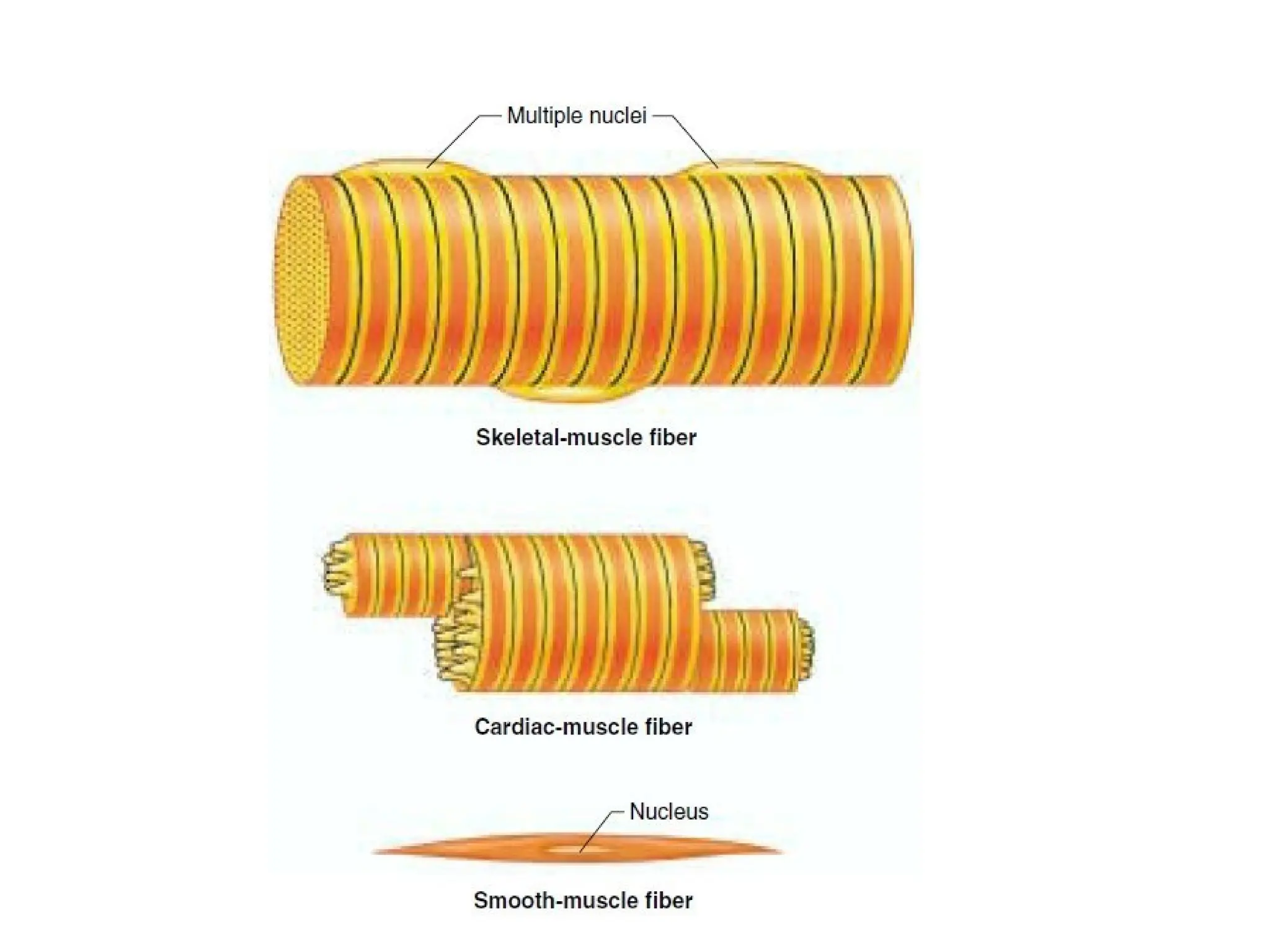
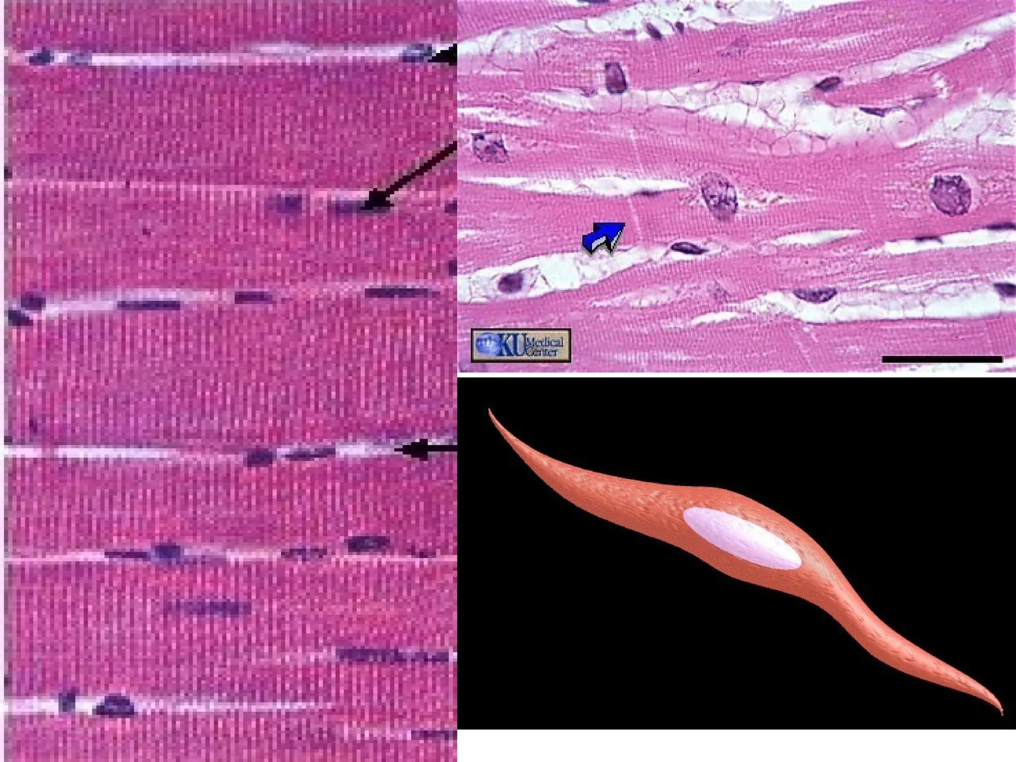
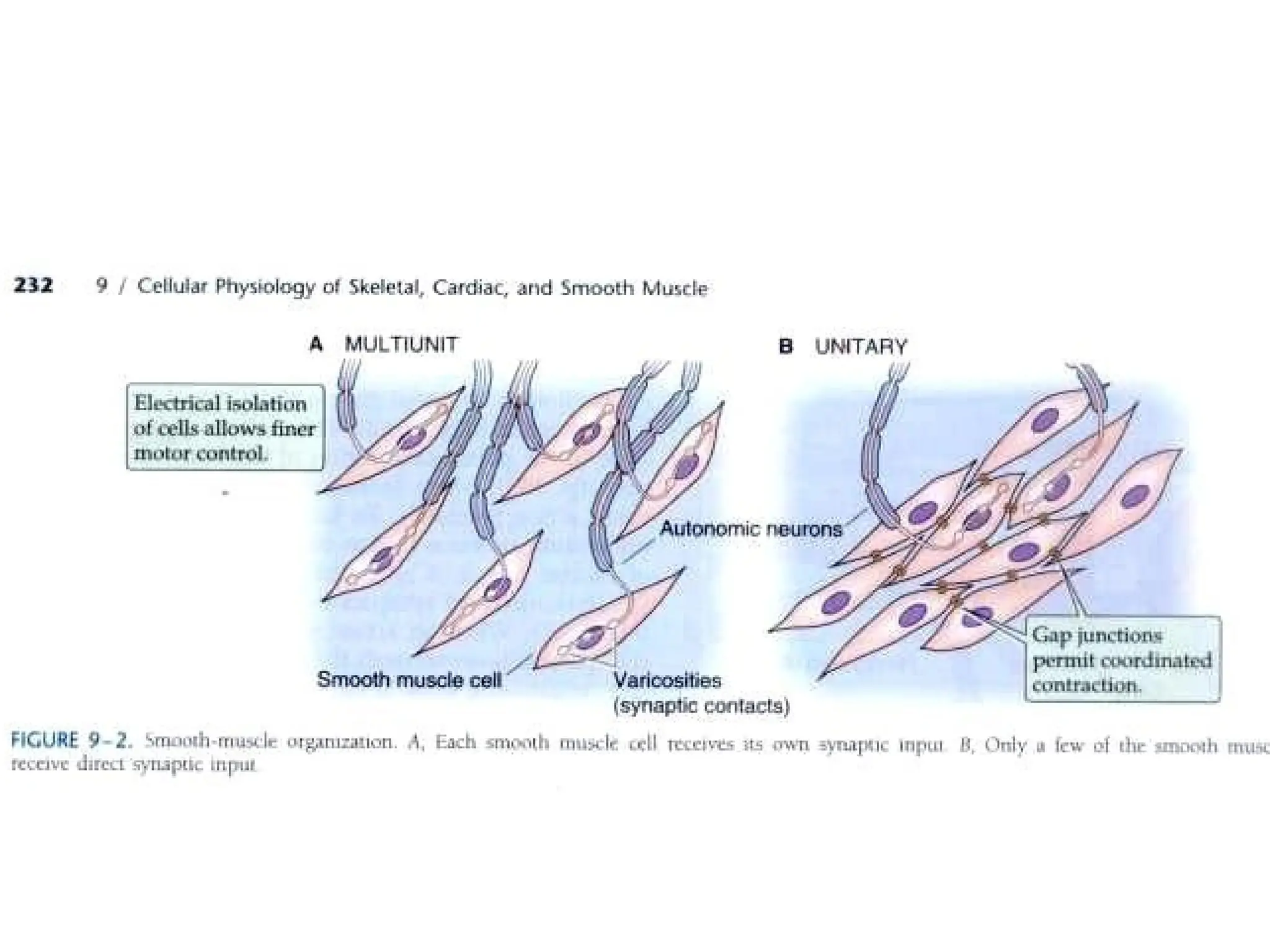
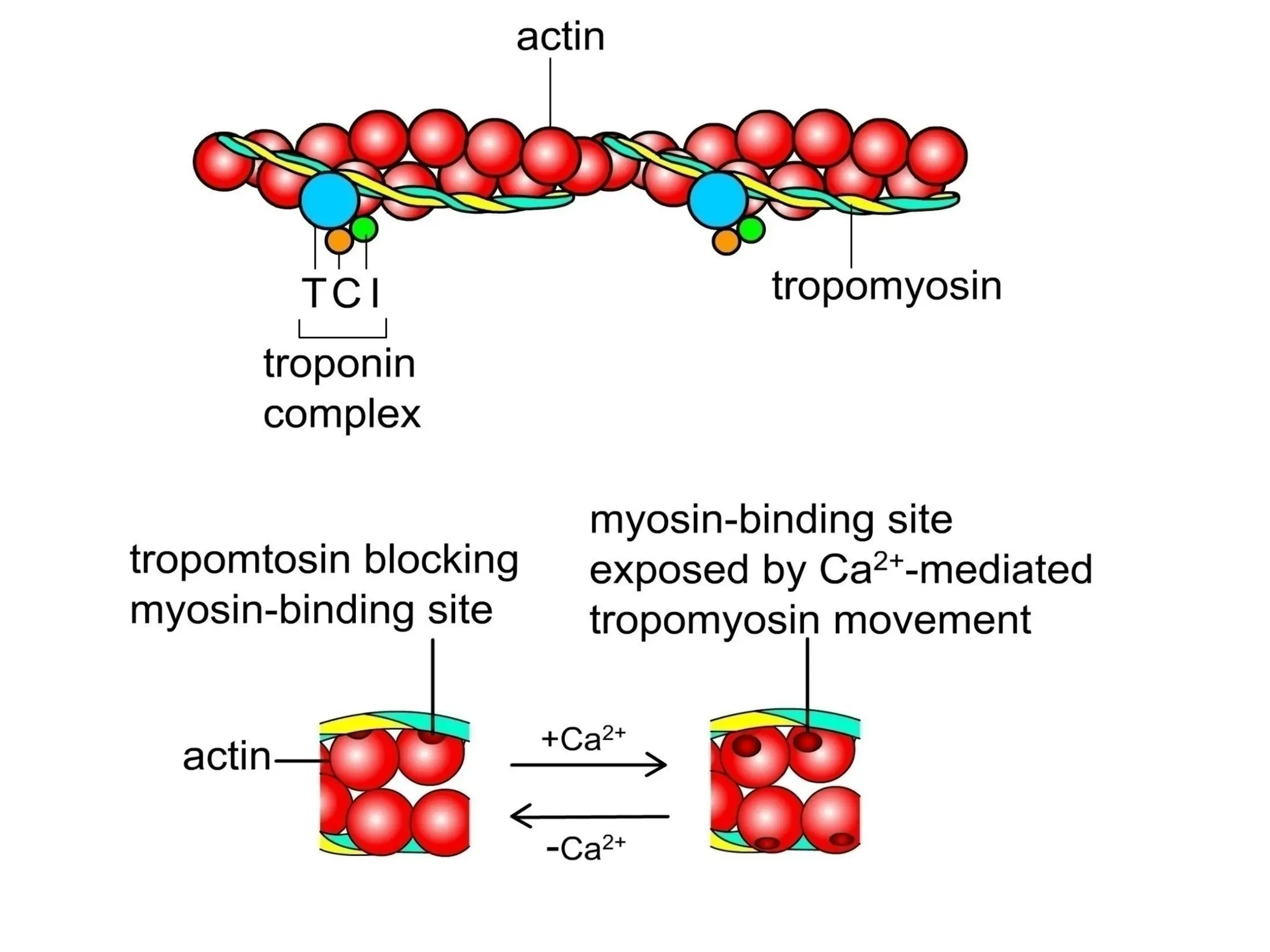
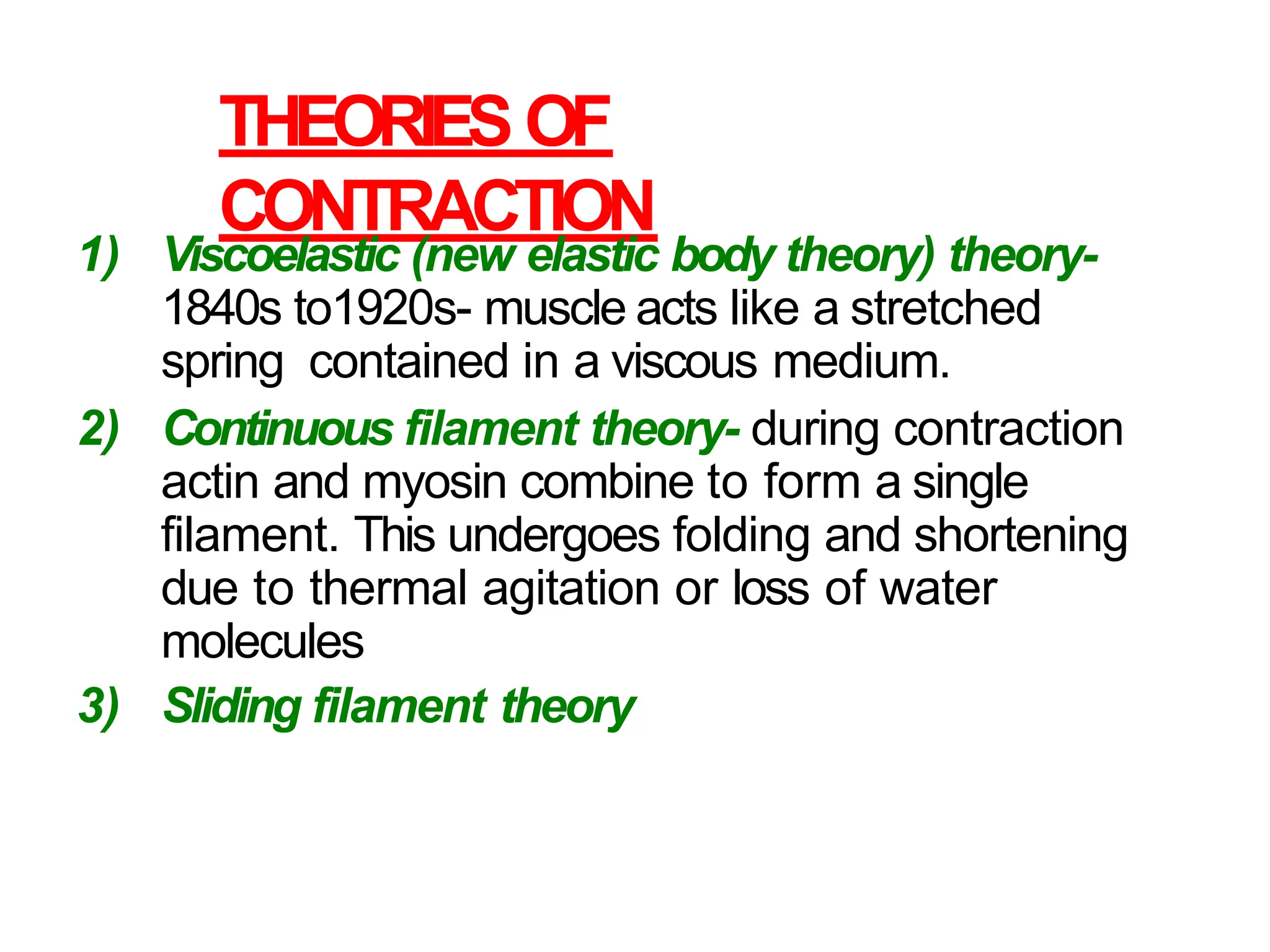
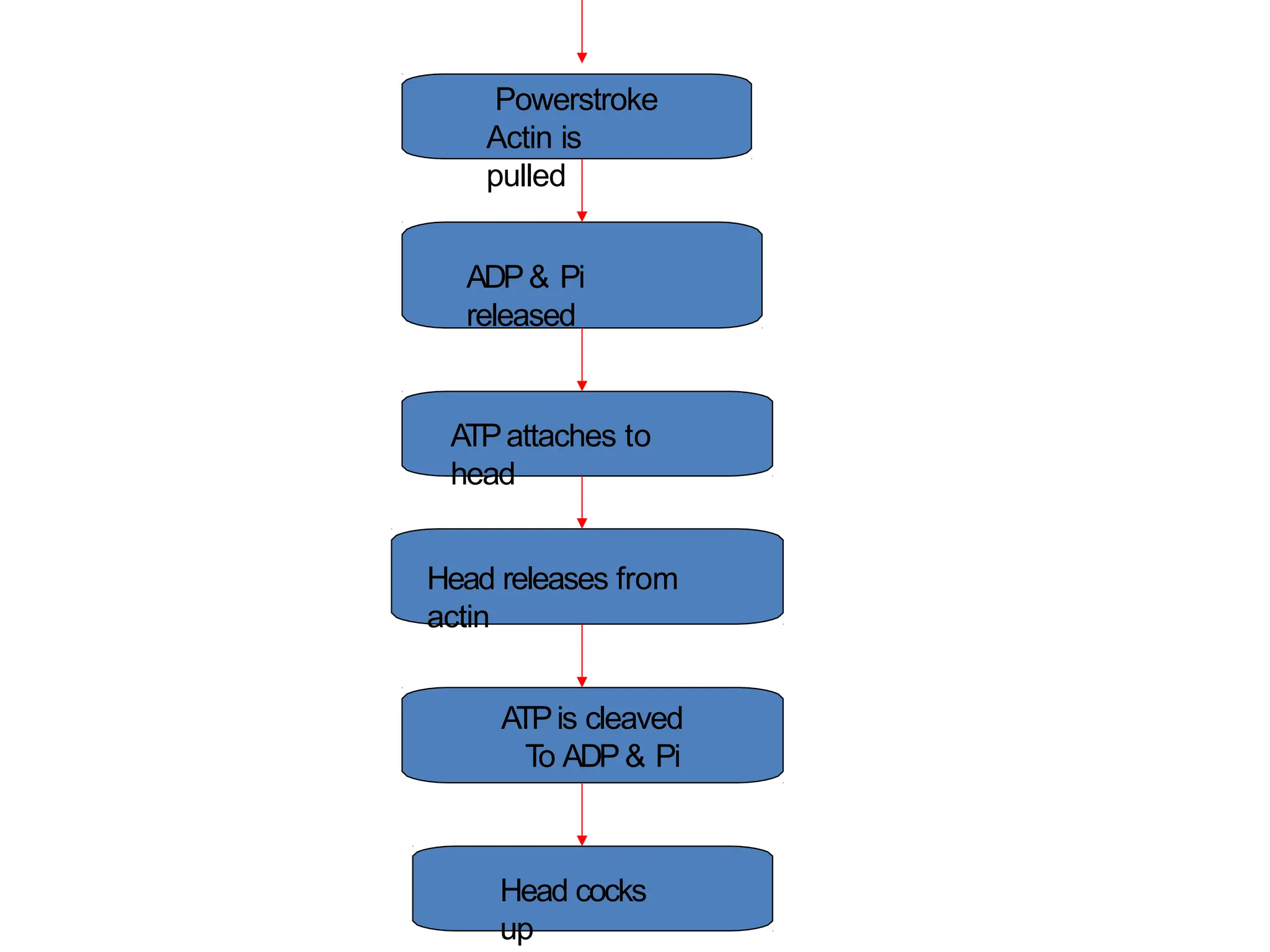
Muscle cells are excitable and capable of transmitting action potentials, converting chemical energy into mechanical responses. There are three types of muscles: skeletal (striated and voluntary), cardiac (less striated and involuntary), and smooth (non-striated and involuntary), each with distinct properties and functions. Muscle contraction theories, including the sliding filament theory, detail how actin and myosin interact during contraction, influenced by factors such as preload, afterload, and the inherent properties of muscle fibers.


![Slow- and Fast-Twitch Fibers
(continued)
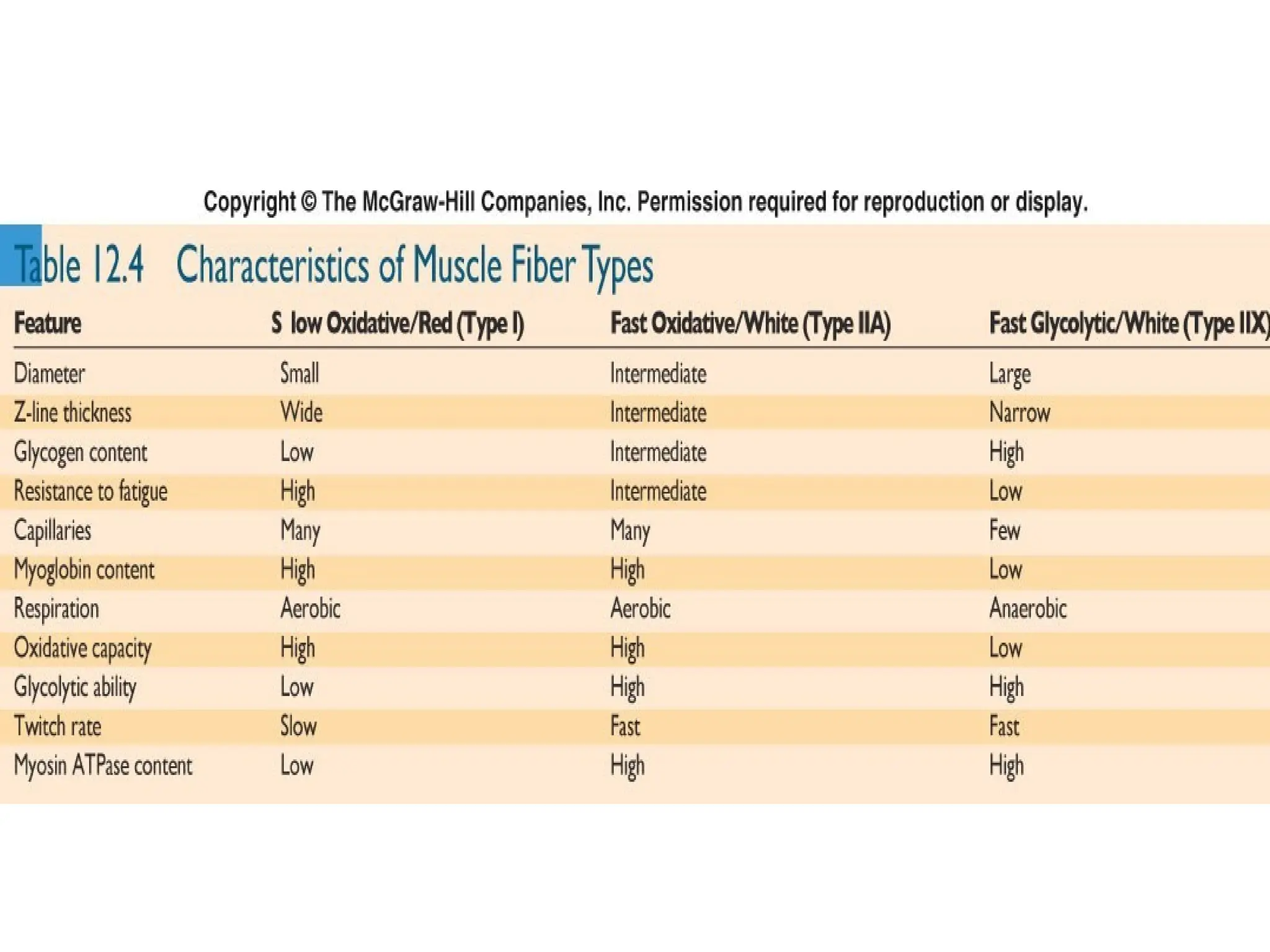
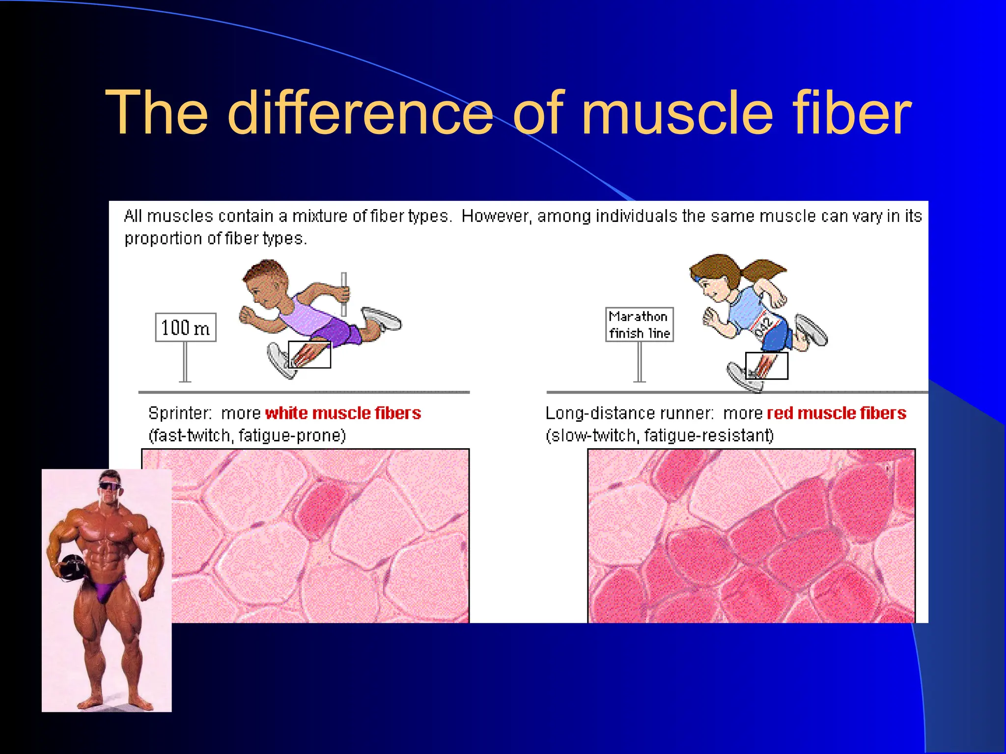
• Slow-twitch (type I fibers):
– Red fibers.
– High oxidative capacity for aerobic
respiration.
– Resistant to fatigue.
– Have rich capillary supply.
– Numerous mitochondria and aerobic
enzymes.
– High [myoglobin].
• Soleus muscle in the leg.
www.freelivedoctor.com](https://image.slidesharecdn.com/skeletalmuscleproperty-241211050149-dc9424a4/75/Skeletal-muscle-property-pptx-properties-of-smooth-muscle-96-2048.jpg)