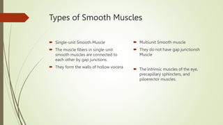
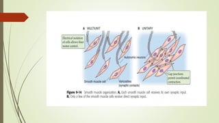
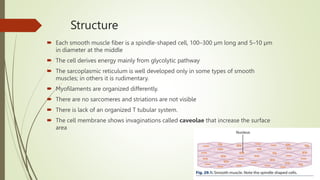
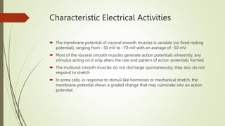
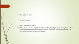
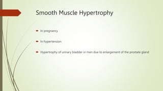
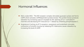
Smooth muscle
By Dr A Amar Sandeep
content
 Types of Smooth Muscles
 Functional Organization
 Innervations of Smooth Muscles
 Electrical Properties
 Mechanism of Contraction
 Mechanical properties
 Smooth Muscle Hypertrophy
 Neural and Hormonal Influences
Types of Smooth Muscles
 Single-unit Smooth Muscle
 The muscle fibers in single-unit
smooth muscles are connected to
each other by gap junctions.
 They form the walls of hollow viscera
 Multiunit Smooth muscle
 They do not have gap junctionsh
Muscle
 The intrinsic muscles of the eye,
precapillary sphincters, and
piloerector muscles.
Functional Organization
 Circular
 Circular and longitudinal
 Circular, longitudinal and oblique
Structure
 Each smooth muscle fiber is a spindle-shaped cell, 100–300 µm long and 5–10 µm
in diameter at the middle
 The cell derives energy mainly from glycolytic pathway
 The sarcoplasmic reticulum is well developed only in some types of smooth
muscles; in others it is rudimentary.
 Myofilaments are organized differently.
 There are no sarcomeres and striations are not visible
 There is lack of an organized T tubular system.
 The cell membrane shows invaginations called caveolae that increase the surface
area
Myofilaments
 Thick, thin and intermediate
 During the sliding-filament mechanism, the gap between the actin filaments
reduces and the shortening force is transmitted through the dense bodies to the
plasma membrane producing contraction of the muscle fiber
Innervations of Smooth Muscles
 Smooth muscles exhibit a spontaneous, slow wave rhythm. Neural stimulation only
modulates (increases or decreases) this basic pattern.
 Branches of the autonomic nervous system innervate the smooth muscles, most of
which are supplied by sympathetic as well as parasympathetic fibers
Electrical Properties
 The smooth muscles react to a variety of stimuli, which may be
 Neural (sympathetic or parasympathetic);
 Hormonal (circulating catecholamines, serotonin, histamine, angiotensin,
vasopressin, oxytocin, estrogen, and progesterone);
 Chemical (hypoxia, hypercapnia, and H+ ); cold; and stretch.
Characteristic Electrical Activities
 The membrane potential of visceral smooth muscles is variable (no fixed resting
potential), ranging from –30 mV to –70 mV with an average of –50 mV.
 Most of the visceral smooth muscles generate action potentials inherently; any
stimulus acting on it only alters the rate and pattern of action potentials formed.
 The multiunit smooth muscles do not discharge spontaneously; they also do not
respond to stretch
 In some cells, in response to stimuli like hormones or mechanical stretch, the
membrane potential shows a graded change that may culminate into an action
potential.
Characteristic Electrical Activities
 Excitatory junction potentials
 Pacemaker potential
 Some smooth muscle cells contract without any change in membrane potential.
Mechanism of Contraction
 Calcium Influx
 Calcium Release
 Binding of Calcium to Calmodulin
 Pumping Back of Calcium to the SR
 Calcium Efflux
Molecular Basis of Contraction & relaxation
 Phasic Contraction
 Tonic Contraction
 Latch-Bridge Mechanism
 When MLCP-induced dephosphorylation of myosin light-chain takes place, it does
not bring about dissociation of myosin from actin until the cytoplasmic calcium
concentration falls below a critical level.
Mechanical properties
 Contractile Response
 Muscle Tone - basic slow wave rhythm
 Length–Tension Relationship - plasticity, the relationship is highly inconsistent.
Smooth Muscle Hypertrophy
 In pregnancy
 In hypertension
 Hypertrophy of urinary bladder in men due to enlargement of the prostate gland
Neural Influences
 Autonomic Control
 1. Parasympathetic stimulation is excitatory, Ach acts by increasing the cytoplasmic
Ca++ through phospholipase C and IP3.
 2. Sympathetic stimulation is inhibitory
 Acting via α receptors, NE increases calcium efflux from the cell. This leads to
decline in cytoplasmic Ca++ and relaxation of the muscle.
 Acting via β receptors, NE stimulates adenylyl cyclase that increases formation of
cAMP, which activates the enzyme cAMP-dependent protein kinase. This protein
kinase as well as cAMP increase calcium uptake by the sarcoplasmic reticulum. This
results in fall in cytoplasmic concentration of Ca++
Hormonal Influences
 Nitric oxide (NO) - The NO-receptor complex stimulates guanylyl cyclase and forms
cGMP, which activates cGMPdependent protein kinase. This protein kinase produces
relaxation by increasing calcium reuptake by SR, opening of Ca++activated K+
channels and decreasing IP3-induced Ca++ release by inhibiting phospholipases C.
 Angiotensin II acting via AT II receptors, vasopressin and endothelin stimulate
contraction of smooth muscles by releasing IP3. Adenosine causes relaxation by
increasing the level of cAMP.
Thank you

Smooth muscle.pptx

  • 1.
    Smooth muscle By DrA Amar Sandeep
  • 2.
    content  Types ofSmooth Muscles  Functional Organization  Innervations of Smooth Muscles  Electrical Properties  Mechanism of Contraction  Mechanical properties  Smooth Muscle Hypertrophy  Neural and Hormonal Influences
  • 3.
    Types of SmoothMuscles  Single-unit Smooth Muscle  The muscle fibers in single-unit smooth muscles are connected to each other by gap junctions.  They form the walls of hollow viscera  Multiunit Smooth muscle  They do not have gap junctionsh Muscle  The intrinsic muscles of the eye, precapillary sphincters, and piloerector muscles.
  • 5.
    Functional Organization  Circular Circular and longitudinal  Circular, longitudinal and oblique
  • 6.
    Structure  Each smoothmuscle fiber is a spindle-shaped cell, 100–300 µm long and 5–10 µm in diameter at the middle  The cell derives energy mainly from glycolytic pathway  The sarcoplasmic reticulum is well developed only in some types of smooth muscles; in others it is rudimentary.  Myofilaments are organized differently.  There are no sarcomeres and striations are not visible  There is lack of an organized T tubular system.  The cell membrane shows invaginations called caveolae that increase the surface area
  • 7.
    Myofilaments  Thick, thinand intermediate  During the sliding-filament mechanism, the gap between the actin filaments reduces and the shortening force is transmitted through the dense bodies to the plasma membrane producing contraction of the muscle fiber
  • 8.
    Innervations of SmoothMuscles  Smooth muscles exhibit a spontaneous, slow wave rhythm. Neural stimulation only modulates (increases or decreases) this basic pattern.  Branches of the autonomic nervous system innervate the smooth muscles, most of which are supplied by sympathetic as well as parasympathetic fibers
  • 9.
    Electrical Properties  Thesmooth muscles react to a variety of stimuli, which may be  Neural (sympathetic or parasympathetic);  Hormonal (circulating catecholamines, serotonin, histamine, angiotensin, vasopressin, oxytocin, estrogen, and progesterone);  Chemical (hypoxia, hypercapnia, and H+ ); cold; and stretch.
  • 10.
    Characteristic Electrical Activities The membrane potential of visceral smooth muscles is variable (no fixed resting potential), ranging from –30 mV to –70 mV with an average of –50 mV.  Most of the visceral smooth muscles generate action potentials inherently; any stimulus acting on it only alters the rate and pattern of action potentials formed.  The multiunit smooth muscles do not discharge spontaneously; they also do not respond to stretch  In some cells, in response to stimuli like hormones or mechanical stretch, the membrane potential shows a graded change that may culminate into an action potential.
  • 11.
  • 13.
     Excitatory junctionpotentials  Pacemaker potential  Some smooth muscle cells contract without any change in membrane potential.
  • 14.
    Mechanism of Contraction Calcium Influx  Calcium Release  Binding of Calcium to Calmodulin  Pumping Back of Calcium to the SR  Calcium Efflux
  • 16.
    Molecular Basis ofContraction & relaxation
  • 18.
     Phasic Contraction Tonic Contraction  Latch-Bridge Mechanism  When MLCP-induced dephosphorylation of myosin light-chain takes place, it does not bring about dissociation of myosin from actin until the cytoplasmic calcium concentration falls below a critical level.
  • 19.
    Mechanical properties  ContractileResponse  Muscle Tone - basic slow wave rhythm  Length–Tension Relationship - plasticity, the relationship is highly inconsistent.
  • 20.
    Smooth Muscle Hypertrophy In pregnancy  In hypertension  Hypertrophy of urinary bladder in men due to enlargement of the prostate gland
  • 21.
    Neural Influences  AutonomicControl  1. Parasympathetic stimulation is excitatory, Ach acts by increasing the cytoplasmic Ca++ through phospholipase C and IP3.  2. Sympathetic stimulation is inhibitory  Acting via α receptors, NE increases calcium efflux from the cell. This leads to decline in cytoplasmic Ca++ and relaxation of the muscle.  Acting via β receptors, NE stimulates adenylyl cyclase that increases formation of cAMP, which activates the enzyme cAMP-dependent protein kinase. This protein kinase as well as cAMP increase calcium uptake by the sarcoplasmic reticulum. This results in fall in cytoplasmic concentration of Ca++
  • 22.
    Hormonal Influences  Nitricoxide (NO) - The NO-receptor complex stimulates guanylyl cyclase and forms cGMP, which activates cGMPdependent protein kinase. This protein kinase produces relaxation by increasing calcium reuptake by SR, opening of Ca++activated K+ channels and decreasing IP3-induced Ca++ release by inhibiting phospholipases C.  Angiotensin II acting via AT II receptors, vasopressin and endothelin stimulate contraction of smooth muscles by releasing IP3. Adenosine causes relaxation by increasing the level of cAMP.
  • 25.