Introduction
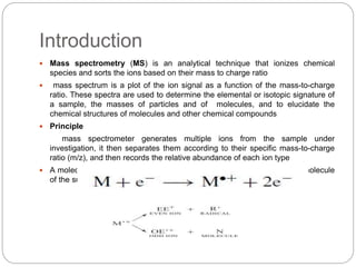
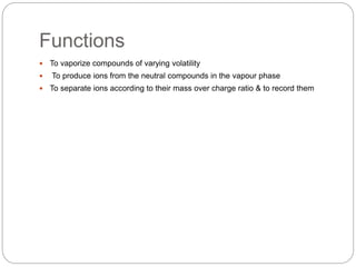
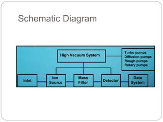
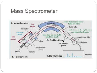
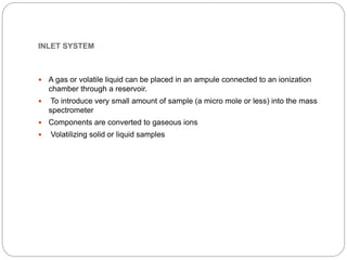
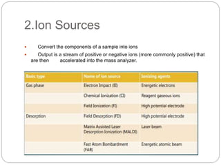
 Mass spectrometry (MS) is an analytical technique that ionizes chemical
species and sorts the ions based on their mass to charge ratio
 mass spectrum is a plot of the ion signal as a function of the mass-to-charge
ratio. These spectra are used to determine the elemental or isotopic signature of
a sample, the masses of particles and of molecules, and to elucidate the
chemical structures of molecules and other chemical compounds
 Principle
mass spectrometer generates multiple ions from the sample under
investigation, it then separates them according to their specific mass-to-charge
ratio (m/z), and then records the relative abundance of each ion type
 A molecular ion results when one electron is removed from the parent molecule
of the substance
By Breena & Anum
Masoood
Mass Spectrometery
Functions
 To vaporize compounds of varying volatility
 To produce ions from the neutral compounds in the vapour phase
 To separate ions according to their mass over charge ratio & to record them
Schematic Diagram
Instrumentation
 Following are components of mass spectrometer
1) inlet system
2) ion sources
3) mass analysers
4) detectors
5) vacuum system
Mass Spectrometer
INLET SYSTEM
 A gas or volatile liquid can be placed in an ampule connected to an ionization
chamber through a reservoir.
 To introduce very small amount of sample (a micro mole or less) into the mass
spectrometer
 Components are converted to gaseous ions
 Volatilizing solid or liquid samples
2.Ion Sources
 Convert the components of a sample into ions
 Output is a stream of positive or negative ions (more commonly positive) that
are then accelerated into the mass analyzer.
Gas Phase
Electron impact Ionization is the most common type of ionization
 The sample is bombarded by electrons which come from a heated filament
 The electrons run in a stream between the cathode and anode
 When the sample passes through the electron stream, the high energy electrons
in the stream knock electrons out of the sample to form ions
Desorption
Matrix Assisted Laser Desorption
 MALDI uses a nitrogen laser to promote ionization of molecules prior to ion
Separation in a mass spectrometer.
 It is usually combined with time of flight (TOF)
 sample has to be dissolved in a matrix that absorbs UV radiation at around the
wavelength (337 nm) produced by the laser.
 The sample solution is mixed with matrix solution on a metal plate and allowed to
dry prior to being introduced into the Instrument. The laser is then directed at the
target plate to promote ionization
 High-molecular-weight compounds as singly charged ions and, in combination
with TOF separation, Measurement of high-molecular-weight species can be
carried out.
 Figure shows the ions generated from coenzyme A and two acyl coas using
MALDITOF in negative ion mode.
Ionisation chamber
Acceleration
 Acceleration is a simple step where the ions are placed between a set of charges
parallel plates
 The ions will then be repelled by one plate and attracted to the other
 There is a slit cut in the plate which the ions are attracted to the force of
attraction and repulsion forces the ions through the slit at an accelerated rate
 The speed of acceleration can be adjusted by changing the charge on the plates.
3.Mass Analyser
 Magnetic Sector Analyzer (MSA) – High resolution, exact mass, original MA
 Quadruple Analyzer (Q) – Low (1 amu) resolution, fast, cheap
 Time-of-Flight Analyzer (TOF) – No upper m/z limit, high throughput
 Ion Trap Mass Analyzer (QSTAR) – Good resolution, all-in-one mass analyzer
 Ion Cyclotron Resonance (FT-ICR) – Highest resolution, exact mass, costly
Ions are deflected by the magnetic field surrounding the instrument which
depends on the mass and charge of the ions
 Ion stream C: The heavier ions are deflected the least
 Ion Stream B: Correct mass ions and charge travel to the detector
 Ion Stream A: The lightest ions are deflected the most
Magnetic sector mass
spectrometry
 The original mass spectrometric technique was based on separation of charged
ions generated in an ion source using a curved magnet. Magnetic sector
instruments are still used.
 In a magnetic sector instrument the ions generated are pushed out of the source
by a repeller potential of same charge as the ion itself (most often positive).
 They are then accelerated in an electric field and travel through an electrostatic
field region so that they are forced to fall into a narrow range of kinetic energies
prior to entering the field of a circular magnet.
 Then they adopt a flight path through the magnetic field depending on their
charge to mass (m/z) ratio; the large ions are
 deflected less by the magnetic field:
Where H is the magnetic field strength, r is the radius of the circular path in which the Ion travels,
and V is the accelerating voltage.
At particular values for H and V only ions of a particular mass adopt a flight path that enables them
to pass through the collector slit and be detected.
Quadrapole mass ANALYZER
 They are generally considerably more compact than magnetic sector instruments
and are commonly found in commercial bench top mass spectrometers
 The heart of a quadrupole instrument is the four parallel cylindrical (originally
hyperbolic) rods that serve as electrodes
 Opposite rods are connected electrically, one pair being attached to the positive
side of a variable dc source and the other pair to the negative terminal
 Variable radio-frequency ac voltages, which are 1800 out of phase, are applied
to each pair of rods.
 To obtain a mass spectrum with this device ions are accelerated into the space
between the rod; by a potential difference of 5 to 10 V
 Meanwhile, the ac and dc voltages on the rods are increased simultaneously
while maintaining their ratio constant. At any given moment, all of the ions except
those having a certain m/z value strike the rods and are converted to neutral
molecules. Thus, only ions having a limited range of m/z values reach the
transducer
Continue…
4.Detectors
 The final element of the mass spectrometer is the detector that records either the
charge induced or the current produced when an ion passes by or hits a surface
 In a scanning instrument, the signal produced in the detector during the course of
the scan versus where the instrument is in the scan (at what m/Q) will produce
a mass spectrum, a record of ions as a function of m/Q.
 Typically, some type of electron multiplier is used, though other detectors
including Fraraday Cups and ion-to-proton detectors are also used
5.Vacuum system
 Diffusion and turbomolecular pumps often used to achieve the high vacuum
necessary for operating many mass spectrometers
 These pumps are used with a rough pump (or forepump) to move gas molecules
from inside a vacuum chamber (a mass spectrometer) to outside the system
 Although the pressures achieved by these two pumping systems are similar, the
cost of equipment, the operating costs, and the procedures and speed of pump
down and vent cycles are quite different
Types
 Diffusion pump
In a diffusion pump, a net direction is achieved by collision of the residual gas
molecules with a directed and confined stream of gasphase molecules of the
pump’s working fluid
 Turbomolecular pump
In a turbomolecular pump, the residual gas molecule collide with the angled
spinning rotors on a turbine shaft
Applications
 Pharmaceutical Industry
Bioavailability studies
Drug metabolism studies,Pharmacokinetics
Characterization of potential drugs
Drug Degradation product analysis
Screening of drug candidates
Identifying drug targets
 Trace Gas Analysis
 Biomolecular characterization
Proteins and peptides
Oligonucleotides
 Environmentalanalysis
Pesicides on fodd
Soil and ground water conatmination
 Space Exploration
 Forensic Toxicology
 Archaeological Dating
mass spectro pres.pptx

mass spectro pres.pptx

  • 1.
    Introduction  Mass spectrometry(MS) is an analytical technique that ionizes chemical species and sorts the ions based on their mass to charge ratio  mass spectrum is a plot of the ion signal as a function of the mass-to-charge ratio. These spectra are used to determine the elemental or isotopic signature of a sample, the masses of particles and of molecules, and to elucidate the chemical structures of molecules and other chemical compounds  Principle mass spectrometer generates multiple ions from the sample under investigation, it then separates them according to their specific mass-to-charge ratio (m/z), and then records the relative abundance of each ion type  A molecular ion results when one electron is removed from the parent molecule of the substance
  • 2.
    By Breena &Anum Masoood Mass Spectrometery
  • 3.
    Functions  To vaporizecompounds of varying volatility  To produce ions from the neutral compounds in the vapour phase  To separate ions according to their mass over charge ratio & to record them
  • 4.
  • 5.
    Instrumentation  Following arecomponents of mass spectrometer 1) inlet system 2) ion sources 3) mass analysers 4) detectors 5) vacuum system
  • 6.
  • 7.
    INLET SYSTEM  Agas or volatile liquid can be placed in an ampule connected to an ionization chamber through a reservoir.  To introduce very small amount of sample (a micro mole or less) into the mass spectrometer  Components are converted to gaseous ions  Volatilizing solid or liquid samples
  • 8.
    2.Ion Sources  Convertthe components of a sample into ions  Output is a stream of positive or negative ions (more commonly positive) that are then accelerated into the mass analyzer.
  • 9.
    Gas Phase Electron impactIonization is the most common type of ionization  The sample is bombarded by electrons which come from a heated filament  The electrons run in a stream between the cathode and anode  When the sample passes through the electron stream, the high energy electrons in the stream knock electrons out of the sample to form ions
  • 10.
    Desorption Matrix Assisted LaserDesorption  MALDI uses a nitrogen laser to promote ionization of molecules prior to ion Separation in a mass spectrometer.  It is usually combined with time of flight (TOF)  sample has to be dissolved in a matrix that absorbs UV radiation at around the wavelength (337 nm) produced by the laser.  The sample solution is mixed with matrix solution on a metal plate and allowed to dry prior to being introduced into the Instrument. The laser is then directed at the target plate to promote ionization  High-molecular-weight compounds as singly charged ions and, in combination with TOF separation, Measurement of high-molecular-weight species can be carried out.  Figure shows the ions generated from coenzyme A and two acyl coas using MALDITOF in negative ion mode.
  • 11.
  • 12.
    Acceleration  Acceleration isa simple step where the ions are placed between a set of charges parallel plates  The ions will then be repelled by one plate and attracted to the other  There is a slit cut in the plate which the ions are attracted to the force of attraction and repulsion forces the ions through the slit at an accelerated rate  The speed of acceleration can be adjusted by changing the charge on the plates.
  • 13.
    3.Mass Analyser  MagneticSector Analyzer (MSA) – High resolution, exact mass, original MA  Quadruple Analyzer (Q) – Low (1 amu) resolution, fast, cheap  Time-of-Flight Analyzer (TOF) – No upper m/z limit, high throughput  Ion Trap Mass Analyzer (QSTAR) – Good resolution, all-in-one mass analyzer  Ion Cyclotron Resonance (FT-ICR) – Highest resolution, exact mass, costly Ions are deflected by the magnetic field surrounding the instrument which depends on the mass and charge of the ions  Ion stream C: The heavier ions are deflected the least  Ion Stream B: Correct mass ions and charge travel to the detector  Ion Stream A: The lightest ions are deflected the most
  • 14.
    Magnetic sector mass spectrometry The original mass spectrometric technique was based on separation of charged ions generated in an ion source using a curved magnet. Magnetic sector instruments are still used.  In a magnetic sector instrument the ions generated are pushed out of the source by a repeller potential of same charge as the ion itself (most often positive).  They are then accelerated in an electric field and travel through an electrostatic field region so that they are forced to fall into a narrow range of kinetic energies prior to entering the field of a circular magnet.  Then they adopt a flight path through the magnetic field depending on their charge to mass (m/z) ratio; the large ions are  deflected less by the magnetic field: Where H is the magnetic field strength, r is the radius of the circular path in which the Ion travels, and V is the accelerating voltage. At particular values for H and V only ions of a particular mass adopt a flight path that enables them to pass through the collector slit and be detected.
  • 15.
    Quadrapole mass ANALYZER They are generally considerably more compact than magnetic sector instruments and are commonly found in commercial bench top mass spectrometers  The heart of a quadrupole instrument is the four parallel cylindrical (originally hyperbolic) rods that serve as electrodes  Opposite rods are connected electrically, one pair being attached to the positive side of a variable dc source and the other pair to the negative terminal  Variable radio-frequency ac voltages, which are 1800 out of phase, are applied to each pair of rods.  To obtain a mass spectrum with this device ions are accelerated into the space between the rod; by a potential difference of 5 to 10 V  Meanwhile, the ac and dc voltages on the rods are increased simultaneously while maintaining their ratio constant. At any given moment, all of the ions except those having a certain m/z value strike the rods and are converted to neutral molecules. Thus, only ions having a limited range of m/z values reach the transducer
  • 16.
  • 17.
    4.Detectors  The finalelement of the mass spectrometer is the detector that records either the charge induced or the current produced when an ion passes by or hits a surface  In a scanning instrument, the signal produced in the detector during the course of the scan versus where the instrument is in the scan (at what m/Q) will produce a mass spectrum, a record of ions as a function of m/Q.  Typically, some type of electron multiplier is used, though other detectors including Fraraday Cups and ion-to-proton detectors are also used
  • 18.
    5.Vacuum system  Diffusionand turbomolecular pumps often used to achieve the high vacuum necessary for operating many mass spectrometers  These pumps are used with a rough pump (or forepump) to move gas molecules from inside a vacuum chamber (a mass spectrometer) to outside the system  Although the pressures achieved by these two pumping systems are similar, the cost of equipment, the operating costs, and the procedures and speed of pump down and vent cycles are quite different Types  Diffusion pump In a diffusion pump, a net direction is achieved by collision of the residual gas molecules with a directed and confined stream of gasphase molecules of the pump’s working fluid  Turbomolecular pump In a turbomolecular pump, the residual gas molecule collide with the angled spinning rotors on a turbine shaft
  • 19.
    Applications  Pharmaceutical Industry Bioavailabilitystudies Drug metabolism studies,Pharmacokinetics Characterization of potential drugs Drug Degradation product analysis Screening of drug candidates Identifying drug targets  Trace Gas Analysis  Biomolecular characterization Proteins and peptides Oligonucleotides  Environmentalanalysis Pesicides on fodd Soil and ground water conatmination  Space Exploration  Forensic Toxicology  Archaeological Dating