SlideShare a Scribd company logo
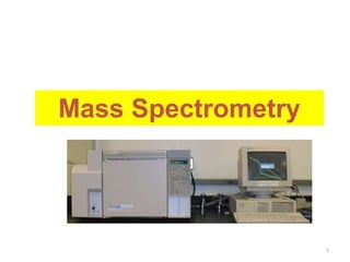
Mass Spectrometry
1
Introduction
 MS is an analytical technique that provides qualitative &
quantitative information, including the mass of molecules &
atoms in samples as well as the molecular structure of
organic and inorganic cpds
– It is an instrument that separates gas phase ionized
atoms, molecules, & fragments of molecules by the
difference in their mass-to-charge ratios.
 In MS, a substance is converted into fragment ions.
2
Introduction…
 The fragments (usually cations) are sorted on the basis of
mass-to-charge ratio, m/z.
 The bulk of the ions usually carry a unit positive charge, thus
m/z is equivalent to the MW of the fragment.
 The analysis of MS information involves the re-assembling of
fragments, working backwards to generate the original
molecule.
3
 A MS needs to perform three functions:
• Creation of ions – e.g. the sample molecules are
subjected to a high energy beam of electrons,
converting some of them to ions
• Separation of ions – as they are accelerated in an
electric field, the ions are separated according to mass-
to-charge ratio (m/z)
• Detection of ions – as each separated population of
ions is generated, the spectrometer needs to qualify
and quantify them
Mass Spectrometer
4
5
Mass Spectrometer…
Inlet
Ion
source
Mass
Analyzer Detector
Data
System
High Vacuum System
Mass Spectrometer Block Diagram
7
Inlet System:
To introduce a very small amount of sample (mol or less) into
the MS that converted to gaseous ions. ( a means for
volatilizing solid or liquid samples is presents).
Ion sources:
Convert the components of a sample into ions.
Mass analyzer
Analogous to grating in an optical spectrometer.
Dispersion is based upon the mass/charge ratios of the analyte
ions rather than upon the wavelength of photons.
Mass Spectrometer…
Detectors:
Convert the beam of ions into an electrical signal that can
then be processed, stored in the memory of a computer
and displayed or recorded in a variety ways.
Vacuum System:
To create low pressure (10-4 to 10-8 torr) in all the instrument
components except the signal processor and readout.
To prevent the ions of interest from colliding with
air molecules.
Mass Spectrometer…
8
9
Ion sources
Starting point: Formation of gaseous analyte Ions.
Methods of ion formation: Two major categories:
1- Gas-phase sources
-The sample is first vaporized and then ionized.
-Restricted to thermally stable compounds of B.pt. < 500 0C.
-Limited to Compounds of MWt’s <103dalton .
2- Desorption sources
 The sample in a solid or liquid state is converted directly into
gaseous ions (not require volatilization of analyte molecules)
 Applicable to nonvolatile and thermally unstable samples
 Applicable to analytes having of 105 dalton or larger.
Ion sources
10
Ionizing agent
Name
Basic Type
Energetic electrons
Electron Impact (E I)
Reagent gaseous ions
Chemical Ionization (CI)
High-potential electrode
Field ionization (FI)
Gas Phase
High-potential electrode
Field desorption (FD)
Desorption
High electric Field
Electrospray ionization (ESI)
Laser beam
Matrix-assisted desorption/ionization
(MALDI)
Fission fragments from
252Cf
Plasma desorption (PD)
Energetic atomic beam
Fast atom bombardment (FAB)
Energetic beam of ions
Secondary ion mass spectrometry (SIMS)
High temperature
Thermospray ionization (TI)
Ion sources
In a mass spectrometer, molecules in the gaseous state under low pressure are
bombarded with a beam of high energy electrons (about 70 electron-volts; eV)
This bombardment can first dislodge one of the electrons of the molecule and
produce a positively charged ion called “The molecular ion”
M + e’ M+• + 2 e’
Molecule high-energy electron molecular ion
The molecular ion contains also an odd number of electrons, thus it is a Free
radical, or generally “Radical Cation”
Electron Ionization
11
• This is done by adding a reagent gas into the ion
source.
• Electrons ionize the reagent gas (e.g. methane).
• These ions are highly reactive and react with the
analyte (XH).
• A molecular ion: or
Chemical Ionization
12
• At the end of this capillary, a fine aerosolis formed by nitrogen gas
flowing along the tip of the capillary.
• Because the mobile phase is volatile, the liquid droplets evaporate by
electrical potential & are flushed away by adrying gas.
• The analyte molecules remain charged & are extracted into the
vacuum area of MS.
• Used for Acidic or
basic analytes
Electrospray Ionization
13
Atmospheric Pressure Chemical Ionization
14
can then react with the analyte (MH):
&
hn
Laser
1. Sample is mixed with matrix (X)
and dried on plate.
2. Laser flash ionizes matrix
molecules.
3. Sample molecules (M) are
ionized by proton transfer:
XH+ + M  MH+ + X.
MH+
MALDI: Matrix Assisted Laser Desorption Ionization
+/- 20 kV Grid (0 V)
Sample plate
• ICP-MS
• The sample constituents are atomized into free
atoms, and some of the free atoms are also
ionized by losing an electron in hot plasma.
Inductively Coupled Plasma
16
17
Mass Analyzers
Mass analyzers separate ions based on their mass-to-charge ratio (m/z)
– Several devices are available for separating ions with different m/z
ratios.
o magnetic-sector,
o quadrupole,
o time-of-flight and
o ion traps are most used.
Mass analyzer should be:
- Capable of distinguishing between minute mass differences.
- Allows passage of sufficient number of ions to yield measurable
currents.
- Key specifications are:
o resolution,
o mass measurement accuracy, and
o sensitivity.
=>
18
Magnetic-sector
•Amount of deflection depends on m/z.
•The detector signal is proportional to the number of ions hitting it.
•By varying the magnetic field, ions of all masses are collected and counted.
• Analyzer tube is surrounded by a magnet whose magnetic field deflects
the positively charged fragments in a curved path.
• At a given magnetic field strength, the degree to which the path is
curved depends on the mass-to-charge ratio (m/e) of the fragment:
• Path of a fragment with a smaller m/e value will bend more than that of
a heavier fragment. In this way, the particles with the same m/z values
can be separated from all the others.
• If a fragment’s path matches the curvature of analyzer tube, the
fragment will pass through the tube and out the ion exit slit (These ions
are said to be in register).
• A collector records the relative number of fragments with a particular
m/e passing through the slit. The more stable the fragment, the more
likely it will make it to the collector.
• Strength of magnetic field is gradually increased, so fragments with
progressively larger m/e values are guided through tube and out the exit
slit.
Mass Analyzers…
19
Quadrupole Mass Analyzer
Uses a combination of RF
and DC voltages to operate
as a mass filter.
• Has four parallel metal
rods.
• Lets one mass pass
through at a time.
• Can scan through all
masses or sit at one
fixed mass.
mass scanning mode
m1
m3
m4 m2
m3
m1
m4
m2
single mass transmission mode
m2 m2 m2 m2
m3
m1
m4
m2
Quadrupoles have variable ion transmission modes
22
Time-of-flight (TOF) Mass Analyzer
•Ions are accelerated in pulses by means of an electric potential imposed
on a back plate right in the back of the ion source.
•The mass of each ion is thus determined based on its flight time to reach
the detector.
• Small ions reach the detector before large ones.
HYPHENATED TECHNIQUES
• Hyphenated Techniques combine
chromatographic and spectral methods to
exploit the advantages of both.
23
• GC-MS: the analytes are ionized under vacuum
conditions e.g. by EI, CI
• LC-MS: ionization is performed at atmospheric
pressure by ESI, APCI
• In both cases sample constituents are separated
by passage through a chromatographic column.
• Each cpd elutes into the mass spectrometer for mass
spectrometric analysis.
• MS - advanced chromatography detector.
Chromatography Coupled with MS
24
Fig. Total ion current chromatogram & mass spectrum for component 3 in a mixture
of four components
25
• Full scan and recording of spectra;
• Selected ion monitoring (SIM);
• Selected reaction monitoring (SRM).
26
The Mass Spectrum?
27
A mass spectrum is a presentation of the masses of the
positively charged ions (peaks) separated on the basis of
mass/charge (m/z) versus their relative concentrations.
Mass Spectrum (cont.)
28
Three types of peaks are observed in a MS Spectrum:
1. Base Peak
2. Molecular Ion Peak
3. Fragment Peaks
Base Peak
The most intense peak (most stable ion) in the mass spectrum
is called the Base Peak.
This Peak is assigned a value of 100%.
The intensities of the other peaks are reported as percentage
of the base peak.
Molecular Ion Peak
– In a Mass Spectrum Molecular Ion Peak is usually the peak
of highest mass number except for the isotope peak.
– It is important to note that there are possibilities and cases
where Base Peak and Molecular Ion Peak for a given
compound are same.
Mass Spectrum (cont.)
29
30
Fragment Peaks
All other peaks obtained and seen in the Mass Spectrum are derived
from Molecular Ion Peak or Base Peak and are thus called Fragment
Peaks.
*Base peak can be also fragment peak
M+ peak
Base peak
F. peak
F. peak
Mass Spectrum (cont.)
Example: Pentane molecule:
Mass spectrum of pentane,
shown as a bar graph and in
tabular form.
Base peak represents fragment
that appears in greatest
abundance. Value of molecular
ion gives molecular mass of the
compound.
Note that:
The way by which the
molecular ion to be
fragmented depends
on the strength of its
bonds and stability of
the fragments.
31
Note that:
1- In some cases the base peak is the molecular ion peak, but in most cases it is
different from that of the molecular ion (according to the relative stability of ions
during fragmentation).
2- Peaks are commonly observed at m/e values one and two units less than the
m/e values of the carbocations because the carbocations can undergo further
fragmentation by losing one or two hydrogen atoms.
3- Small peak that occurs at m/e 73 (0.52%) is known as M+1 peak to indicate
that “It is one mass unit greater than the molecular ion(M+.),
The M+1 peak appear because most of elements have more than one naturally
occurring isotope.
32
Principal stable isotops of common elements:
From the table :
•For C, H, N and Si the principal heavier isotope is M+1.
•For O, Si, S, Cl and Br , the principal heavier isotope is the M+2
•For F and I, there is no heavier isotopes (not affect M+1 or M+2)
33
Some guides to determine molecular formula of
organic compounds:
 Nitrogen rule:
– If molecular ion (M+) is even number, compound must contain an
even number of N atoms (zero is considered as even number)
 Number of Carbon atoms:
– Relative abundance of M+1 peak can be used for determination of
number of carbons assuming that silicon and large number of
nitrogens (more than 3 N) are not present.
No of Carbons = Relative abundance of M+1/ 1.10
 Relative abundance of M+2 peak indicates the presence (or
absence) of Oxygen (0.2), Silicon (3.35),Sulfer (4.4), Chlorine
(32.5) & Bromine (98.0)
34
Some guides to determine molecular formula of
organic compounds…
 Molecular formula after this can be established by adding
the suitable number of hydrogens and oxygens if necessary.
 The index of hydrogen deficiency (Due to double and triple
bonds and cyclization) can be calculated for the compounds
containing C, H, N, O, S & halogens from the formula:
Index = Carbons - Hydrogens/2 – Halogens/2 + Nitrogens/2 +1
– Divalent atoms like O and S are not counted in formula. The index
mainly used to indicate the number of double bonds.
35
High Resolution Mass Spectroscopy:
• Resolution: A measure of how well a mass
spectrometer separates ions of different mass.
– Low resolution: Refers to instruments capable of
separating only ions that differ in nominal mass;
that is ions that differ by at least 1 or more atomic
mass units (amu).
– High resolution: Refers to instruments capable of
separating ions that differ in mass by as little as
0.0001 amu.
36
High Resolution Mass Spectroscopy…
• Example:
– A molecule with mass of 44 could be C3H8, C2H4O, CO2, or
CN2H4.
– Thus, high resolution mass spectrometer is capable of
measuring mass with a high accuracy so can easily
distinguish among these 4 molecules.
C3H8 C2H4O CO2 CN2H4
44.06260 44.02620 43.98983 44.03740
37
Fragmentation rules in MS
1. Intensity of M.+ is Larger for linear chain than for branched compound
2. Intensity of M.+ decrease with Increasing M.W.
3. Cleavage is favored at branching
 reflecting the Increased stability of the carbocations
 This is a consequence of the increased stability of a 30 carbocation over a 20 ,
which in turn is more stable than a 10 one.
R
R”
CH
R’
Loss of Largest Subst. Is most favored
38
C
H3
CH3
C
H3
C
H3
CH3
C
H3
C
H3
MW=170
M.+ is absent with heavy branching
Fragmentation occur at branching: largest fragment loss
Branched alkanes
39
Fragmentation rules in MS…
4. Aromatic Rings, Double bond, Cyclic
structures stabilize M.+
5. Double bond favor Allylic Cleavage
 Resonance – Stabilized Cation
C
H2
+
CH CH2 R
- R
.
C
H2
+
CH CH2
C
H2 CH CH2
+
40
41
Fragmentation rules in MS….
6. a) Saturated Rings lose a Alkyl Chain (case
of branching)
b) Unsaturated Rings  Retro-Diels-Alder rxn
(Two Bond Cleavage)
CH2
CH2
CH2
CH2
+
+ . + .
R
+ .
+
-R
.
42
Fragmentation rules in MS….
7. In alkyl-substituted aromatic compounds,
cleavage is very probable at the bond β to the
ring.
– Giving the resonance-stabilized benzyl ion or more
likely, the tropylium ion:
Tropylium ion 43
8. C-C Next to Heteroatom cleave leaving
the charge on the Heteroatom
Fragmentation rules in MS….
44
9- Cleavage associated with elimination of small, stable, neutral molecules
such as carbon monoxide, olefines, water, ammonia, ketones or alcohols,
often occur with some sort of rearrangement.
 One important example is the so-called McLafferty rearrangement (Two
step fragmentation or two bond cleavage)
 To undergo a McLafferty rearrangement, a molecule must possess an
 Appropriately located heteroatom
 A П -system (usually a double bond), and
 An abstractable hydrogen atom g to the C = O system
45
Fragmentation rules in MS….
Pharmaceutical analysis
Bioavailability studies
Drug metabolism studies, pharmacokinetics
Characterization of potential drugs
Drug degradation product analysis
Screening of drug candidates
Identifying drug targets
Biomolecule characterization
Proteins and peptides
Oligonucleotides
Environmental analysis
Pesticides on foods
Soil and groundwater contamination
Forensic analysis/clinical
Applications of Mass Spectrometry
MS/MS
MS/MS
+
+
+ +
+
1 peptide
selected for
MS/MS
The masses of all
the pieces give an
MS/MS spectrum
Peptide
mixture
Have only masses
to start
Interpretation of an MS/MS spectrum to derive
structural information is analogous to solving
a puzzle
+
+
+ +
+
Use the fragment ion masses as specific pieces of
the puzzle to help piece the intact molecule back
together

More Related Content

Similar to Mass spectrometry instrument for analysing mass

Mass.pptx instrumentation, principle, theory
Mass.pptx instrumentation, principle, theoryMass.pptx instrumentation, principle, theory
Mass.pptx instrumentation, principle, theory
Dr. Vijaya Barge
 
MASS SPECTROSCOPY.pptx by Pranav Lendhey
MASS SPECTROSCOPY.pptx by Pranav LendheyMASS SPECTROSCOPY.pptx by Pranav Lendhey
MASS SPECTROSCOPY.pptx by Pranav Lendhey
36PranavLendhey
 
mass spectro pres.pptx
mass spectro pres.pptxmass spectro pres.pptx
mass spectro pres.pptx
breenaawan
 
Mass spectroscopy
Mass spectroscopyMass spectroscopy
Mass spectroscopy
aqsa ayoub
 
Mass spectrometry
Mass spectrometryMass spectrometry
Mass spectrometry
AMRITHA K.T.K
 
Mass spectroscopy
Mass spectroscopyMass spectroscopy
Mass spectroscopy
Aliya Firdous
 
Mass spectrometry.pptx
Mass spectrometry.pptxMass spectrometry.pptx
Mass spectrometry.pptx
Prarthana Sharma
 
Mass spectrometry
Mass spectrometryMass spectrometry
Mass spectrometry
Dr. Krishna Swamy. G
 
Mass spectrometry
Mass spectrometryMass spectrometry
Mass spectrometry
Sadiq Rahim
 
Mass Spectrometry
Mass SpectrometryMass Spectrometry
Mass Spectrometry
Burdwan University
 
Mass Spectroscopy (Instrumentation & Spectral analysis ).pptx
Mass Spectroscopy (Instrumentation & Spectral analysis ).pptxMass Spectroscopy (Instrumentation & Spectral analysis ).pptx
Mass Spectroscopy (Instrumentation & Spectral analysis ).pptx
Aquib Siddiqui
 
mass shruti
mass shrutimass shruti
mass shruti
Shruti Verma
 
Presentation on mass spectroscopy
Presentation on mass spectroscopyPresentation on mass spectroscopy
Presentation on mass spectroscopy
smita shelke
 
Lc -ms seminar
Lc -ms seminarLc -ms seminar
Lc -ms seminar
NIDHIYJ
 
MS.pptx
MS.pptxMS.pptx
MS.pptx
GeletaGalataa
 
Mass assingment
Mass  assingmentMass  assingment
Mass assingment
arikatlakalyani
 
Mass spectrometry detail to up
Mass spectrometry detail to upMass spectrometry detail to up
Mass spectrometry detail to up
Shouvik kumar Nandy
 
mass spectrOSCOPY
 mass spectrOSCOPY mass spectrOSCOPY
Mass Spectrometry
Mass SpectrometryMass Spectrometry
Mass Spectrometry
University of Allahabad
 
Mass spectroscopy for M Sc I Chemistry SPPU
Mass spectroscopy for M Sc I Chemistry SPPUMass spectroscopy for M Sc I Chemistry SPPU
Mass spectroscopy for M Sc I Chemistry SPPU
siraj174
 

Similar to Mass spectrometry instrument for analysing mass (20)

Mass.pptx instrumentation, principle, theory
Mass.pptx instrumentation, principle, theoryMass.pptx instrumentation, principle, theory
Mass.pptx instrumentation, principle, theory
 
MASS SPECTROSCOPY.pptx by Pranav Lendhey
MASS SPECTROSCOPY.pptx by Pranav LendheyMASS SPECTROSCOPY.pptx by Pranav Lendhey
MASS SPECTROSCOPY.pptx by Pranav Lendhey
 
mass spectro pres.pptx
mass spectro pres.pptxmass spectro pres.pptx
mass spectro pres.pptx
 
Mass spectroscopy
Mass spectroscopyMass spectroscopy
Mass spectroscopy
 
Mass spectrometry
Mass spectrometryMass spectrometry
Mass spectrometry
 
Mass spectroscopy
Mass spectroscopyMass spectroscopy
Mass spectroscopy
 
Mass spectrometry.pptx
Mass spectrometry.pptxMass spectrometry.pptx
Mass spectrometry.pptx
 
Mass spectrometry
Mass spectrometryMass spectrometry
Mass spectrometry
 
Mass spectrometry
Mass spectrometryMass spectrometry
Mass spectrometry
 
Mass Spectrometry
Mass SpectrometryMass Spectrometry
Mass Spectrometry
 
Mass Spectroscopy (Instrumentation & Spectral analysis ).pptx
Mass Spectroscopy (Instrumentation & Spectral analysis ).pptxMass Spectroscopy (Instrumentation & Spectral analysis ).pptx
Mass Spectroscopy (Instrumentation & Spectral analysis ).pptx
 
mass shruti
mass shrutimass shruti
mass shruti
 
Presentation on mass spectroscopy
Presentation on mass spectroscopyPresentation on mass spectroscopy
Presentation on mass spectroscopy
 
Lc -ms seminar
Lc -ms seminarLc -ms seminar
Lc -ms seminar
 
MS.pptx
MS.pptxMS.pptx
MS.pptx
 
Mass assingment
Mass  assingmentMass  assingment
Mass assingment
 
Mass spectrometry detail to up
Mass spectrometry detail to upMass spectrometry detail to up
Mass spectrometry detail to up
 
mass spectrOSCOPY
 mass spectrOSCOPY mass spectrOSCOPY
mass spectrOSCOPY
 
Mass Spectrometry
Mass SpectrometryMass Spectrometry
Mass Spectrometry
 
Mass spectroscopy for M Sc I Chemistry SPPU
Mass spectroscopy for M Sc I Chemistry SPPUMass spectroscopy for M Sc I Chemistry SPPU
Mass spectroscopy for M Sc I Chemistry SPPU
 

Recently uploaded

(June 12, 2024) Webinar: Development of PET theranostics targeting the molecu...
(June 12, 2024) Webinar: Development of PET theranostics targeting the molecu...(June 12, 2024) Webinar: Development of PET theranostics targeting the molecu...
(June 12, 2024) Webinar: Development of PET theranostics targeting the molecu...
Scintica Instrumentation
 
HOW DO ORGANISMS REPRODUCE?reproduction part 1
HOW DO ORGANISMS REPRODUCE?reproduction part 1HOW DO ORGANISMS REPRODUCE?reproduction part 1
HOW DO ORGANISMS REPRODUCE?reproduction part 1
Shashank Shekhar Pandey
 
waterlessdyeingtechnolgyusing carbon dioxide chemicalspdf
waterlessdyeingtechnolgyusing carbon dioxide chemicalspdfwaterlessdyeingtechnolgyusing carbon dioxide chemicalspdf
waterlessdyeingtechnolgyusing carbon dioxide chemicalspdf
LengamoLAppostilic
 
NuGOweek 2024 Ghent programme overview flyer
NuGOweek 2024 Ghent programme overview flyerNuGOweek 2024 Ghent programme overview flyer
NuGOweek 2024 Ghent programme overview flyer
pablovgd
 
Mending Clothing to Support Sustainable Fashion_CIMaR 2024.pdf
Mending Clothing to Support Sustainable Fashion_CIMaR 2024.pdfMending Clothing to Support Sustainable Fashion_CIMaR 2024.pdf
Mending Clothing to Support Sustainable Fashion_CIMaR 2024.pdf
Selcen Ozturkcan
 
23PH301 - Optics - Optical Lenses.pptx
23PH301 - Optics  -  Optical Lenses.pptx23PH301 - Optics  -  Optical Lenses.pptx
23PH301 - Optics - Optical Lenses.pptx
RDhivya6
 
Equivariant neural networks and representation theory
Equivariant neural networks and representation theoryEquivariant neural networks and representation theory
Equivariant neural networks and representation theory
Daniel Tubbenhauer
 
ESR spectroscopy in liquid food and beverages.pptx
ESR spectroscopy in liquid food and beverages.pptxESR spectroscopy in liquid food and beverages.pptx
ESR spectroscopy in liquid food and beverages.pptx
PRIYANKA PATEL
 
Immersive Learning That Works: Research Grounding and Paths Forward
Immersive Learning That Works: Research Grounding and Paths ForwardImmersive Learning That Works: Research Grounding and Paths Forward
Immersive Learning That Works: Research Grounding and Paths Forward
Leonel Morgado
 
The cost of acquiring information by natural selection
The cost of acquiring information by natural selectionThe cost of acquiring information by natural selection
The cost of acquiring information by natural selection
Carl Bergstrom
 
Eukaryotic Transcription Presentation.pptx
Eukaryotic Transcription Presentation.pptxEukaryotic Transcription Presentation.pptx
Eukaryotic Transcription Presentation.pptx
RitabrataSarkar3
 
11.1 Role of physical biological in deterioration of grains.pdf
11.1 Role of physical biological in deterioration of grains.pdf11.1 Role of physical biological in deterioration of grains.pdf
11.1 Role of physical biological in deterioration of grains.pdf
PirithiRaju
 
8.Isolation of pure cultures and preservation of cultures.pdf
8.Isolation of pure cultures and preservation of cultures.pdf8.Isolation of pure cultures and preservation of cultures.pdf
8.Isolation of pure cultures and preservation of cultures.pdf
by6843629
 
The debris of the ‘last major merger’ is dynamically young
The debris of the ‘last major merger’ is dynamically youngThe debris of the ‘last major merger’ is dynamically young
The debris of the ‘last major merger’ is dynamically young
Sérgio Sacani
 
SAR of Medicinal Chemistry 1st by dk.pdf
SAR of Medicinal Chemistry 1st by dk.pdfSAR of Medicinal Chemistry 1st by dk.pdf
SAR of Medicinal Chemistry 1st by dk.pdf
KrushnaDarade1
 
快速办理(UAM毕业证书)马德里自治大学毕业证学位证一模一样
快速办理(UAM毕业证书)马德里自治大学毕业证学位证一模一样快速办理(UAM毕业证书)马德里自治大学毕业证学位证一模一样
快速办理(UAM毕业证书)马德里自治大学毕业证学位证一模一样
hozt8xgk
 
Sciences of Europe journal No 142 (2024)
Sciences of Europe journal No 142 (2024)Sciences of Europe journal No 142 (2024)
Sciences of Europe journal No 142 (2024)
Sciences of Europe
 
Sharlene Leurig - Enabling Onsite Water Use with Net Zero Water
Sharlene Leurig - Enabling Onsite Water Use with Net Zero WaterSharlene Leurig - Enabling Onsite Water Use with Net Zero Water
Sharlene Leurig - Enabling Onsite Water Use with Net Zero Water
Texas Alliance of Groundwater Districts
 
Pests of Storage_Identification_Dr.UPR.pdf
Pests of Storage_Identification_Dr.UPR.pdfPests of Storage_Identification_Dr.UPR.pdf
Pests of Storage_Identification_Dr.UPR.pdf
PirithiRaju
 
Authoring a personal GPT for your research and practice: How we created the Q...
Authoring a personal GPT for your research and practice: How we created the Q...Authoring a personal GPT for your research and practice: How we created the Q...
Authoring a personal GPT for your research and practice: How we created the Q...
Leonel Morgado
 

Recently uploaded (20)

(June 12, 2024) Webinar: Development of PET theranostics targeting the molecu...
(June 12, 2024) Webinar: Development of PET theranostics targeting the molecu...(June 12, 2024) Webinar: Development of PET theranostics targeting the molecu...
(June 12, 2024) Webinar: Development of PET theranostics targeting the molecu...
 
HOW DO ORGANISMS REPRODUCE?reproduction part 1
HOW DO ORGANISMS REPRODUCE?reproduction part 1HOW DO ORGANISMS REPRODUCE?reproduction part 1
HOW DO ORGANISMS REPRODUCE?reproduction part 1
 
waterlessdyeingtechnolgyusing carbon dioxide chemicalspdf
waterlessdyeingtechnolgyusing carbon dioxide chemicalspdfwaterlessdyeingtechnolgyusing carbon dioxide chemicalspdf
waterlessdyeingtechnolgyusing carbon dioxide chemicalspdf
 
NuGOweek 2024 Ghent programme overview flyer
NuGOweek 2024 Ghent programme overview flyerNuGOweek 2024 Ghent programme overview flyer
NuGOweek 2024 Ghent programme overview flyer
 
Mending Clothing to Support Sustainable Fashion_CIMaR 2024.pdf
Mending Clothing to Support Sustainable Fashion_CIMaR 2024.pdfMending Clothing to Support Sustainable Fashion_CIMaR 2024.pdf
Mending Clothing to Support Sustainable Fashion_CIMaR 2024.pdf
 
23PH301 - Optics - Optical Lenses.pptx
23PH301 - Optics  -  Optical Lenses.pptx23PH301 - Optics  -  Optical Lenses.pptx
23PH301 - Optics - Optical Lenses.pptx
 
Equivariant neural networks and representation theory
Equivariant neural networks and representation theoryEquivariant neural networks and representation theory
Equivariant neural networks and representation theory
 
ESR spectroscopy in liquid food and beverages.pptx
ESR spectroscopy in liquid food and beverages.pptxESR spectroscopy in liquid food and beverages.pptx
ESR spectroscopy in liquid food and beverages.pptx
 
Immersive Learning That Works: Research Grounding and Paths Forward
Immersive Learning That Works: Research Grounding and Paths ForwardImmersive Learning That Works: Research Grounding and Paths Forward
Immersive Learning That Works: Research Grounding and Paths Forward
 
The cost of acquiring information by natural selection
The cost of acquiring information by natural selectionThe cost of acquiring information by natural selection
The cost of acquiring information by natural selection
 
Eukaryotic Transcription Presentation.pptx
Eukaryotic Transcription Presentation.pptxEukaryotic Transcription Presentation.pptx
Eukaryotic Transcription Presentation.pptx
 
11.1 Role of physical biological in deterioration of grains.pdf
11.1 Role of physical biological in deterioration of grains.pdf11.1 Role of physical biological in deterioration of grains.pdf
11.1 Role of physical biological in deterioration of grains.pdf
 
8.Isolation of pure cultures and preservation of cultures.pdf
8.Isolation of pure cultures and preservation of cultures.pdf8.Isolation of pure cultures and preservation of cultures.pdf
8.Isolation of pure cultures and preservation of cultures.pdf
 
The debris of the ‘last major merger’ is dynamically young
The debris of the ‘last major merger’ is dynamically youngThe debris of the ‘last major merger’ is dynamically young
The debris of the ‘last major merger’ is dynamically young
 
SAR of Medicinal Chemistry 1st by dk.pdf
SAR of Medicinal Chemistry 1st by dk.pdfSAR of Medicinal Chemistry 1st by dk.pdf
SAR of Medicinal Chemistry 1st by dk.pdf
 
快速办理(UAM毕业证书)马德里自治大学毕业证学位证一模一样
快速办理(UAM毕业证书)马德里自治大学毕业证学位证一模一样快速办理(UAM毕业证书)马德里自治大学毕业证学位证一模一样
快速办理(UAM毕业证书)马德里自治大学毕业证学位证一模一样
 
Sciences of Europe journal No 142 (2024)
Sciences of Europe journal No 142 (2024)Sciences of Europe journal No 142 (2024)
Sciences of Europe journal No 142 (2024)
 
Sharlene Leurig - Enabling Onsite Water Use with Net Zero Water
Sharlene Leurig - Enabling Onsite Water Use with Net Zero WaterSharlene Leurig - Enabling Onsite Water Use with Net Zero Water
Sharlene Leurig - Enabling Onsite Water Use with Net Zero Water
 
Pests of Storage_Identification_Dr.UPR.pdf
Pests of Storage_Identification_Dr.UPR.pdfPests of Storage_Identification_Dr.UPR.pdf
Pests of Storage_Identification_Dr.UPR.pdf
 
Authoring a personal GPT for your research and practice: How we created the Q...
Authoring a personal GPT for your research and practice: How we created the Q...Authoring a personal GPT for your research and practice: How we created the Q...
Authoring a personal GPT for your research and practice: How we created the Q...
 

Mass spectrometry instrument for analysing mass

  • 2. Introduction  MS is an analytical technique that provides qualitative & quantitative information, including the mass of molecules & atoms in samples as well as the molecular structure of organic and inorganic cpds – It is an instrument that separates gas phase ionized atoms, molecules, & fragments of molecules by the difference in their mass-to-charge ratios.  In MS, a substance is converted into fragment ions. 2
  • 3. Introduction…  The fragments (usually cations) are sorted on the basis of mass-to-charge ratio, m/z.  The bulk of the ions usually carry a unit positive charge, thus m/z is equivalent to the MW of the fragment.  The analysis of MS information involves the re-assembling of fragments, working backwards to generate the original molecule. 3
  • 4.  A MS needs to perform three functions: • Creation of ions – e.g. the sample molecules are subjected to a high energy beam of electrons, converting some of them to ions • Separation of ions – as they are accelerated in an electric field, the ions are separated according to mass- to-charge ratio (m/z) • Detection of ions – as each separated population of ions is generated, the spectrometer needs to qualify and quantify them Mass Spectrometer 4
  • 7. 7 Inlet System: To introduce a very small amount of sample (mol or less) into the MS that converted to gaseous ions. ( a means for volatilizing solid or liquid samples is presents). Ion sources: Convert the components of a sample into ions. Mass analyzer Analogous to grating in an optical spectrometer. Dispersion is based upon the mass/charge ratios of the analyte ions rather than upon the wavelength of photons. Mass Spectrometer…
  • 8. Detectors: Convert the beam of ions into an electrical signal that can then be processed, stored in the memory of a computer and displayed or recorded in a variety ways. Vacuum System: To create low pressure (10-4 to 10-8 torr) in all the instrument components except the signal processor and readout. To prevent the ions of interest from colliding with air molecules. Mass Spectrometer… 8
  • 9. 9 Ion sources Starting point: Formation of gaseous analyte Ions. Methods of ion formation: Two major categories: 1- Gas-phase sources -The sample is first vaporized and then ionized. -Restricted to thermally stable compounds of B.pt. < 500 0C. -Limited to Compounds of MWt’s <103dalton . 2- Desorption sources  The sample in a solid or liquid state is converted directly into gaseous ions (not require volatilization of analyte molecules)  Applicable to nonvolatile and thermally unstable samples  Applicable to analytes having of 105 dalton or larger. Ion sources
  • 10. 10 Ionizing agent Name Basic Type Energetic electrons Electron Impact (E I) Reagent gaseous ions Chemical Ionization (CI) High-potential electrode Field ionization (FI) Gas Phase High-potential electrode Field desorption (FD) Desorption High electric Field Electrospray ionization (ESI) Laser beam Matrix-assisted desorption/ionization (MALDI) Fission fragments from 252Cf Plasma desorption (PD) Energetic atomic beam Fast atom bombardment (FAB) Energetic beam of ions Secondary ion mass spectrometry (SIMS) High temperature Thermospray ionization (TI) Ion sources
  • 11. In a mass spectrometer, molecules in the gaseous state under low pressure are bombarded with a beam of high energy electrons (about 70 electron-volts; eV) This bombardment can first dislodge one of the electrons of the molecule and produce a positively charged ion called “The molecular ion” M + e’ M+• + 2 e’ Molecule high-energy electron molecular ion The molecular ion contains also an odd number of electrons, thus it is a Free radical, or generally “Radical Cation” Electron Ionization 11
  • 12. • This is done by adding a reagent gas into the ion source. • Electrons ionize the reagent gas (e.g. methane). • These ions are highly reactive and react with the analyte (XH). • A molecular ion: or Chemical Ionization 12
  • 13. • At the end of this capillary, a fine aerosolis formed by nitrogen gas flowing along the tip of the capillary. • Because the mobile phase is volatile, the liquid droplets evaporate by electrical potential & are flushed away by adrying gas. • The analyte molecules remain charged & are extracted into the vacuum area of MS. • Used for Acidic or basic analytes Electrospray Ionization 13
  • 14. Atmospheric Pressure Chemical Ionization 14 can then react with the analyte (MH): &
  • 15. hn Laser 1. Sample is mixed with matrix (X) and dried on plate. 2. Laser flash ionizes matrix molecules. 3. Sample molecules (M) are ionized by proton transfer: XH+ + M  MH+ + X. MH+ MALDI: Matrix Assisted Laser Desorption Ionization +/- 20 kV Grid (0 V) Sample plate
  • 16. • ICP-MS • The sample constituents are atomized into free atoms, and some of the free atoms are also ionized by losing an electron in hot plasma. Inductively Coupled Plasma 16
  • 17. 17 Mass Analyzers Mass analyzers separate ions based on their mass-to-charge ratio (m/z) – Several devices are available for separating ions with different m/z ratios. o magnetic-sector, o quadrupole, o time-of-flight and o ion traps are most used. Mass analyzer should be: - Capable of distinguishing between minute mass differences. - Allows passage of sufficient number of ions to yield measurable currents. - Key specifications are: o resolution, o mass measurement accuracy, and o sensitivity.
  • 18. => 18 Magnetic-sector •Amount of deflection depends on m/z. •The detector signal is proportional to the number of ions hitting it. •By varying the magnetic field, ions of all masses are collected and counted.
  • 19. • Analyzer tube is surrounded by a magnet whose magnetic field deflects the positively charged fragments in a curved path. • At a given magnetic field strength, the degree to which the path is curved depends on the mass-to-charge ratio (m/e) of the fragment: • Path of a fragment with a smaller m/e value will bend more than that of a heavier fragment. In this way, the particles with the same m/z values can be separated from all the others. • If a fragment’s path matches the curvature of analyzer tube, the fragment will pass through the tube and out the ion exit slit (These ions are said to be in register). • A collector records the relative number of fragments with a particular m/e passing through the slit. The more stable the fragment, the more likely it will make it to the collector. • Strength of magnetic field is gradually increased, so fragments with progressively larger m/e values are guided through tube and out the exit slit. Mass Analyzers… 19
  • 20. Quadrupole Mass Analyzer Uses a combination of RF and DC voltages to operate as a mass filter. • Has four parallel metal rods. • Lets one mass pass through at a time. • Can scan through all masses or sit at one fixed mass.
  • 21. mass scanning mode m1 m3 m4 m2 m3 m1 m4 m2 single mass transmission mode m2 m2 m2 m2 m3 m1 m4 m2 Quadrupoles have variable ion transmission modes
  • 22. 22 Time-of-flight (TOF) Mass Analyzer •Ions are accelerated in pulses by means of an electric potential imposed on a back plate right in the back of the ion source. •The mass of each ion is thus determined based on its flight time to reach the detector. • Small ions reach the detector before large ones.
  • 23. HYPHENATED TECHNIQUES • Hyphenated Techniques combine chromatographic and spectral methods to exploit the advantages of both. 23
  • 24. • GC-MS: the analytes are ionized under vacuum conditions e.g. by EI, CI • LC-MS: ionization is performed at atmospheric pressure by ESI, APCI • In both cases sample constituents are separated by passage through a chromatographic column. • Each cpd elutes into the mass spectrometer for mass spectrometric analysis. • MS - advanced chromatography detector. Chromatography Coupled with MS 24
  • 25. Fig. Total ion current chromatogram & mass spectrum for component 3 in a mixture of four components 25
  • 26. • Full scan and recording of spectra; • Selected ion monitoring (SIM); • Selected reaction monitoring (SRM). 26
  • 27. The Mass Spectrum? 27 A mass spectrum is a presentation of the masses of the positively charged ions (peaks) separated on the basis of mass/charge (m/z) versus their relative concentrations.
  • 28. Mass Spectrum (cont.) 28 Three types of peaks are observed in a MS Spectrum: 1. Base Peak 2. Molecular Ion Peak 3. Fragment Peaks Base Peak The most intense peak (most stable ion) in the mass spectrum is called the Base Peak. This Peak is assigned a value of 100%. The intensities of the other peaks are reported as percentage of the base peak.
  • 29. Molecular Ion Peak – In a Mass Spectrum Molecular Ion Peak is usually the peak of highest mass number except for the isotope peak. – It is important to note that there are possibilities and cases where Base Peak and Molecular Ion Peak for a given compound are same. Mass Spectrum (cont.) 29
  • 30. 30 Fragment Peaks All other peaks obtained and seen in the Mass Spectrum are derived from Molecular Ion Peak or Base Peak and are thus called Fragment Peaks. *Base peak can be also fragment peak M+ peak Base peak F. peak F. peak Mass Spectrum (cont.)
  • 31. Example: Pentane molecule: Mass spectrum of pentane, shown as a bar graph and in tabular form. Base peak represents fragment that appears in greatest abundance. Value of molecular ion gives molecular mass of the compound. Note that: The way by which the molecular ion to be fragmented depends on the strength of its bonds and stability of the fragments. 31
  • 32. Note that: 1- In some cases the base peak is the molecular ion peak, but in most cases it is different from that of the molecular ion (according to the relative stability of ions during fragmentation). 2- Peaks are commonly observed at m/e values one and two units less than the m/e values of the carbocations because the carbocations can undergo further fragmentation by losing one or two hydrogen atoms. 3- Small peak that occurs at m/e 73 (0.52%) is known as M+1 peak to indicate that “It is one mass unit greater than the molecular ion(M+.), The M+1 peak appear because most of elements have more than one naturally occurring isotope. 32
  • 33. Principal stable isotops of common elements: From the table : •For C, H, N and Si the principal heavier isotope is M+1. •For O, Si, S, Cl and Br , the principal heavier isotope is the M+2 •For F and I, there is no heavier isotopes (not affect M+1 or M+2) 33
  • 34. Some guides to determine molecular formula of organic compounds:  Nitrogen rule: – If molecular ion (M+) is even number, compound must contain an even number of N atoms (zero is considered as even number)  Number of Carbon atoms: – Relative abundance of M+1 peak can be used for determination of number of carbons assuming that silicon and large number of nitrogens (more than 3 N) are not present. No of Carbons = Relative abundance of M+1/ 1.10  Relative abundance of M+2 peak indicates the presence (or absence) of Oxygen (0.2), Silicon (3.35),Sulfer (4.4), Chlorine (32.5) & Bromine (98.0) 34
  • 35. Some guides to determine molecular formula of organic compounds…  Molecular formula after this can be established by adding the suitable number of hydrogens and oxygens if necessary.  The index of hydrogen deficiency (Due to double and triple bonds and cyclization) can be calculated for the compounds containing C, H, N, O, S & halogens from the formula: Index = Carbons - Hydrogens/2 – Halogens/2 + Nitrogens/2 +1 – Divalent atoms like O and S are not counted in formula. The index mainly used to indicate the number of double bonds. 35
  • 36. High Resolution Mass Spectroscopy: • Resolution: A measure of how well a mass spectrometer separates ions of different mass. – Low resolution: Refers to instruments capable of separating only ions that differ in nominal mass; that is ions that differ by at least 1 or more atomic mass units (amu). – High resolution: Refers to instruments capable of separating ions that differ in mass by as little as 0.0001 amu. 36
  • 37. High Resolution Mass Spectroscopy… • Example: – A molecule with mass of 44 could be C3H8, C2H4O, CO2, or CN2H4. – Thus, high resolution mass spectrometer is capable of measuring mass with a high accuracy so can easily distinguish among these 4 molecules. C3H8 C2H4O CO2 CN2H4 44.06260 44.02620 43.98983 44.03740 37
  • 38. Fragmentation rules in MS 1. Intensity of M.+ is Larger for linear chain than for branched compound 2. Intensity of M.+ decrease with Increasing M.W. 3. Cleavage is favored at branching  reflecting the Increased stability of the carbocations  This is a consequence of the increased stability of a 30 carbocation over a 20 , which in turn is more stable than a 10 one. R R” CH R’ Loss of Largest Subst. Is most favored 38
  • 39. C H3 CH3 C H3 C H3 CH3 C H3 C H3 MW=170 M.+ is absent with heavy branching Fragmentation occur at branching: largest fragment loss Branched alkanes 39
  • 40. Fragmentation rules in MS… 4. Aromatic Rings, Double bond, Cyclic structures stabilize M.+ 5. Double bond favor Allylic Cleavage  Resonance – Stabilized Cation C H2 + CH CH2 R - R . C H2 + CH CH2 C H2 CH CH2 + 40
  • 41. 41
  • 42. Fragmentation rules in MS…. 6. a) Saturated Rings lose a Alkyl Chain (case of branching) b) Unsaturated Rings  Retro-Diels-Alder rxn (Two Bond Cleavage) CH2 CH2 CH2 CH2 + + . + . R + . + -R . 42
  • 43. Fragmentation rules in MS…. 7. In alkyl-substituted aromatic compounds, cleavage is very probable at the bond β to the ring. – Giving the resonance-stabilized benzyl ion or more likely, the tropylium ion: Tropylium ion 43
  • 44. 8. C-C Next to Heteroatom cleave leaving the charge on the Heteroatom Fragmentation rules in MS…. 44
  • 45. 9- Cleavage associated with elimination of small, stable, neutral molecules such as carbon monoxide, olefines, water, ammonia, ketones or alcohols, often occur with some sort of rearrangement.  One important example is the so-called McLafferty rearrangement (Two step fragmentation or two bond cleavage)  To undergo a McLafferty rearrangement, a molecule must possess an  Appropriately located heteroatom  A П -system (usually a double bond), and  An abstractable hydrogen atom g to the C = O system 45 Fragmentation rules in MS….
  • 46. Pharmaceutical analysis Bioavailability studies Drug metabolism studies, pharmacokinetics Characterization of potential drugs Drug degradation product analysis Screening of drug candidates Identifying drug targets Biomolecule characterization Proteins and peptides Oligonucleotides Environmental analysis Pesticides on foods Soil and groundwater contamination Forensic analysis/clinical Applications of Mass Spectrometry
  • 47. MS/MS MS/MS + + + + + 1 peptide selected for MS/MS The masses of all the pieces give an MS/MS spectrum Peptide mixture Have only masses to start
  • 48. Interpretation of an MS/MS spectrum to derive structural information is analogous to solving a puzzle + + + + + Use the fragment ion masses as specific pieces of the puzzle to help piece the intact molecule back together