05/17/2025
SKELETAL SYSTEM
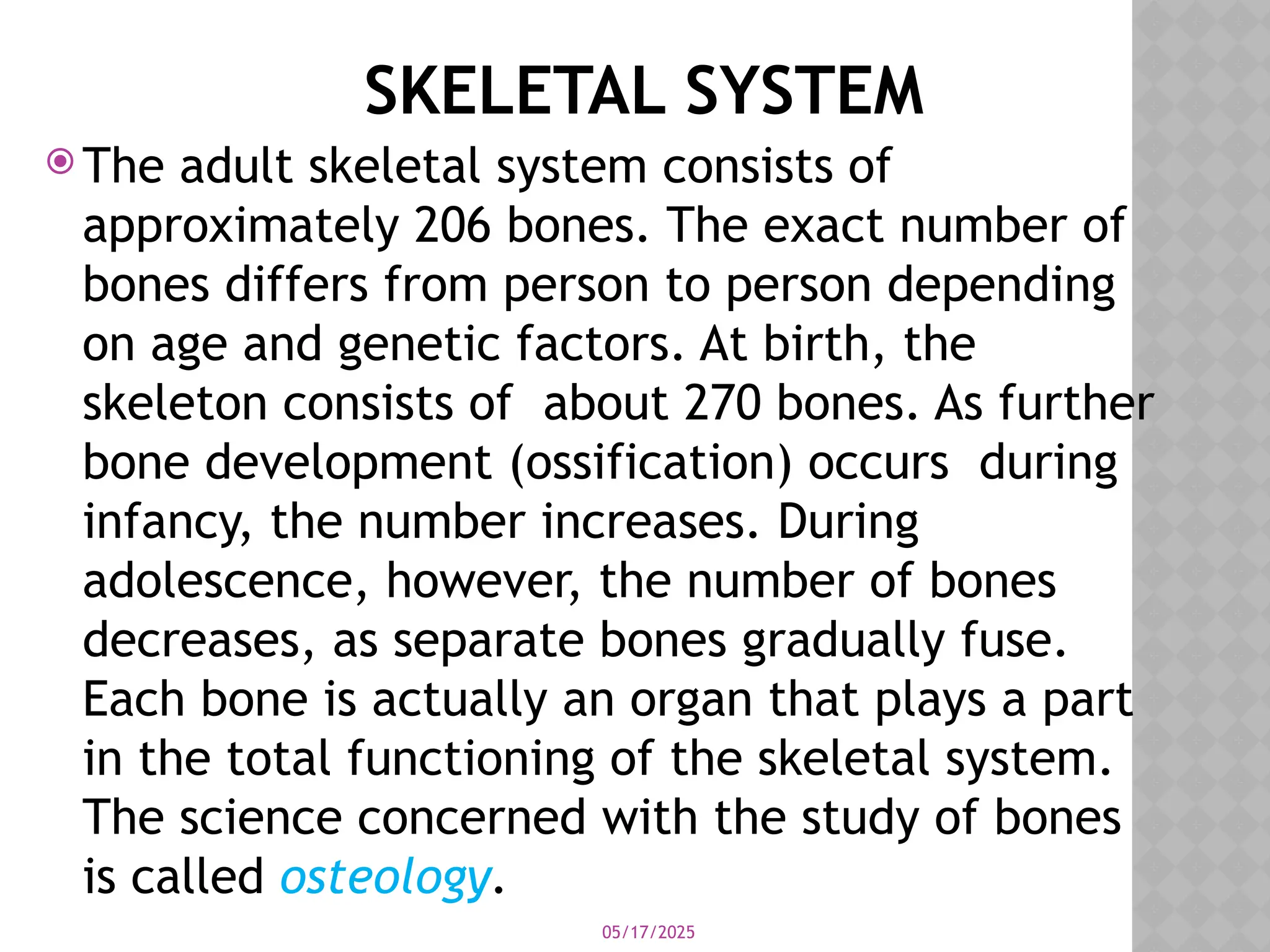
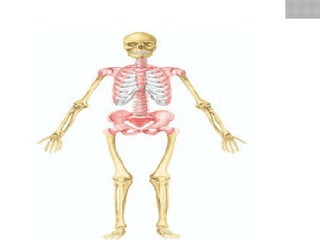
Theadult skeletal system consists of
approximately 206 bones. The exact number of
bones differs from person to person depending
on age and genetic factors. At birth, the
skeleton consists of about 270 bones. As further
bone development (ossification) occurs during
infancy, the number increases. During
adolescence, however, the number of bones
decreases, as separate bones gradually fuse.
Each bone is actually an organ that plays a part
in the total functioning of the skeletal system.
The science concerned with the study of bones
is called osteology.
05/17/2025
Some adults haveextra bones within the
sutures (joints) of the skull called sutural
(wormian) bones. Additional bones may
develop in tendons in response to stress as
the tendons repeatedly move across a joint.
Bones formed this way are called sesamoid
bones. Sesamoid bones, like the sutural
bones, vary in number. The patellae
(“kneecaps”) are two sesamoid bones all
people have.
5.
05/17/2025
the skeletonis divided into axial and
appendicular portions
The axial skeleton consists of the bones that
form the axis of the body and support and
protect the organs of the head, neck, and
trunk. The components of the axial skeleton
are as follows:
6.
05/17/2025
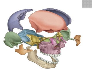
1. Skull.The skull consists of two sets of
bones: the cranial bones that form the
cranium, and the facial bones that support
the eyes and nose and form the bony
framework of the oral cavity.
2. Auditory ossicles. Three auditory ossicles
(“ear bones”) are present in the middle-ear
chamber of each ear and serve to transmit
sound impulses.
05/17/2025
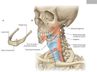
3. Hyoidbone. The hyoid bone is located
above the larynx (“voice box”) and below
the mandible (“jawbone”). It supports the
tongue and assists in swallowing.
4. Vertebral column. The vertebral column
(“backbone”) consists of 26 individual bones
separated by cartilaginous intervertebral
discs. In the pelvic region, several vertebrae
are fused to form the sacrum,
9.
05/17/2025
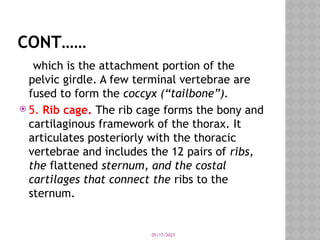
CONT……
which is theattachment portion of the
pelvic girdle. A few terminal vertebrae are
fused to form the coccyx (“tailbone”).
5. Rib cage. The rib cage forms the bony and
cartilaginous framework of the thorax. It
articulates posteriorly with the thoracic
vertebrae and includes the 12 pairs of ribs,
the flattened sternum, and the costal
cartilages that connect the ribs to the
sternum.
05/17/2025
The appendicularskeleton is composed of
the bones of the upper and lower extremities
and the bony girdles that anchor the
appendages to the axial skeleton
1. Pectoral girdle. The paired scapulae
(shoulder blades) and clavicles (collarbones)
are the appendicular components of the
pectoral girdle,
12.
05/17/2025
CONT…..
The primaryfunction of the pectoral girdle is
to provide attachment for the muscles that
move the brachium (arm) and antebrachium
(forearm).
2. Upper extremities. Each upper extremity
contains a proximal humerus within the
brachium, an ulna and radius within the
antebrachium, the carpal bones, the
metacarpal bones, and the phalanges
(“finger bones”) of the hand.
13.
05/17/2025
3. Pelvicgirdle. The two ossa coxae
(“hipbones”)
The pelvic girdle supports the weight of the
body through the vertebral column and
protects the viscera within the pelvic cavity.
14.
05/17/2025
4. Lowerextremities. Each lower extremity
contains a proximal femur (“thighbone”)
within the thigh, a tibia shinbone and fibula
within the leg, the tarsal bones, the
metatarsal bones, and the phalanges (“toe
bones”) of the foot. In addition, the patella
“kneecap”) is located on the anterior surface
of the knee joint, between the thigh and leg.
15.
05/17/2025
FUNCTIONS
OF THE SKELETALSYSTEM
The bones of the skeleton perform the
mechanical functions of support, protection,
and leverage for body movement and the
metabolic functions of hemopoiesis and
storage of fat and minerals.
1) Support. 2)protection.3) Body
movement.
4) Hemopoiesis. 5) Fat storage. 6) Mineral
storage.
16.
05/17/2025
BONE STRUCTURE
Eachbone has a characteristic shape and
diagnostic surface features that indicate its
functional relationship to other bones,
muscles, and to the body structure as a
whole.
17.
05/17/2025
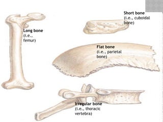
SHAPES OF BONES
The bones of the skeleton are grouped on the
basis of shape into four principal categories:
long bones, short bones, flat bones, and
irregular bones
1. Long bones. Most of the bones of the
upper and lower extremities are of this type
(e.g., the humerus, radius, ulna, metacarpal
bones, femur, tibia, fibula, metatarsal
bones, and phalanges).
05/17/2025
2. Shortbones. Short bones are somewhat
cube-shaped and are found in the wrist and
ankle where they transfer forces of
movement.
3. Flat bones. Flat bones have a broad
surface for muscle attachment or protection
of underlying organs (e.g., the cranial bones,
ribs, and bones of the shoulder girdle).
20.
05/17/2025
4. Irregularbones. Irregular bones have
varied shapes and many surface features for
muscle attachment or articulation (e.g., the
vertebrae and certain bones of the skull).
Bone tissue is organized as compact (dense)
bone or spongy (cancellous) bone, and most
bones have both types.
21.
05/17/2025
SURFACE FEATURES OFBONE
Structure Description and
Example
Articulating Surfaces
Condyle: A large, rounded articulating
knob (the occipital condyle of the occipital
bone)
Facet: A flattened or shallow articulating
surface (the costal facet of a thoracic
vertebra)
Head: A prominent, rounded articulating
end of a bone (the head of the femur)
22.
05/17/2025
CONTI………
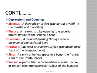
Depressions andOpenings
alveolus : A deep pit or socket (the dental alveoli in
the maxilla and mandible)
Fissure: A narrow, slitlike opening (the superior
orbital fissure of the sphenoid bone)
Foramen: A rounded opening through a bone
magnum of the occipital bone
Fossa: A flattened or shallow surface (the mandibular
fossa of the temporal bone)
Sinus: A cavity or hollow space in a bone (the frontal
sinus of the frontal bone)
Sulcus: A groove that accommodates a vessel, nerve,
or tendon (the intertubercular sulcus of the humerus
23.
05/17/2025
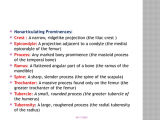
Nonarticulating Prominences:
Crest : A narrow, ridgelike projection (the iliac crest )
Epicondyle: A projection adjacent to a condyle (the medial
epicondyle of the femur)
Process: Any marked bony prominence (the mastoid process
of the temporal bone)
Ramus: A flattened angular part of a bone (the ramus of the
mandible)
Spine: A sharp, slender process (the spine of the scapula)
Trochanter: A massive process found only on the femur (the
greater trochanter of the femur)
Tubercle: A small, rounded process (the greater tubercle of
the humerus)
Tuberosity: A large, roughened process (the radial tuberosity
of the radius)
24.
05/17/2025
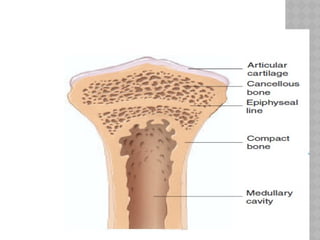
STRUCTURE OF ATYPICAL LONG
BONE
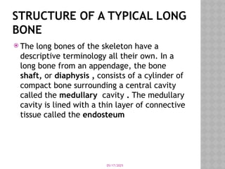
The long bones of the skeleton have a
descriptive terminology all their own. In a
long bone from an appendage, the bone
shaft, or diaphysis , consists of a cylinder of
compact bone surrounding a central cavity
called the medullary cavity . The medullary
cavity is lined with a thin layer of connective
tissue called the endosteum
25.
05/17/2025
CONT..
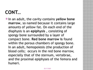
In anadult, the cavity contains yellow bone
marrow, so named because it contains large
amounts of yellow fat. On each end of the
diaphysis is an epiphysis , consisting of
spongy bone surrounded by a layer of
compact bone. Red bone marrow is found
within the porous chambers of spongy bone.
In an adult, hemopoiesis (the production of
blood cells; occurs in the red bone marrow,
especially that of the sternum, vertebrae,
and the proximal epiphyses of the femora and
humeri.
26.
05/17/2025
. Articularcartilage, which is composed of
thin hyaline cartilage, caps each epiphysis
and facilitates joint movement. Along the
diaphysis are nutrient foramina—small
openings into the bone that allow nutrient
vessels to pass into the bone for nourishment
of the living tissue. Between the diaphysis
and epiphysis is a cartilaginous epiphyseal
plate—a region of mitotic activity that is
responsible for linear bone growth.
27.
05/17/2025
epiphyseal linereplaces the plate and final
ossification occurs between the epiphysis and
the diaphysis. A periosteum of dense
regular connective tissue covers the surface
of the bone, except over the articular
cartilage. This highly vascular layer serves as
a place for a tendon-muscle attachment and
is responsible for (increase in width). The
periosteum is secured to the bone by
perforating (Sharpey’s) fibers, composed of
bundles of collagenous fibers.
28.
05/17/2025
BONE TISSUE
Bonetissue is composed of several types of
bone cells embedded in a matrix of ground
substance, inorganic salts (calcium and
phosphorus), and collagenous fibers. Bone
cells and ground substance give bone
flexibility and strength; the inorganic salts
give it hardness
29.
05/17/2025
BONE CELLS
Thereare five principal types of bone cells
contained within bone tissue.
Osteogenic cells are found in the bone
tissues in contact with the endosteum and
the periosteum.
These cells respond to trauma, such as a
fracture, by giving rise to bone-forming cells
(osteoblasts) and bone-destroying cells
(osteoclasts).
30.
05/17/2025
Osteoblasts:are bone-formingcells that
synthesize and secrete unmineralized ground
substance. They are abundant in areas of high
metabolism within bone, such as under the
periosteum and bordering the medullary
cavity.
Osteocytes: are mature bone cells derived
from osteoblasts that have secreted bone
tissue around themselves. Osteocytes
maintain healthy bone tissue by secreting
enzymes and influencing bone mineral
content
31.
05/17/2025
Osteoclasts :are large multinuclear cells
that enzymatically break down bone tissue,
releasing calcium, magnesium,and other
minerals to the blood. These cells are
important in bone growth, remodeling, and
healing.
32.
05/17/2025
Bone-lining cells:are derived from
osteoblasts along the surface of most bones
in the adult skeleton. These cells are thought
to regulate the movement of calcium and
phosphate into and out of bone matrix.
33.
05/17/2025
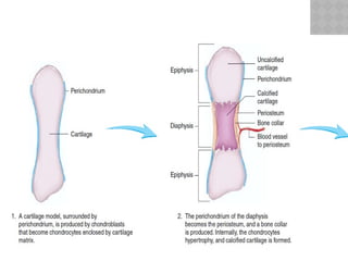
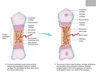
BONE GROWTH
Inmost bone development, a cartilaginous
model is gradually replaced by bone tissue
during endochondral bone formation
05/17/2025
DEVELOPMENT OF BONE
Bone formation, or ossification, begins at
about the fourth week of embryonic
development, but ossification centers cannot
be readily observed until about the tenth
week.
38.
05/17/2025
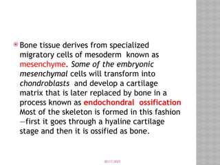
Bone tissuederives from specialized
migratory cells of mesoderm known as
mesenchyme. Some of the embryonic
mesenchymal cells will transform into
chondroblasts and develop a cartilage
matrix that is later replaced by bone in a
process known as endochondral ossification
Most of the skeleton is formed in this fashion
—first it goes through a hyaline cartilage
stage and then it is ossified as bone.
39.
05/17/2025
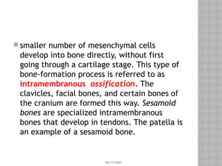
smaller numberof mesenchymal cells
develop into bone directly, without first
going through a cartilage stage. This type of
bone-formation process is referred to as
intramembranous ossification. The
clavicles, facial bones, and certain bones of
the cranium are formed this way. Sesamoid
bones are specialized intramembranous
bones that develop in tendons. The patella is
an example of a sesamoid bone.
05/17/2025
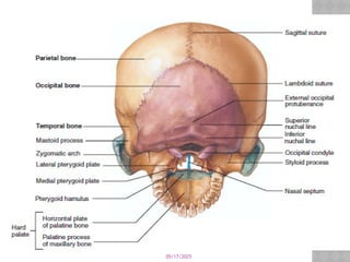
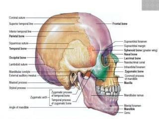
SKULL
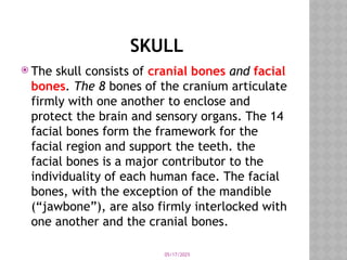
The skullconsists of cranial bones and facial
bones. The 8 bones of the cranium articulate
firmly with one another to enclose and
protect the brain and sensory organs. The 14
facial bones form the framework for the
facial region and support the teeth. the
facial bones is a major contributor to the
individuality of each human face. The facial
bones, with the exception of the mandible
(“jawbone”), are also firmly interlocked with
one another and the cranial bones.
42.
05/17/2025
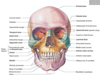
The skullhas several cavities. The cranial
cavity is the largest, with an approximate
capacity of 1,300 to 1,350 cc. The nasal
cavity is formed by both cranial and facial
bones and is partitioned into two chambers,
or nasal fossae, by a nasal septum of bone
and cartilage. Four sets of paranasal
sinuses, located within the bones
surrounding the nasal area, communicate via
ducts into the nasal cavity.
43.
05/17/2025
inner-ear cavitiesare positioned inferior to
the cranial cavity and house the organs of
hearing and balance. The two orbits for the
eyeballs are formed by facial and cranial
bones. The oral, or buccal cavity, which is
only partially formed by bone, is completely
within the facial region
44.
05/17/2025
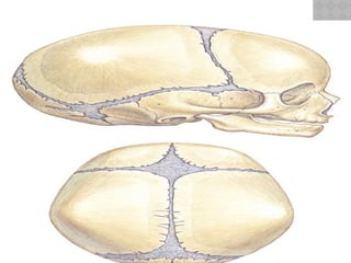
During fetaldevelopment and infancy, the
bones of the cranium are separated by
fibrous unions. There are also six large areas
of connective tissue membrane that cover
the gaps between the developing bones.
These membranous sheets are called
fontanels, meaning “little fountains.
45.
05/17/2025
1. Anterior(frontal) fontanel. is diamond-
shaped and is the most prominent. It is
located on the anteromedian portion of the
skull.
2. Posterior (occipital) fontanel. is
positioned at the back of the skull on the
median line. It is also diamond-shaped, but
smaller than the anterior fontanel.
46.
05/17/2025
3. Anterolateral(sphenoid) fontanels: The
paired anterolateral fontanels are found on
both sides of the skull, directly lateral to the
anterior fontanel. They are relatively small
and irregularly shaped.
4. Posterolateral (mastoid) fontanels: The
paired posterolateral fontanels, also
irregularly shaped, are located on the
posterolateral sides of the skull.
47.
05/17/2025
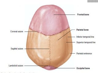
A prominentsagittal suture extends the
anteroposterior median length of the skull
between the anterior and posterior
fontanels. A coronal suture extends from the
anterior fontanel to the anterolateral
fontanel. A lambdoid suture extends from the
posterior fontanel to the posterolateral
fontanel. A squamous suture connects the
posterolateral fontanel to the anterolateral
fontanel.