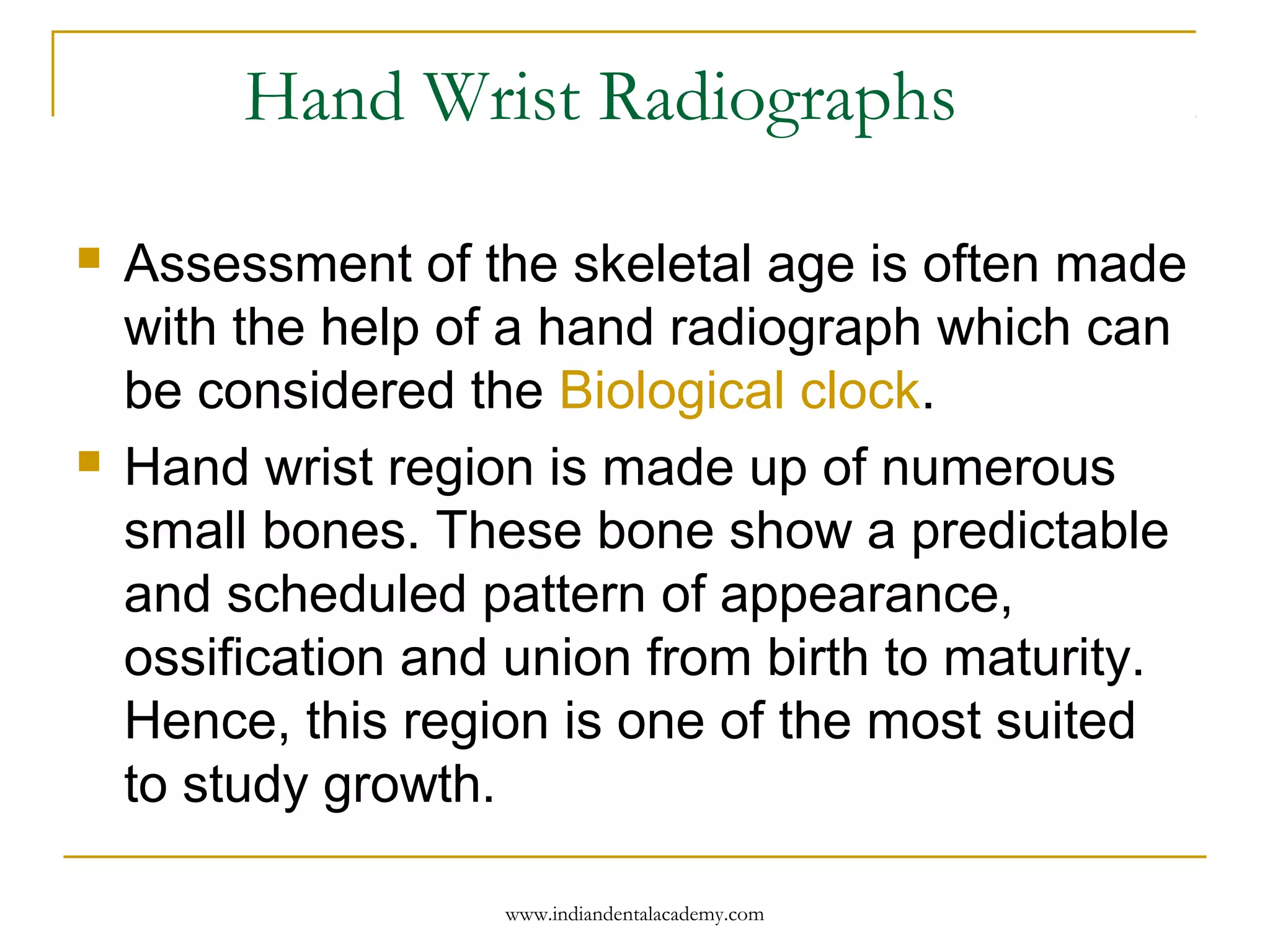
![Maturation Assessment by Hagg and
Taranger
Analyzed from radiograph taken between the
ages of 6 and 18 years, by assessing of the
ossification of the ulnar sesamoid of the
metacarpophalangeal joint of first finger.
Certain specified stages of 3 epiphyseal bone
-Middle and distal phalanges of third finger
[MP3 and DP3] and distal epiphysis of
Radius.
www.indiandentalacademy.com](https://image.slidesharecdn.com/skeletalmaturityindicator11-160512121130/75/Skeletal-maturity-indicator-11-13-2048.jpg)
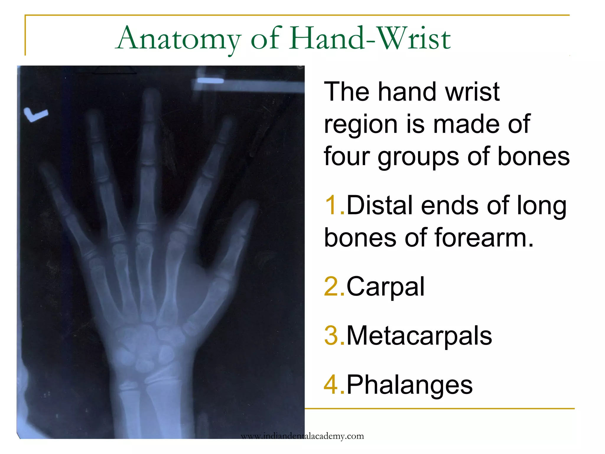
![Sesamoid
Sesamoid is usually attained during the
acceleration period of the pubertal growth
spurt [onset of peak height velocity]
www.indiandentalacademy.com](https://image.slidesharecdn.com/skeletalmaturityindicator11-160512121130/75/Skeletal-maturity-indicator-11-14-2048.jpg)
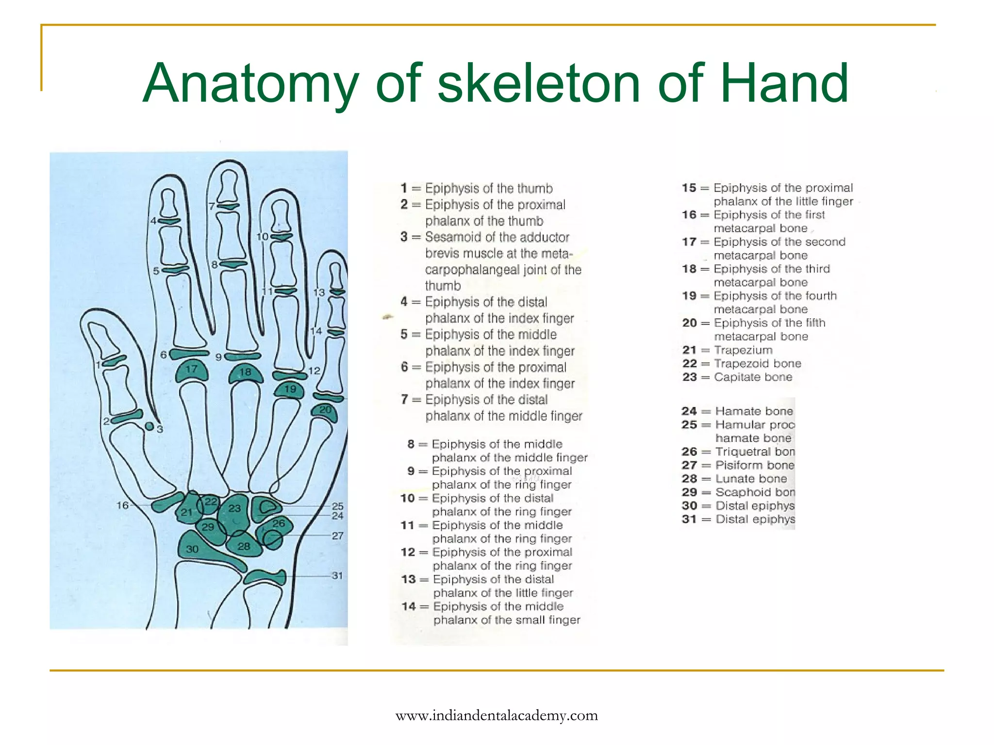
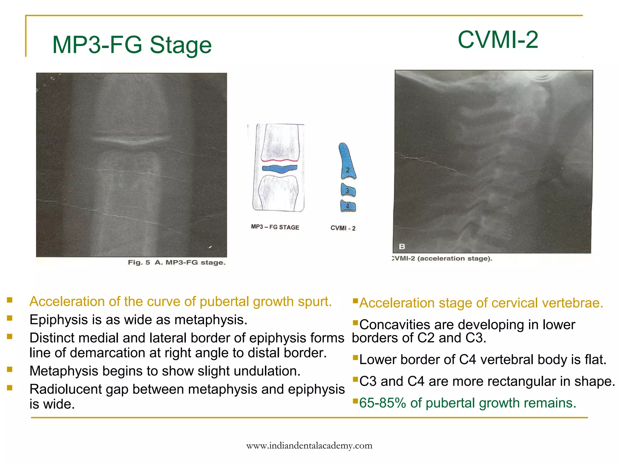
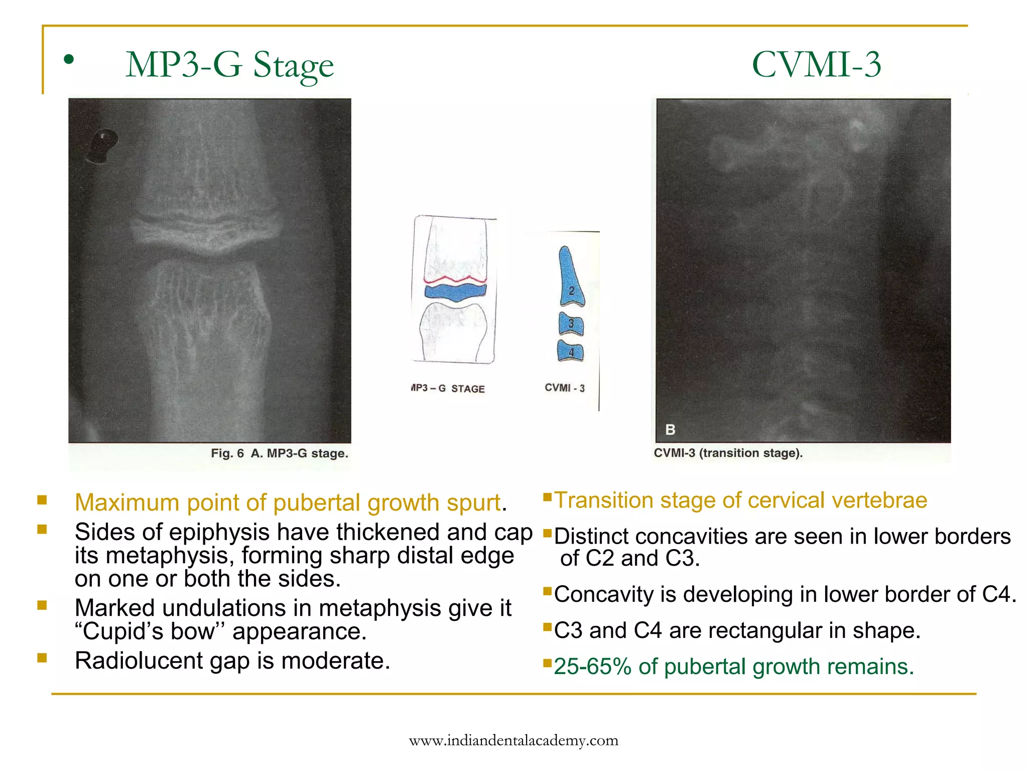
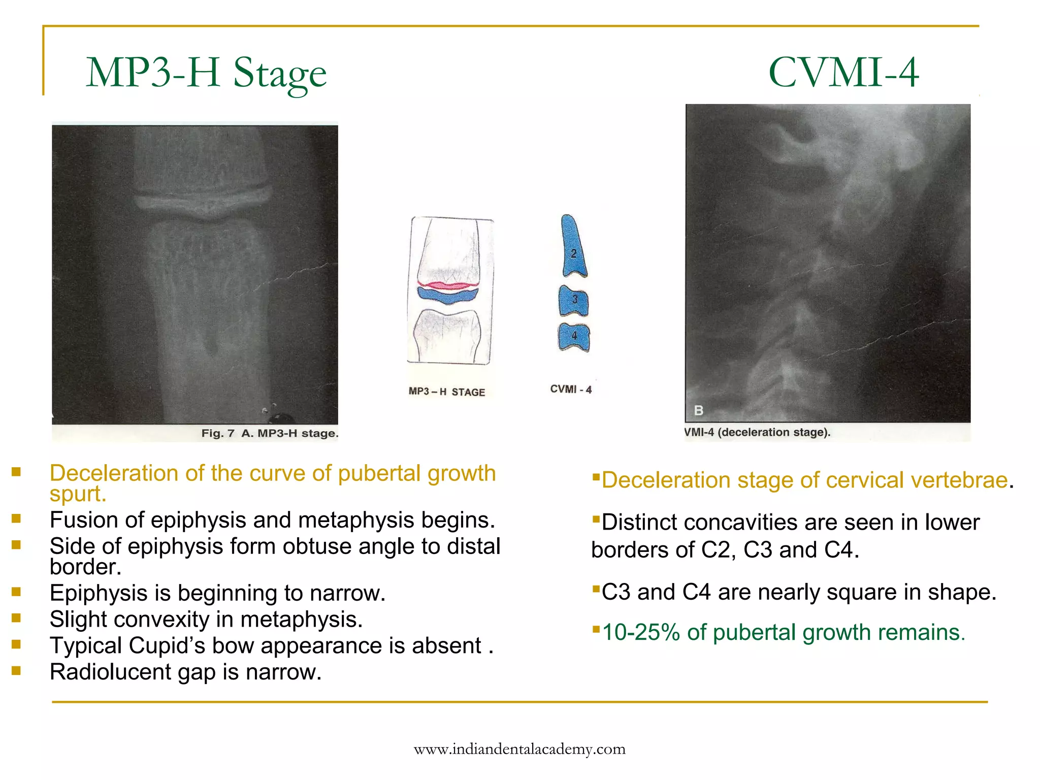
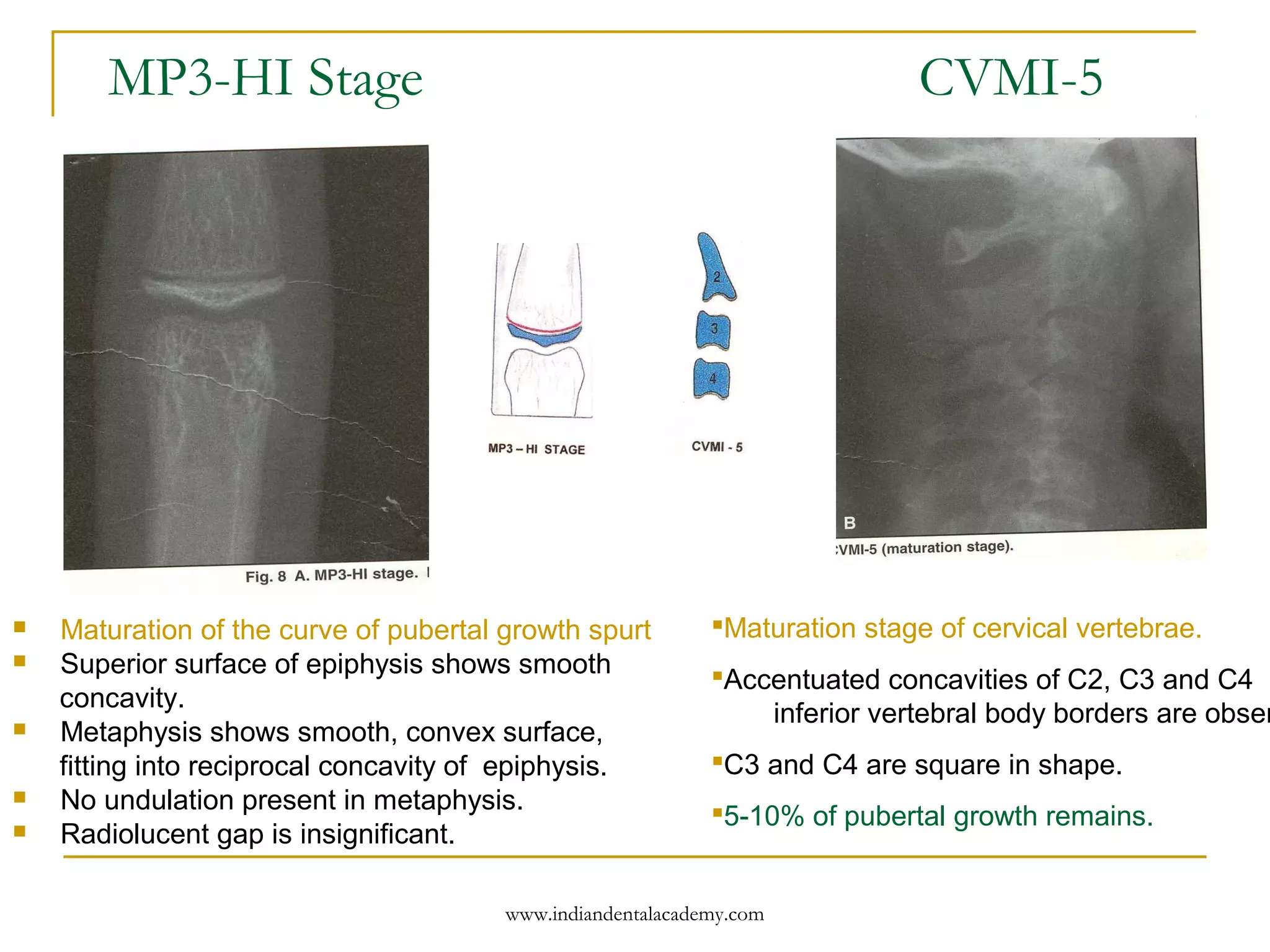
![Modified MP3 Cervical Vertebrae
MP3-F Stage
Start of the curve of pubertal growth spurt .
Epiphysis is as wide as metaphysis
End of epiphysis are tapered and rounded.
Radiolucent gap [cartilageous epiphyseal growth
plate] between epiphysis and metaphysis is wide.
Initiation stage of cervical vertebrae
C2,C3 and C4 inferior vertebral body
borders are flat.
Superior vertebral borders are tapered
from posterior to anterior [wedge shape]
80-100% of pubertal growth remains.
CVMI-1
www.indiandentalacademy.com](https://image.slidesharecdn.com/skeletalmaturityindicator11-160512121130/75/Skeletal-maturity-indicator-11-15-2048.jpg)
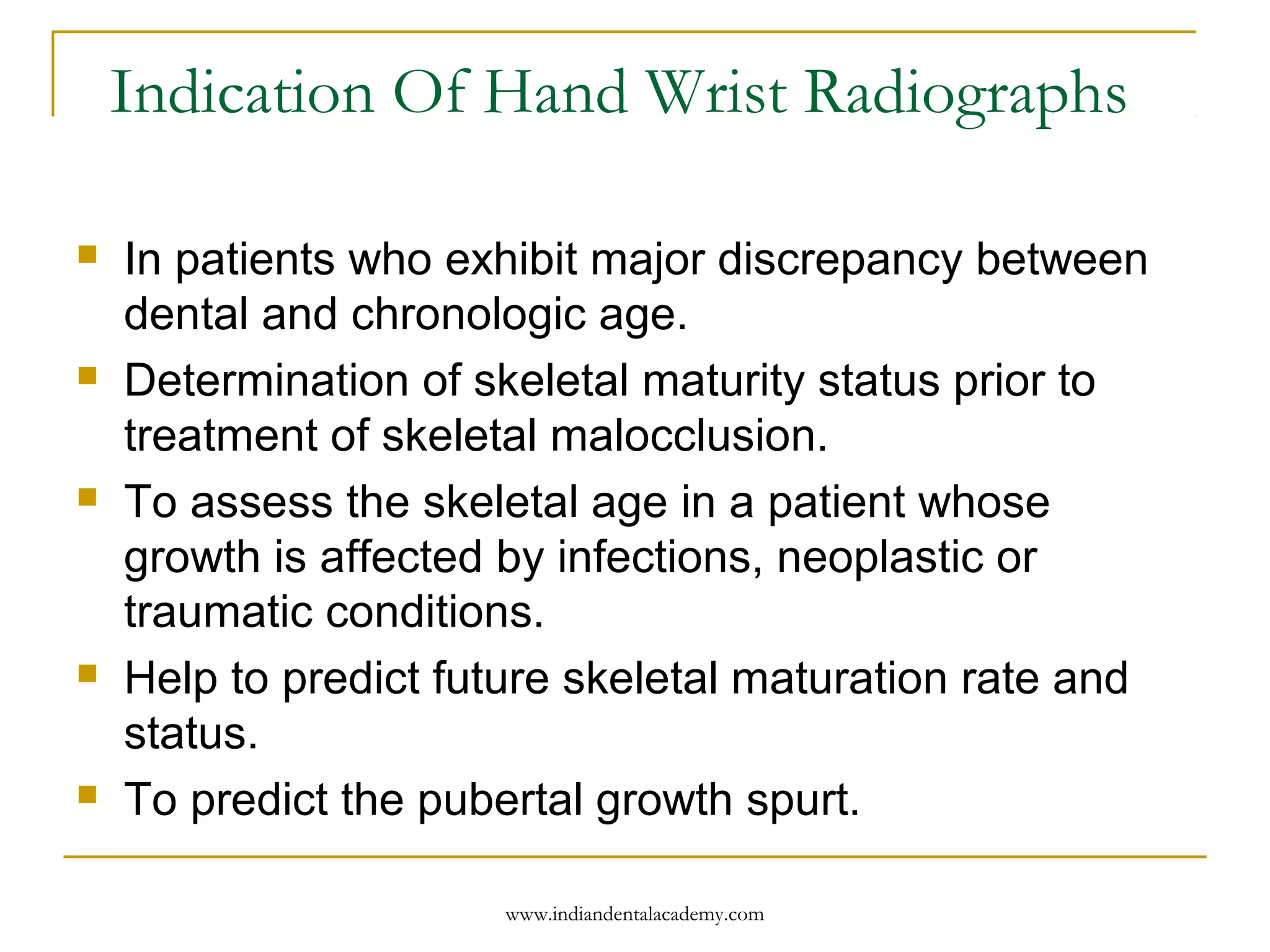
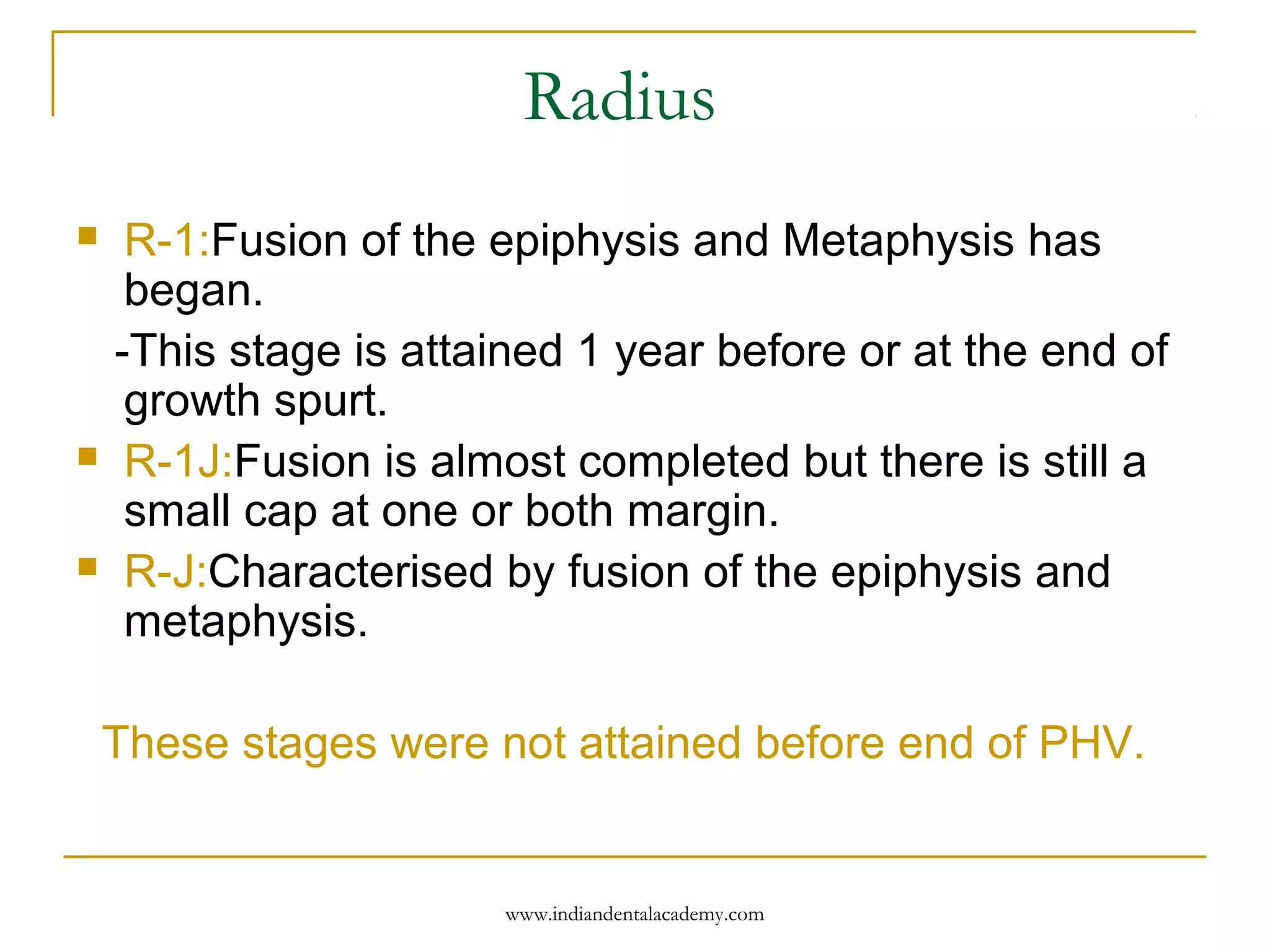
![Third finger distal phalanx
DP3-1:Fusion of Epiphysis and Metaphysis is
completed.
-This is attained during the deceleration period
of pubertal growth spurt [ end of PHV] .
www.indiandentalacademy.com](https://image.slidesharecdn.com/skeletalmaturityindicator11-160512121130/75/Skeletal-maturity-indicator-11-21-2048.jpg)

The document discusses skeletal maturity indicators that are crucial for assessing biological age and planning orthodontic treatments. It details methods for evaluating skeletal age, including radiographic assessments of the hand-wrist and cervical vertebrae, and explains various stages of bone maturation. Accurate skeletal age assessment helps in determining treatment timing, evaluating prognosis, and understanding the effects of genetics and environment on growth.


