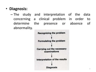
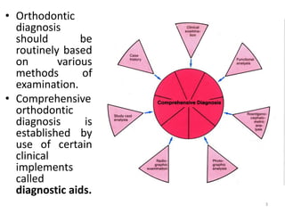
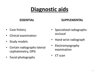
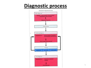
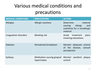
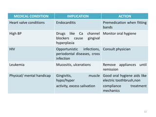
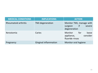
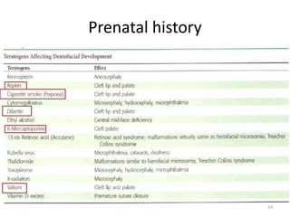
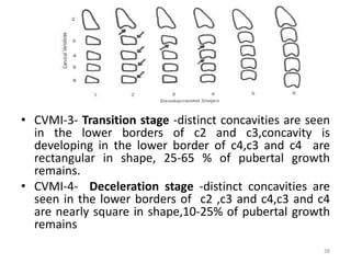
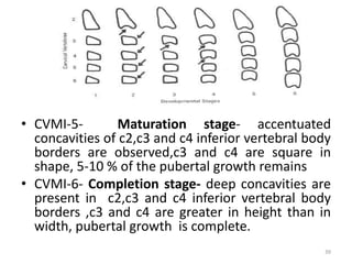
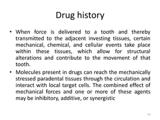
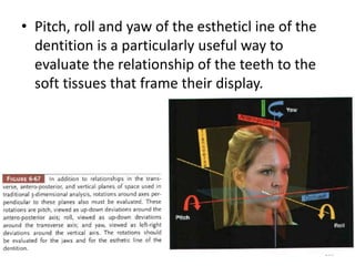
SlideShare a Scribd company logo
ORTHODONTIC
DIAGNOSIS
By :
Dr. Anjali Rajeshkumar Jaiswal
1st Year Post Graduate Student
1
• Diagnosis:
– The study and interpretation of the data
concerning a clinical problem in order to
determine the presence or absence of
abnormality.
2
• Orthodontic
diagnosis
should be
routinely based
on various
methods of
examination.
• Comprehensive
orthodontic
diagnosis is
established by
use of certain
clinical
implements
called
diagnostic aids.
3
Diagnostic aids
ESSENTIAL
• Case history
• Clinical examination
• Study models
• Certain radiographs-lateral
cephalometry, OPG
• Facial photographs
SUPPLEMENTAL
• Specialized radiographs-
occlusal
• Hand wrist radiograph
• Electromyography
examination
• CT scan
4
Diagnostic process
5
• The process of orthodontic diagnosis lends
itself well to the PROBLEM ORIENTED
APPAROACH.
• The diagnosis and treatment planning are
made in a series of logical steps.
• The first two steps constitute diagnosis.
They are:
1. Development of an adequate diagnostic
database
2. Formulation of a problem list – the diagnosis –
from the database.
6
• For orthodontic purposes, the database may
be thought of as derived from three major
sources:
(1) patient questioning,
(2) clinical examination of the patient, and
(3) evaluation of diagnostic records, including dental
casts, radiographs, and photographs.
7
Questionnaire/Interview
• Chief concern
• Medical and Dental history
• Physical growth evaluation
• Social and behavioral evaluation
8
Chief concern
• There are three major reasons for patient
concern about the alignment and occlusion of
the teeth:
– Impaired dentofacial esthetics that can lead to
psychosocial problems,
– Impaired function, and a
– Desire to enhance dentofacial esthetics and
thereby the quality of life
9
Medical history
• A careful medical and dental history is needed
for orthodontic patients both to provide a
proper background for understanding the
patient’s overall situation and to evaluate
specific orthodontically related concerns.
• Two areas deserve a special attention
– Trauma resulting in TMJ injuries
– Medication of any type for systemic diseases
10
Various medical conditions and
precautions
MEDICAL CONDITIONS IMPLICATIONS ACTION
Allergies Allergic reactions Determine material
causing allergy and
substitute for a nonallergic
material
Coagulation disorders Bleeding risk Avoid treatment plans
involving extractions
Diabetes Periodontal breakdown Monitor adequate control
of the disease, consult
periodontist
Epilepsy Medication causing gingival
hypertrophy
Monitor excellent plaque
control
11
MEDICAL CONDITION IMPLICATION ACTION
Heart valve conditions Endocarditis Premedication when fitting
bands
High BP Drugs like Ca channel
blockers cause gingival
hyperplasia
Monitor oral hygiene
HIV Opportunistic infections,
periodontal diseases, cross
infection
Consult physician
Leukemia Mucositis, ulcerations Remove appliances until
remission
Physical/ mental handicap Gingivitis, muscle
hypo/hyper
activity, excess salivation
Good oral hygiene aids like
electric toothbrush,non
compliance treatment
mechanics
12
MEDICAL CONDITIONS IMPLICATIONS ACTION
Rheumatoid arthritis TMJ degeneration Monitor TMJ, manage with
surgeon if severe
degeneration
Xerostomia Caries Monitor for loose
appliance, consider
fluoride rinses
Pregnancy Gingival inflammation Monitor oral hygiene
13
Prenatal history
14
Dental history
• Information about age of eruption and
exfoliation
• Earlier treatment received by the patient –
shows the attitude towards treatment
• Previous extraction – Amount of damage to
the cortical plates, resorption & remodeling
status of the bone at the extraction site
15
Physical growth evaluation
• Rapid growth during the adolescent growth
spurt facilitates tooth movement, but growth
modification may not be possible in a child
who is beyond the peak of the growth spurt.
Name of spurt Female Male
Infantile/ childhood
growth spurt
3yrs 3yrs
Mixed dentition/ juvenile
growth spurt
6-7yrs 7-9yrs
Pre-pubertal/ adolescent
growth spurt
11-12yrs 14-15yrs
16
Growth chart
Used to follow a child over time to
evaluate whether there is an
unexpected change in growth
pattern.
• Ask questions about how rapidly
the child has grown recently
• Secondary sexual characteristics
• Height - weight records and the
child’s progress on standard
growth charts can be obtained
from the child’s pediatrician
17
18
Hand wrist radiograph
• The ossification and development of the
carpal bones of the wrist, the metacarpals of
the hands, and the phalanges of the fingers
form a chronology of skeletal development
• The state of ossification of ulnar sesamoid or
hamate bones can be used to obtain an
estimate of the timing of the adolescent
growth spurt.
19
Anatomy of hand wrist
20
Bjork, Grace and Brown method
• Divided skeletal development into 9 stages.
• Each stage represents the level of skeletal
maturity.
• Chronologic ages were given by Schopf in
1978.
21
• Stage 1—(male-10.6y
female-8.1)– the
epiphyis and
diaphysis of the
proximal phalanx of
index finger are
equal. Occurs approx
3 years before the
peak of pubertal
growth spurt
22
• Stage 2-(male-12y
female-8.1) –the
epiphysis and
diaphysis of the
middle phalanx of
the middle finger are
equal.
23
• Stage 3 (Male-12.6y Female-9.6y)—1.hamular
process of the hamate exhibits ossification
2.Ossification of pisiform 3.The epiphysis and
diaphysis of radius are equal
24
• Stage 4 (Male-13 y
Female-1o.6y)-initial
mineralization of the
ulnar sesamoid of the
thumb.
• Increased ossification
of the hamular process
of the hamate bone.
• Marks the beginning
of pubertal growth
spurt.
25
• Stage 5----(male-
14y; female-11y)-
marks the peak of
the pubertal spurt.
• Capping of the
diaphysis by the
epiphysis is seen in
middle phalanx of
the third finger,
Proximal phalanx
of the thumb and
In radius.
26
Stage 6 (male-
15y;female-13y)
• Signifies the end
of pubertal
growth spurt
• Union between
diaphysis and
epiphysis of distal
phalanx of middle
finger.
27
• Stage 7—
(male-15.9
female- 13.9)
• Union
between
epiphysis and
diaphysis
proximal
phalanx of
little finger
28
• Stage 8 –
(male-15.9
female-13.9)
• Fusion between
epiphysis and
diaphysis of
middle phalanx
of the third
finger
29
• Stage 9
(Male-18.5y
Female-16y)
• Fusion
between
epiphysis
and
diaphysis of
the radius
30
Fishman Skeletal Maturity Assessment
• It uses four stage and six anatomical site
located on thumb, third finger ,fifth finger and
radius.
• 11 discrete adolescent maturity indicators
covering the entire period of adolescent
growth, are found on these sites.
31
• 4 stages of bone maturation are used in this
method. They are-
1. Epiphysis equal in width to diaphysis
2. Appearance of adductor sesamoid of the thumb
3. Capping of epiphysis
4. Fusion of epiphysis
32
• SMI 1- 3rd finger proximal phalanx
shows equal width of the
epiphysis and diaphysis
• SMI 2-width of the epiphysis
equal to that of the diaphysis in
the middle phalanx of 3rd finger
• SMI 3-width of the epiphysis
equal to that of the diaphysis in
the middle phalanx of 5th finger
• SMI 4-appearance of adductor
sesamoid of the thumb
• SMI 5-capping of the epiphysis
seen in the distal phalanx of 3rd
finger
33
• SMI 6- capping of the epiphysis seen
in the middle phalanx of 3rd finger
• SMI 7- capping of the epiphysis seen
in the middle phalanx of 5th finger
• SMI 8-fusion of epiphysis and
diaphysis in the distal phalanx of 3rd
finger
• SMI 9- fusion of epiphysis and
diaphysis in the proximal phalanx of
3rd finger
• SMI 10- fusion of epiphysis and
diaphysis in the middle phalanx of 3rd
finger
• SMI 11- fusion of epiphysis and
diaphysis in the radius.
34
35
Vertebrae Maturity Indicators- Hassel
and Farman
36
• CVMI-1- Initiation stage- c2,c3 and c4 inferior
vertebral bodies are flat, superior vertebral bodies are
tapered from posterior to anterior(wedge shaped), 80-
100 % of growth remains
• CVMI-2- Acceleration stage -concavities are
developing in the lower borders of c2 and c3,lower
border of c4 vertebral body is flat,c3 and c4 are more
rectangular in shape,65- 85 % of growth remains.
37
• CVMI-3- Transition stage -distinct concavities are seen
in the lower borders of c2 and c3,concavity is
developing in the lower border of c4,c3 and c4 are
rectangular in shape, 25-65 % of pubertal growth
remains.
• CVMI-4- Deceleration stage -distinct concavities are
seen in the lower borders of c2 ,c3 and c4,c3 and c4
are nearly square in shape,10-25% of pubertal growth
remains
38
• CVMI-5- Maturation stage- accentuated
concavities of c2,c3 and c4 inferior vertebral body
borders are observed,c3 and c4 are square in
shape, 5-10 % of the pubertal growth remains
• CVMI-6- Completion stage- deep concavities are
present in c2,c3 and c4 inferior vertebral body
borders ,c3 and c4 are greater in height than in
width, pubertal growth is complete.
39
Social and behavioral evaluation
1. The patient’s motivation for the treatment
2. What he or she expects from the treatment
3. How cooperative or uncooperative the
patient is likely to be.
42
Clinical examination
• Clinical findings are the prerequisite for the
correct assessment and interpretation of the
quantitative analysis, i.e. the overall general
and the specific clinical findings, which serve
as the foundation of treatment decisions.
43
Overall general evaluation
• BODY TYPE
– Ectomorphic: tall & thin
– Mesomorphic: average build
– Endomorphic: short and fat
• BUILD
– Asthenic :thin built and usually possess narrow dental arches
– Plethoric/pyknic: obese built & generally have broad dental
arches
– Athletic : neither thin nor obese ; normally built and normal
dental arches
44
45
• GAIT
– Act of walking or locomotion, it is the way person walks.
– Abnormalities of gait are usually associated with
neuromuscular disorder which may have a dental
correlation.
• HEIGHT & WEIGHT
– They give a clue to the physical maturation & growth of
the patient, which may have dentofacial correlation.
• POSTURE
– This refers to the way a person stands.
– Abnormal posture can predispose to malocclusion due to
alteration in maxillo-mandibular relationship.
46
47
Cephalic and facial examination
• The shape of the head and facial structures
are assessed, measurements can be evaluated
according to the cephalic index of the head
and the morphologic facial index.
48
Cephalic Index
Maximum skull width
CI = _________________________
Maximum skull length
MESOCEPHALIC :Average
shape of the head-Normal
dental arch.
DOLICOCEPHALIC :Long &
narrow head-Narrow
dental arch.
BRACHYCEPHALIC: Broad
& short head-Broad
dental arch.
49
Morphologic facial index
Morphologic facial height
MFI = _______________________________
Bi zygomatic width.
Leptoprosopic :
long & narrow face
Mesoprosopic:
Average
face/Normal face
Euryprosopic : Broad
& short face
50
Examination of soft tissues
• Extra oral:
– Forehead
– Nose
– Lips
– Chin
• Intra oral:
– Tongue
– Lip and cheek frenal attachments
– Gingiva
– Oral and palatal mucosa
51
Forehead-size
52
Forehead-slope
FLAT PROTRUDING STEEP
53
Nose-size
Normal case
H:V=2:1
Microrhinic type Large
54
Nasal-contour
Straight Convex Crooked
55
Nostrils
Width of nostril= approximately 70% of the length of nose
Usually oval and
Bilaterally symmetrical
Slight nasal anomaly
With wide nostrils
Cartilagenous septal
deviation
56
Vertical lip relationship
Upper lip length - 1/3rd of lower facial height
Lower lip length - 2/3rd of lower facial height
57
Lip morphology
Harmonious lip profile Short upper lip Eversion of
mucosal part
58
Horizontal lip profile
Protrusion of
Lower lip
Normal case Marked retrusion of
Lower lip
59
Lip step- Korkhaus
Positive lip step Normal Negative lip step
60
Nasolabial angle
Acute Normal Obtuse
61
Chin formation and contour
Normal Protruded chin Retruded chin
62
Tongue-shape
63
Tongue-size
64
Tongue-length
65
Lingual frenum
66
Labial frenum
67
Blanch test
68
Gingiva
Healthy Gingiva Thin Fragile Gingiva
Fribrous Gingiva 69
Palatal mucosa
70
Evaluation of facial and dental
appearance
• Should be done in three steps:
1. Macro esthetics – face in all three plane of space
2. Mini esthetics – the smile framework
3. Micro esthetics – the teeth
71
Macro esthetics – frontal examination
72
73
74
75
76
Macro esthetics – profile analysis
1. Establishing whether the jaws are proportionately positioned in the
anteroposterior plane of space
77
78
2. Evaluation of lip posture and incisor prominence.
79
80
3. Re-evaluation of vertical facial proportions, and evaluation of mandibular plane
angle
82
Mini esthetics – tooth lip relationship
83
• It is important to evaluate not only the
characteristics of the face, but the relationship
of the dentition to the face. This can begin
with an examination of symmetry, in which it
is particularly important to note the
relationship of the dental midline of each arch
to the skeletal midline of that jaw
84
85
86
87
Micro esthetics
88
89
90
91
92
Drug history
• When force is delivered to a tooth and thereby
transmitted to the adjacent investing tissues, certain
mechanical, chemical, and cellular events take place
within these tissues, which allow for structural
alterations and contribute to the movement of that
tooth.
• Molecules present in drugs can reach the mechanically
stressed paradental tissues through the circulation and
interact with local target cells. The combined effect of
mechanical forces and one or more of these agents
may be inhibitory, additive, or synergistic
93
• NSAIDs:
– Inhibition of the inflammatory reaction produced
by PGs slows the tooth movement.
– The levels of matrix metalloproteinases (MMP9
and MMP2) were found to be increased, along
with elevated collagenase activity, followed by a
reduction in procollagen synthesis which is
essential for bone and periodontal remodeling.
– The whole process is controlled by inhibition of
cyclooxygenase (COX) activity, leading to altered
vascular and extravascular matrix remodeling,
causing a reduction in the pace of the tooth
movement.
94
• Eg : aspirin, indimithacin, imidaxole,
flurbiprofen
• In such cases, a specific COX-2 inhibitor, a drug
with no effect on PGE2 synthesis, can be
prescribed.
• Because it selectively blocks COX-2 enzyme
and not COX-1 enzyme, the drug can be safely
employed during orthodontic
mechanotherapy, without causing negative
effects on tooth movement.
95
• Bisphosphonates:
– Cause a rise in intracellular calcium levels in
osteoclastic-like cell line, reduction of osteoclastic
activity, prevention of osteoclastic development
from hematopoietic precursors, and production of
an osteoclast inhibitory factor.
– BPNs can inhibit orthodontic tooth movement and
delay the orthodontic treatment.
96
• Oral contraceptives:
– Estrogen is considered to be the most important
hormone affecting the bone metabolism in
women.
– It inhibits the production of various cytokines
which are involved in bone resorption by
stimulating osteoclast formation and osteoclast
bone resorption.
– It decreases the velocity of tooth movement.
97
• Calcitonin:
– Calcitonin inhibits bone resorption by direct action
on osteoclasts, decreasing their ruffled surface
which forms contact with resorptive pit.
– It is considered to inhibit the tooth movement;
consequently, delay in orthodontic treatment can
be expected.
98
• Corticosteroids:
– The main effect of corticosteroid on bone tissue is
direct inhibition of osteoblastic function and thus
decreases total bone formation.
– Corticosteroids increase the rate of tooth
movement, and since new bone formation can be
difficult in a treated patient, they decrease the
stability of tooth movement and stability of
orthodontic treatment in general.
99
• Immunomodulatory drugs:
– Immunomodulatory drugs modulate nuclear
factor kappa - Beta , tyrosine kinases in signaling
pathway, IL - 6, MMPs and PGE2, all of which are
essential for the bone remodeling process.
– Cyclosporine A: produce severe gingival
hyperplasia, making orthodontic treatment and
maintenance of oral hygiene difficult.
– Anticancer drugs: are known to produce damage
to precursor cells involved in bone remodeling
process, thereby complicating tooth movement.
100
• Anticonvulsants:
– It induces gingival hyperplasia, , making
application of orthodontic mechanics and
maintaining oral hygiene difficult.
– If used during pregnancy, it can produce fetal
hydantoin syndrome characterized by hypoplastic
phalanges, cleft palate, hare lip, and microcephaly.
101
• Fluorides:
– Fluoride is one of the trace elements having an
effect on tissue metabolism.
– Sodium fluoride has been shown to inhibit the
osteoclastic activity and reduce the number of
active osteoclasts, thus delay orthodontic tooth
movement and increase the time of orthodontic
treatment.
102
Mental attitude of patients
• MM House Classification (1950)
– Philosophical
– Exacting
– Hysterical
– Indifferent
103
• Philosophical:
– The best mental attitude for denture acceptance is the
philosophical type.
– This patient is rationale, sensible, calm and composed
in different situations.
– These patients are willing to rely on the dentist’s
advice for diagnosis and treatment.
• Exacting:
– The exacting patient may have all of good attributes of
the philosophical patients; however he may require
extreme care, effort and patience on the part of
dentist.
– This patient is methodical, precise, and accurate and
at times makes severe demands.
– Once satisfied an exacting patient may become the
practioner’s greatest supporter
104
• Hysterical:
– The hysterical type is emotionally unstable, excitable
and excessively apprehensive.
– These patients submit to treatment alas a last resort,
have negative attitude, are often in poor health, are
poorly adjusted, often appear exacting but with
unfounded complaints, and have unrealistic
expectations.
• Indifferent:
– The indifferent type of patients presents a
questionable or unfavorable prognosis.
– He is apathetic and uninterested and lacks motivation.
– He pays no attention to instructions, will not co-
operate, and is prone to blame the dentist for poor
dental health.
105
Growth changes in soft tissue
• Upper lip length:
– Mean: males- 23.8mm
females- 20.1mm
– From 7 to 18yrs, increased from 19.8mm to
22.5mm in males, and 19.1mm to 20.2mm in
females.
– Average increment 2.7mm in males, 1.15mm in
females.
106
• Nose height:
– At 7 years, nearly same in both sexes.
– Rapid increase between 7 to 8 years, slowed down
between 8 to 11yrs, with pubertal acceleration
during 14 to 17 yrs.
• Chin:
– Soft tissue thickness at the level of soft tissue
pogonion showed total increase of 2.7mm in
males and 2mm in females.
107
• Nasolabial angle:
– Decreased from 7 yrs to 18 yrs in both sexes.
– Males: from 107.89.4 degrees to 105.89
degrees
– Females: from 114.79.5 degrees to 110.710.9
degrees
• Mentolabial angle:
– Males: 125.38.4 degrees at 7yrs to 125.112.9
degrees at 18yrs
– Females: 136.111.6 degrees at 7 yrs to
127.112.9 degrees at 18 yrs.
108
• Lip tonicity:
– Normal lip: minimal tonicity
– Hypertonic lip: firm and redder
– Hypotonic lip: flaccid
• Upper lip:
– With lip tension- contour flattens.
– Flaccid lips form an accentuated curve with the vermilion
lip area showing an accentuation of curve.
– The flaccid lip generally is thick (12 to 20 mm from
anterior vermilion to labial incisor) giving the lip the
appearance of being too far forward relative to the teeth.
• Lower lip:
– When deeply curved, the lower lip is flaccid in character
(Class II, vertical maxillary deficiency).
– When flattened, the lower lip demonstrates tension of
tissues (Class III). 109
• Hyperactive mentalis:
– Deep mentolabial sulcus is characteristic of
hyperactive mentalis muscle.
– It impedes forward development of the anterior
alveolar process of the mandible.
– Occurs together with lip sucking or lip thrust.
– Causes puckering of chin
– Seen in Class II div 1 cases
110
Functional Analysis
• Functional analysis constitutes a considerable
part of the clinical examination.
• Three most important aspects are:
1. Examination of postural rest position and
maximum intercuspation
2. Examination of temporomandibular joint
3. Examination of orofacial dysfunction
111
1. Examination of postural rest position and
maximum intercuspation
i. Determination of postural rest position
ii. Registration of postural rest position
iii. Evaluation of the relationship: postural rest
position and habitual occlusion, in three planes
of space
112
113
Determination of postural rest position
• When the mandible is in the postural resting
position, it is usually 2-3mm below and behind
the centric occlusion- referred to as freeway
space or interocclusal clearance
• Methods:
– Phonetic method
– Command method
– Non-command method
– Combined methods
Speculum – A.M. Schwarz
114
115
Registration of postural rest position
• Extraoral direct method:
– Registration by means of skin reference points
• Extraoral indirect method:
– Roentgenocephalometric registration
– Kinesiographic registration – Jankelson (1984)
116
117
118
119
121
Evaluation of the relationship: postural rest
position and habitual occlusion
• It is analyzed three dimensionally in the
sagittal, vertical and frontal planes
• Closing movement divided into two phases:
– Free phase
– Articular phase
122
• When closing, mandible may undergo
rotational and sliding movement:
– Pure rotational movement
– Rotational movement with anterior sliding
component
– Rotational movement with posterior sliding
component
123
Evaluation in sagittal plane
124
Class II malocclusions
125
Class III malocclusion
126
Mandibular prognathism- Pseudo
forced bite
127
Evaluation in vertical plane
• The freeway space is assessed.
• Important with deep overbite cases.
• According to Hotz and Muhlemann:
– True deep bite
– Pseudo deep bite
128
Evaluation in transverse plane
• Position of midline is observed.
• Relevant is cases with posterior unilateral
crossbite.
• Two types of skeletal mandibular deviation:
1. Laterognathy
2. Lateroclusion
129
Laterognathy
130
Lateroclusion
131
Examination of temporomandibular
joint
132
Auscultation and palpation
133
Examination of muscles of mastication
134
Functional analysis:
Movements of mandible
135
• Deviations from the normal mandibular
movements are a result of asynchronic muscle
contractions, malocclusions, etc.
• Deviations are the first signs of initial
temporomandibular joint problems.
• “C” and “S” types of deviations are typical
signs of functional disturbances.
136
Radiographic examination
• Radiographs taken in habitual occlusion
and/or in open-mouth position are suitable
for examination.
• When analyzing radiographs, following
findings are registered:
– Position of condyle in relation to fossa,
– Width of joint space,
– Change in shape and structure of condylar head
and/or mandibular fossa.
137
138
Examination of orofacial dysfunctions
• Swallowing
• Tongue
• Speech
• Lips
• Respiration
139
Swallowing
Normal Swallow
Retained
Infantile
Swallow
140
Stages Of
Deglutition
1. Oral Phase
2. Pharyngeal Phase
3. Esophageal Phase
141
Tongue thrust
142
143
Lips dysfunction
144
Lip habits
145
Speech dysfunction
• Tongue and lip dysfunctions lead to speech
difficulties, as the normal relationship is
hampered and pronunciation is affected.
146
Mouth breathing
147
Tongue posture in oronasal respiration
148
Examination of breathing mode
• Various methods:
– Mirror test
– Cotton pledget test
– Water holding test
– Observation of nostrils
149
150
THANKYOU
151
152
153
154
155
Orthodontic classification
• Classification has traditionally been an
important tool in the diagnosis-treatment
planning procedure.
• Classification can be viewed as the (orderly)
reduction of the database to a list of the
patient's problems.
156
Classification of malocclusion
• Any deviations from normal occlusion can be
termed as malocclusion, which may vary from
a very slight deviation of a tooth position in
the arch to a significant malpositioning of a
group of teeth or jaws.
157
158
Purpose of Classification :
• Classification helps in diagnosis and planning
treatment for the patients.
• It helps in visualizing and understanding the
problems associated with that malocclusion.
• It helps in communicating the problem.
• Comparison of various malocclusion is made
easy by classification.
Classification
Intra arch
Individual
tooth
malpositions
Inter arch
Dental Skeletal
159
Individual tooth malpositions
• Mesial inclination or tipping
• Distal inclination or tipping
• Lingual inclination or tipping
160
Labial tipping
Infra occlusion
Supra occlusion
161
Rotations
• Mesiolingual/distolabial
• Distolingual/mesiolabial
• Transposition
162
Malrelation of dental arches
Sagittal plane malocclusion
• Pre normal occlusion- lower arch is more
forwardly placed
• Post normal occlusion- lower arch is more
distally placed.
163
Vertical plane malocclusion
Deep bite Open bite
164
Transverse plane malocclusion
• cross bite
165
ANGLE’S CLASSIFICATION
• NORMAL OCCLUSION
• CLASS I MALOCCLUSION
• CLASS II MALOCCLUSION
• CLASS III MALOCCLUSION
166
Normal occlusion
• Normal antero-posterior relationship
between maxilla and mandible.
• Normal molar relationship
• Line of occlusion is a smooth, continuous and
symmetric catenary curve.
167
Class I malocclusion
Class I molar relationship
Mesiobuccal cusp of the maxillary first
molar occludes in the buccal groove of
the mandibular 1st permanent molar
168
• Crowding, spacing, rotations missing tooth etc.
• Normal skeletal and normal muscle relationship
• Class I bimaxillary protrusion– normal class I
relationship but dentition of both the arches are
forwardly placed in relation to facial profile
169
Angle’s class II malocclusion
• Class II molar relationship- disto buccal cusp
of the upper first permanent molar occludes
in the buccal groove of the lower 1st molar
• It is sub classified into
class II division 1
class II division 2
class II subdivision
170
Class II div 1
• Class II molar relation
• Proclined upper incisors –increased overjet
• Presence of abnormal muscle activity-
characterstic feature
• Altered tongue positon- accentuates
narrowing of upper arch
• Lip trap- lower lip cushions the palatal aspect
of the upper teeth
171
172
Class II div 2
• Class II molar relation
• Lingually inclined upper central incisors
• Labially tipped lateral incisors overlapping
the centrals
• Normal perioral muscle activity
• Abnormal backward path of closure
173
174
Class II subdivision
• Class II molar relation on one side and class I
on other
175
Angles’ Class III malocclusion
Class III molar relationship-
Mesiobuccal cusp of maxillary first
Molar occludes in the interdental
Space between the distal cusp of
mandibular first molar and second
Molar.
• Classified into-
True class iii
Pseudo class iii
176
TRUE CLASS III
• Class III molar relation
• Lower incisors lingually inclined
• Lower tongue posture- narrow upper arch
177
PSEUDO CLASS III
• Caused by forward movement of the mandible-
postural or habitual class iii
• Causes of pseudo class iii:-
Occlusal prematurity
Loss of deciduous molars
Large adenoids
178
CLASS III SUBDIVISION
• CLASS III MOLAR RELATION ON ONE SIDE AND
CLASS I RELATION ON THE OTHER
179
Angle’s line of occlusion:
When the teeth are in normal occlusion the line of
greatest occlusal contact will be found to pass over the
mesial and distal inclined planes of the buccal cusps of
the molars and bicuspids and the cutting edges of the
cuspids and incisors of the lower arch, and along the
sulcus between the buccal and lingual cusps of the upper
molars and bicuspids, hence forward, crossing the lingual
ridge of the cuspids and the marginal ridges of the
incisors at a point about one-third the length of their
crowns from their cutting-edges.
180
DRAWBACKS OF ANGLE’S
CLASSIFICATION
• First permanent molar not a fixed point
• Classification is not possible if first molars are
missing
• Malocclusion is considered only in a-p direction
• Individual tooth malocclusion is not considered
• No differentiation between skeletal and dental
malocclusion
• No clue about etiology
181
182
• Modifications by Martin Dewey
Class I malocclusion with-
Type I: Crowded anterior teeth.
Type II: Protrusive maxillary incisors.
Type III: Anterior crossbite.
Type IV: Posterior crossbite.
Type V: Mesial drifting of permanent molar.
183
• Class III with
Type I:Viewed separately, arches are normal,
in occlusion- Edge-to-edge incisor relationship.
Type II:Crowding& lingual relation of
mandibular incisors to maxillary incisors.
Type III:Crowding&cross bite relationship of
maxillary incisor.
184
• Lischer’s modifications – Introduced terms-
1. ‘Neutrocclusion’- Angle’s Class I.
2. ‘Distocclusion’ – Angle’s Class II.
3. ‘Mesiocclusion’ – Angle’s Class III.
He also added the suffix ‘version’
1. Buccoversion.
2. Linguoversion.
3. Supraversion.
4. Infraversion.
185
5. Mesioversion.
6. Distoversion.
7. Axiversion.
8. Torsiversion.
9. Transversion.
186
Simon’s Classification:
• Introduced in 1930s
• Related the teeth to the
rest of the face &
cranium in all three
dimensions.
• Related dental arches to
3 anthropologic planes-
1. The Frankfurt horizontal
plane.
2. The orbital plane.
3. The midsagittal plane.
187
• The vertical relationship (Frankfurt’s plane)-
1. Plane is from porion to orbitale.
2. Closer the dental arch to this plane – attraction.
3. Away from this plane – abstraction.
• The Anteroposterior relationship (Orbital plane)-
1. Perpendicular to the Frankfurt plane.
2. Passes through distal third of upper canine –
‘Simon’s law of the canine’.
3. Further from this plane- Protraction.
4. Closer to this plane – Retraction.
188
• The mediolateral relationship (Midsagittal
plane): It is in the transverse direction.
1. Away from this plane – Distraction.
2. Close to this plane – Contraction.
Advantages of Simon’s classification-
1. Orients the dental arches to the facial
skeleton.
2. Malpositions of teeth & osseous dysplasia
are separated.
189
Ackermann - Proffit Classification
• Developed in 1960s.
• Combination of two schemes – the Angle
classification & the Venn diagram.
• The Venn diagram offers visual
demonstration to the complex interrelated
variables.
190
191
• Classification by groups-
Group 1 – Alignment & symmetry.
Group 2 – Profile.
Group 3 – Lateral or transverse deviations.
Group 4 – Sagittal or anteroposterior deviations.
Group 5 – Vertical deviations.
Group 6, 7, 8 & 9 from interlocking subsets.
192
• Method of application of the classification-
Diagnostic information required.
Step I – Analysis of alignment & symmetry.
 Alignment
 Possibilities are – ideal, crowding, spacing,
mutilated.
Step II – Analysis of profile.
 Anterior or posterior divergence
 Lips – convex, straight, concave.
Step III – Analysis of transverse plane.
 Can be dentoalveolar or skeletal.
 Maxillary or mandibular used to indicate the
jaw involved.
193
Step IV – Analysis of sagittal plane.
 Class
 Angle’s classification applied.
 Can be dentoalveolar or skeletal.
Step V – Analysis of vertical dimension.
 Bite depth
 Can be dentoalveolar or skeletal.
 The possibilities are – Anterior openbite, anterior
deep bite, posterior openbite, posterior collapsed
bite.
194
Advantages-
1. Very comprehensive.
2. Both skeletal & dental aspects considered.
3. Adaptable to computer processing.
Disadvantages-
1. Etiology not taken into consideration.
2. Analysis is essentially static.
195
E.g. of the clinical application of this classification
– Group 9 indicates –
Alignment – both arches crowded.
Profile – Posterior divergent/convex.
Type – Maxillary palatal crossbite, bilaterally,
skeletal & dental.
Class - Class I, excessive overjet, Class II skeletal.
Bite – Openbite, skeletal.
Additions to the Five-Characteristics
Classification System
• Two things particularly help more thorough
analysis:
(I)evaluating the orientation of the esthetic line of
the dentition, which is related to but different
from Angle's functional line of occlusion, and
(2) supplementing the traditional three-dimensional
description of facial and dental relationships with
rotational characteristics around each plane of
space
196
l. Esthetic line of the dentition.
• In modern analysis,a nother curved line characterizing
the appearanceo f the dentition is important, the one
that is seen when evaluating anterior tooth display
(Figure 6-66).
• This line, the estheticl ine of the dentition, follows the
facial edges of the maxillary anterior and posterior
teeth. The orientation of this line, like the orientation
of the head and jaws,i s bestd escribedw hen the
rotational axeso f pitch, roll and yaw are consideredi n
addition to transversea, nteroposterior and vertical
planes of space.
197
2. Pitch, roll, and yaw in systematic
description
• A complete description, however, requires
consideration of both translation
(forward/backward, up/down, right left) in
three-dimensionasl pacea nd rotation about
three perpendicular axes (pitch, roll and yaw).
• The introduction of rotational axes into
systematic description of dentofacial traits
significantly improves the precisiono f the
description.
198
• Pitch, roll and yaw of the estheticl ine of the
dentition is a particularly useful way to
evaluate the relationship of the teeth to the
soft tissues that frame their display.
199
200
Bennet’s Classification
Based on etiology –
1. Class I – Abnormal position of one or more
teeth due to local causes.
2. Class II – Abnormal formation of either arch
due to developmental defects.
3. Class III – Abnormal relationship between U/L
arches
- Between either arches and facial contour.
- Correlated abnormal formation of either
arch.
Skeletal classification
• It derives basis from the classic Angle’s
classification and Strang’s interpretation of
the former.
201
Skeletal class I: Orthognathic face
Important features may be:
• 1. Straight profile
• 2. Normal ANB angle : 2°
 2
• 3. Normal facial angle
(Downs): 82• to 95•
(mean 87.3D)
• 4. Angle of convexity
(Downs): +10• to -8.5
(mean 0)
202
Skeletal class II: Retrognathic face
• That may be due to
prognathic maxilla or
retrognathic mandible
Important features may be:
• 1. Convex profile
• 2. Increased ANB
• 3. Reduced facial angle
• 4. Increased angle of
convexity
• 5. Severe backward rotation
of the mandible may also be
present.
203
Skeletal class III: Prognathic face
• That may be due to
prognathic mandible or
retrognathic maxilla
Important features may be:
• 1. Concave profile
• 2. Prominent chin
• 3. Decreased ANB
• 4. Increased facial angle
• 5. Reduced angle of
convexity.
204
205
British Standard Classification
• Incisor classification – Ballard & Wayman (1964).
• Forms the basis of British standard classification.
Class I – Lower incisor edges preclude with or lie immediately below
the cingulum of the upper central incisors.
Class II – Incisor edges lie posterior to the cingulum.
Div 1 – Increase in overjet & proclination of upper central incisors.
Div 2 – Upper central incisors are retroclined.
Class III – Incisor edges lie anterior to cingulum.
Katz premolar classification
• Given by Morton Katz (1992)
• Shifted the focus from the molars, canines and
incisors to the region of premolars for the
purpose of classifying malocclusion.
206
• Premolar class I:
– It is identified when the most anterior upper
premolar fits exactly into the embrasure created by
the distal contact of the most anterior lower premolar.
– This definition applies when a full complement of
premolars are present, i.e. whether one upper
premolar opposes two lower premolars, or two upper
premolars oppose one lower premolar, or whether
only one premolar is present in each quadrant
207
• Premolar class II: Here the most anterior
upper premolar is occluding mesial of the
embrasure created by the distal contact of the
most anterior lower premolar. The
measurement has a (+) sign
208
• Premolar class III: Here the most anterior
upper premolar ls occluding distal of the
embrasure created by the distal contact of the
most anterior lower premolar. The
measurement has a (-) sign.
209
• Advantages
• The advantages of the premolar classification
system are:
1. This system provides a quantitative treatment
objective that is needed to attain excellent buccal
occlusion.
2. It provides some flexibility in terms of finishing a
case in functional class II or class III buccal
occlusion, while keeping buccal interdigitation as
the prime goal.
3. In deciduous and mixed dentition cases,
emphasis is shifted from the permanent first
molars to the region of current importance, i.e.
deciduous molar region.
210
• Disadvantages
• The disadvantages of this system are:
1. Premolars are commonly missing, malformed
or supernumerary, hence measurement is not
always possible.
2. Severely rotated and ectopically erupted
premolars present problems.
3. No consideration for the facial balance and
aesthetics.
211
Classification in
primary dentition:
Baum (1959)
212
213
References:
• Contemporary orthodontics. William R. Profit,
Henry W. Fields, David M. Sarver. 4th and 5th
edition
• Color Atlas of Dental Medicine: Orthodontics
Diagnosis. Rakosi, Jonas, Graber.
• Orthodontics diagnosis and management of
malocclusion and dentofacial deformity. Om
Prakash Kharbanda. 1st edition.
214
References:
• Grave, K.C., Brown, T. Skeletal ossification and
the adolescent growth spurt. AJO: 69. 6. 611-
615, 1976
• Hassel., B. Farman., A.G. Skeletal maturity
evaluation using cervical vertebrae, AJO-DO .
Jan 58-66. 1995.
215

More Related Content

What's hot

Diagnosis in orthodontics /certified fixed orthodontic courses by Indian den...
Diagnosis in orthodontics  /certified fixed orthodontic courses by Indian den...Diagnosis in orthodontics  /certified fixed orthodontic courses by Indian den...
Diagnosis in orthodontics /certified fixed orthodontic courses by Indian den...
Indian dental academy
 
Treatment of crowding in permanent dentition
Treatment of crowding in permanent dentitionTreatment of crowding in permanent dentition
Treatment of crowding in permanent dentition
Cing Sian Dal
 
Skeletal maturity index
Skeletal maturity indexSkeletal maturity index
Skeletal maturity index
Sk Aziz Ikbal
 
Tweeds analysis & wits appraisal / dental crown & bridge courses
Tweeds analysis & wits appraisal / dental crown & bridge coursesTweeds analysis & wits appraisal / dental crown & bridge courses
Tweeds analysis & wits appraisal / dental crown & bridge courses
Indian dental academy
 
Facial asymmetry in orthodontics
Facial asymmetry in orthodonticsFacial asymmetry in orthodontics
Facial asymmetry in orthodontics
Abhidnya Madansure
 
orthodontic diagnosis - Examination history
orthodontic diagnosis - Examination historyorthodontic diagnosis - Examination history
orthodontic diagnosis - Examination history
Waqar Jeelani
 
Extra oral examination /certified fixed orthodontic courses by Indian dental ...
Extra oral examination /certified fixed orthodontic courses by Indian dental ...Extra oral examination /certified fixed orthodontic courses by Indian dental ...
Extra oral examination /certified fixed orthodontic courses by Indian dental ...
Indian dental academy
 
Hand wrist radiographs
Hand wrist radiographsHand wrist radiographs
Hand wrist radiographs
Indian dental academy
 
smile analysis in Orthodontics
smile analysis in Orthodonticssmile analysis in Orthodontics
smile analysis in Orthodontics
Waqar Jeelani
 
Treatment Planning in Orthodontics
Treatment Planning in OrthodonticsTreatment Planning in Orthodontics
Treatment Planning in Orthodontics
Cing Sian Dal
 
THE USE OF HAND AND WRIST RADIOGRAPH, OPG AND CEPHALOMETRIC RADIOGRAPH FOR TH...
THE USE OF HAND AND WRIST RADIOGRAPH, OPG AND CEPHALOMETRIC RADIOGRAPH FOR TH...THE USE OF HAND AND WRIST RADIOGRAPH, OPG AND CEPHALOMETRIC RADIOGRAPH FOR TH...
THE USE OF HAND AND WRIST RADIOGRAPH, OPG AND CEPHALOMETRIC RADIOGRAPH FOR TH...
Aghimien Osaronse
 
Pre & post surgical orthodontics /certified fixed orthodontic courses by Indi...
Pre & post surgical orthodontics /certified fixed orthodontic courses by Indi...Pre & post surgical orthodontics /certified fixed orthodontic courses by Indi...
Pre & post surgical orthodontics /certified fixed orthodontic courses by Indi...
Indian dental academy
 
Smile in orthodontics
Smile in orthodonticsSmile in orthodontics
Smile in orthodontics
Jicky Rajan
 
Skeletal maturity indicators
Skeletal maturity indicators Skeletal maturity indicators
Skeletal maturity indicators
Moosa Ahmed
 
Clinical facial analysis (cfa) for orthodontists
Clinical facial analysis (cfa) for orthodontistsClinical facial analysis (cfa) for orthodontists
Clinical facial analysis (cfa) for orthodontists
University of Sydney and Edinbugh
 
Model analysis.ppt
Model analysis.pptModel analysis.ppt
Model analysis.ppt
DentalYoutube
 
Curve of spee /certified fixed orthodontic courses by Indian dental academy
Curve of spee /certified fixed orthodontic courses by Indian dental academy Curve of spee /certified fixed orthodontic courses by Indian dental academy
Curve of spee /certified fixed orthodontic courses by Indian dental academy
Indian dental academy
 
Mc namara analysis
Mc namara  analysisMc namara  analysis
Mc namara analysis
stanly stan
 
cast analysis
cast analysiscast analysis
cast analysis
Waqar Jeelani
 
Growth rotations in orthodontics
Growth rotations  in orthodonticsGrowth rotations  in orthodontics
Growth rotations in orthodontics
Mohammed Basheer Naha
 

What's hot (20)

Diagnosis in orthodontics /certified fixed orthodontic courses by Indian den...
Diagnosis in orthodontics  /certified fixed orthodontic courses by Indian den...Diagnosis in orthodontics  /certified fixed orthodontic courses by Indian den...
Diagnosis in orthodontics /certified fixed orthodontic courses by Indian den...
 
Treatment of crowding in permanent dentition
Treatment of crowding in permanent dentitionTreatment of crowding in permanent dentition
Treatment of crowding in permanent dentition
 
Skeletal maturity index
Skeletal maturity indexSkeletal maturity index
Skeletal maturity index
 
Tweeds analysis & wits appraisal / dental crown & bridge courses
Tweeds analysis & wits appraisal / dental crown & bridge coursesTweeds analysis & wits appraisal / dental crown & bridge courses
Tweeds analysis & wits appraisal / dental crown & bridge courses
 
Facial asymmetry in orthodontics
Facial asymmetry in orthodonticsFacial asymmetry in orthodontics
Facial asymmetry in orthodontics
 
orthodontic diagnosis - Examination history
orthodontic diagnosis - Examination historyorthodontic diagnosis - Examination history
orthodontic diagnosis - Examination history
 
Extra oral examination /certified fixed orthodontic courses by Indian dental ...
Extra oral examination /certified fixed orthodontic courses by Indian dental ...Extra oral examination /certified fixed orthodontic courses by Indian dental ...
Extra oral examination /certified fixed orthodontic courses by Indian dental ...
 
Hand wrist radiographs
Hand wrist radiographsHand wrist radiographs
Hand wrist radiographs
 
smile analysis in Orthodontics
smile analysis in Orthodonticssmile analysis in Orthodontics
smile analysis in Orthodontics
 
Treatment Planning in Orthodontics
Treatment Planning in OrthodonticsTreatment Planning in Orthodontics
Treatment Planning in Orthodontics
 
THE USE OF HAND AND WRIST RADIOGRAPH, OPG AND CEPHALOMETRIC RADIOGRAPH FOR TH...
THE USE OF HAND AND WRIST RADIOGRAPH, OPG AND CEPHALOMETRIC RADIOGRAPH FOR TH...THE USE OF HAND AND WRIST RADIOGRAPH, OPG AND CEPHALOMETRIC RADIOGRAPH FOR TH...
THE USE OF HAND AND WRIST RADIOGRAPH, OPG AND CEPHALOMETRIC RADIOGRAPH FOR TH...
 
Pre & post surgical orthodontics /certified fixed orthodontic courses by Indi...
Pre & post surgical orthodontics /certified fixed orthodontic courses by Indi...Pre & post surgical orthodontics /certified fixed orthodontic courses by Indi...
Pre & post surgical orthodontics /certified fixed orthodontic courses by Indi...
 
Smile in orthodontics
Smile in orthodonticsSmile in orthodontics
Smile in orthodontics
 
Skeletal maturity indicators
Skeletal maturity indicators Skeletal maturity indicators
Skeletal maturity indicators
 
Clinical facial analysis (cfa) for orthodontists
Clinical facial analysis (cfa) for orthodontistsClinical facial analysis (cfa) for orthodontists
Clinical facial analysis (cfa) for orthodontists
 
Model analysis.ppt
Model analysis.pptModel analysis.ppt
Model analysis.ppt
 
Curve of spee /certified fixed orthodontic courses by Indian dental academy
Curve of spee /certified fixed orthodontic courses by Indian dental academy Curve of spee /certified fixed orthodontic courses by Indian dental academy
Curve of spee /certified fixed orthodontic courses by Indian dental academy
 
Mc namara analysis
Mc namara  analysisMc namara  analysis
Mc namara analysis
 
cast analysis
cast analysiscast analysis
cast analysis
 
Growth rotations in orthodontics
Growth rotations  in orthodonticsGrowth rotations  in orthodontics
Growth rotations in orthodontics
 

Similar to Orthodontic diagnosis

Skeetal maturity indicators
Skeetal maturity indicatorsSkeetal maturity indicators
Skeetal maturity indicators
Shweta Dhope
 
Value of routine ultrasound examination at 35–37 weeks’ gestation in diagnosi...
Value of routine ultrasound examination at 35–37 weeks’ gestation in diagnosi...Value of routine ultrasound examination at 35–37 weeks’ gestation in diagnosi...
Value of routine ultrasound examination at 35–37 weeks’ gestation in diagnosi...
Võ Tá Sơn
 
Skeletal maturity.ppt
Skeletal maturity.pptSkeletal maturity.ppt
Skeletal maturity.ppt
DrSureshKumarK
 
Cvm
CvmCvm
Skeletal maturity indicators
Skeletal maturity indicatorsSkeletal maturity indicators
Skeletal maturity indicators
Ronald Lall
 
Orthodontic diagnosis
Orthodontic diagnosisOrthodontic diagnosis
Orthodontic diagnosis
Miliya Parveen
 
Case history
Case historyCase history
Case history
Dr. Dhruvi Pandya
 
General examination ms 2020
General examination ms 2020General examination ms 2020
General examination ms 2020
cardilogy
 
Bone age assessent.pptx
Bone age  assessent.pptxBone age  assessent.pptx
Bone age assessent.pptx
ssuser2dcad1
 
Case history
Case historyCase history
Case history
Dr Himanshu Arora
 
Pbl 1 preop assesment
Pbl 1   preop assesmentPbl 1   preop assesment
Pbl 1 preop assesment
Dharma Ferns
 
Bone age assessent
Bone age  assessentBone age  assessent
Bone age assessent
Magdy Shafik M. Ramadan
 
Diagnosis and treatment planning for complete dentures by dr brajendra singh ...
Diagnosis and treatment planning for complete dentures by dr brajendra singh ...Diagnosis and treatment planning for complete dentures by dr brajendra singh ...
Diagnosis and treatment planning for complete dentures by dr brajendra singh ...
RISHIRAJ COLLEGE OF DENTAL SCIENCES AND RESEARCH CENTRE.
 
Collins Endometriosis TVS Radiopgrahics 2019.pptx
Collins Endometriosis TVS Radiopgrahics 2019.pptxCollins Endometriosis TVS Radiopgrahics 2019.pptx
Collins Endometriosis TVS Radiopgrahics 2019.pptx
JanOlofsson4
 
Diagnostic aids in orthodontics
Diagnostic aids in  orthodonticsDiagnostic aids in  orthodontics
Diagnostic aids in orthodontics
MMCDSR , Haryana
 
IMAGING REQUEST FORMS IRF ANALYSIS 2021
IMAGING REQUEST FORMS IRF ANALYSIS 2021IMAGING REQUEST FORMS IRF ANALYSIS 2021
IMAGING REQUEST FORMS IRF ANALYSIS 2021
JUDE AVOROSI
 
Orthodontic diagnosis
Orthodontic diagnosisOrthodontic diagnosis
Orthodontic diagnosis
santhoshikayithi
 
Clinical Summary and reasoning Format in Practice.pptx
Clinical Summary and reasoning Format in Practice.pptxClinical Summary and reasoning Format in Practice.pptx
Clinical Summary and reasoning Format in Practice.pptx
UmarAliyuSaadu
 
BONE AGE ESTIMATION
BONE AGE ESTIMATIONBONE AGE ESTIMATION
BONE AGE ESTIMATION
neerajkumar800736
 
orthodontic diagnosis for general practitioners.docx
orthodontic diagnosis  for general practitioners.docxorthodontic diagnosis  for general practitioners.docx
orthodontic diagnosis for general practitioners.docx
Dr.Mohammed Alruby
 

Similar to Orthodontic diagnosis (20)

Skeetal maturity indicators
Skeetal maturity indicatorsSkeetal maturity indicators
Skeetal maturity indicators
 
Value of routine ultrasound examination at 35–37 weeks’ gestation in diagnosi...
Value of routine ultrasound examination at 35–37 weeks’ gestation in diagnosi...Value of routine ultrasound examination at 35–37 weeks’ gestation in diagnosi...
Value of routine ultrasound examination at 35–37 weeks’ gestation in diagnosi...
 
Skeletal maturity.ppt
Skeletal maturity.pptSkeletal maturity.ppt
Skeletal maturity.ppt
 
Cvm
CvmCvm
Cvm
 
Skeletal maturity indicators
Skeletal maturity indicatorsSkeletal maturity indicators
Skeletal maturity indicators
 
Orthodontic diagnosis
Orthodontic diagnosisOrthodontic diagnosis
Orthodontic diagnosis
 
Case history
Case historyCase history
Case history
 
General examination ms 2020
General examination ms 2020General examination ms 2020
General examination ms 2020
 
Bone age assessent.pptx
Bone age  assessent.pptxBone age  assessent.pptx
Bone age assessent.pptx
 
Case history
Case historyCase history
Case history
 
Pbl 1 preop assesment
Pbl 1   preop assesmentPbl 1   preop assesment
Pbl 1 preop assesment
 
Bone age assessent
Bone age  assessentBone age  assessent
Bone age assessent
 
Diagnosis and treatment planning for complete dentures by dr brajendra singh ...
Diagnosis and treatment planning for complete dentures by dr brajendra singh ...Diagnosis and treatment planning for complete dentures by dr brajendra singh ...
Diagnosis and treatment planning for complete dentures by dr brajendra singh ...
 
Collins Endometriosis TVS Radiopgrahics 2019.pptx
Collins Endometriosis TVS Radiopgrahics 2019.pptxCollins Endometriosis TVS Radiopgrahics 2019.pptx
Collins Endometriosis TVS Radiopgrahics 2019.pptx
 
Diagnostic aids in orthodontics
Diagnostic aids in  orthodonticsDiagnostic aids in  orthodontics
Diagnostic aids in orthodontics
 
IMAGING REQUEST FORMS IRF ANALYSIS 2021
IMAGING REQUEST FORMS IRF ANALYSIS 2021IMAGING REQUEST FORMS IRF ANALYSIS 2021
IMAGING REQUEST FORMS IRF ANALYSIS 2021
 
Orthodontic diagnosis
Orthodontic diagnosisOrthodontic diagnosis
Orthodontic diagnosis
 
Clinical Summary and reasoning Format in Practice.pptx
Clinical Summary and reasoning Format in Practice.pptxClinical Summary and reasoning Format in Practice.pptx
Clinical Summary and reasoning Format in Practice.pptx
 
BONE AGE ESTIMATION
BONE AGE ESTIMATIONBONE AGE ESTIMATION
BONE AGE ESTIMATION
 
orthodontic diagnosis for general practitioners.docx
orthodontic diagnosis  for general practitioners.docxorthodontic diagnosis  for general practitioners.docx
orthodontic diagnosis for general practitioners.docx
 

Recently uploaded

8 Surprising Reasons To Meditate 40 Minutes A Day That Can Change Your Life.pptx
8 Surprising Reasons To Meditate 40 Minutes A Day That Can Change Your Life.pptx8 Surprising Reasons To Meditate 40 Minutes A Day That Can Change Your Life.pptx
8 Surprising Reasons To Meditate 40 Minutes A Day That Can Change Your Life.pptx
Holistified Wellness
 
Cardiac Assessment for B.sc Nursing Student.pdf
Cardiac Assessment for B.sc Nursing Student.pdfCardiac Assessment for B.sc Nursing Student.pdf
Cardiac Assessment for B.sc Nursing Student.pdf
shivalingatalekar1
 
Ear and its clinical correlations By Dr. Rabia Inam Gandapore.pptx
Ear and its clinical correlations By Dr. Rabia Inam Gandapore.pptxEar and its clinical correlations By Dr. Rabia Inam Gandapore.pptx
Ear and its clinical correlations By Dr. Rabia Inam Gandapore.pptx
Dr. Rabia Inam Gandapore
 
Osteoporosis - Definition , Evaluation and Management .pdf
Osteoporosis - Definition , Evaluation and Management .pdfOsteoporosis - Definition , Evaluation and Management .pdf
Osteoporosis - Definition , Evaluation and Management .pdf
Jim Jacob Roy
 
Vestibulocochlear Nerve by Dr. Rabia Inam Gandapore.pptx
Vestibulocochlear Nerve by Dr. Rabia Inam Gandapore.pptxVestibulocochlear Nerve by Dr. Rabia Inam Gandapore.pptx
Vestibulocochlear Nerve by Dr. Rabia Inam Gandapore.pptx
Dr. Rabia Inam Gandapore
 
Adhd Medication Shortage Uk - trinexpharmacy.com
Adhd Medication Shortage Uk - trinexpharmacy.comAdhd Medication Shortage Uk - trinexpharmacy.com
Adhd Medication Shortage Uk - trinexpharmacy.com
reignlana06
 
The Best Ayurvedic Antacid Tablets in India
The Best Ayurvedic Antacid Tablets in IndiaThe Best Ayurvedic Antacid Tablets in India
The Best Ayurvedic Antacid Tablets in India
Swastik Ayurveda
 
Histololgy of Female Reproductive System.pptx
Histololgy of Female Reproductive System.pptxHistololgy of Female Reproductive System.pptx
Histololgy of Female Reproductive System.pptx
AyeshaZaid1
 
REGULATION FOR COMBINATION PRODUCTS AND MEDICAL DEVICES.pptx
REGULATION FOR COMBINATION PRODUCTS AND MEDICAL DEVICES.pptxREGULATION FOR COMBINATION PRODUCTS AND MEDICAL DEVICES.pptx
REGULATION FOR COMBINATION PRODUCTS AND MEDICAL DEVICES.pptx
LaniyaNasrink
 
Tests for analysis of different pharmaceutical.pptx
Tests for analysis of different pharmaceutical.pptxTests for analysis of different pharmaceutical.pptx
Tests for analysis of different pharmaceutical.pptx
taiba qazi
 
Artificial Intelligence Symposium (THAIS)
Artificial Intelligence Symposium (THAIS)Artificial Intelligence Symposium (THAIS)
Artificial Intelligence Symposium (THAIS)
Josep Vidal-Alaball
 
Ketone bodies and metabolism-biochemistry
Ketone bodies and metabolism-biochemistryKetone bodies and metabolism-biochemistry
Ketone bodies and metabolism-biochemistry
Dhayanithi C
 
share - Lions, tigers, AI and health misinformation, oh my!.pptx
share - Lions, tigers, AI and health misinformation, oh my!.pptxshare - Lions, tigers, AI and health misinformation, oh my!.pptx
share - Lions, tigers, AI and health misinformation, oh my!.pptx
Tina Purnat
 
TEST BANK For An Introduction to Brain and Behavior, 7th Edition by Bryan Kol...
TEST BANK For An Introduction to Brain and Behavior, 7th Edition by Bryan Kol...TEST BANK For An Introduction to Brain and Behavior, 7th Edition by Bryan Kol...
TEST BANK For An Introduction to Brain and Behavior, 7th Edition by Bryan Kol...
rightmanforbloodline
 
Diabetic nephropathy diagnosis treatment
Diabetic nephropathy diagnosis treatmentDiabetic nephropathy diagnosis treatment
Diabetic nephropathy diagnosis treatment
arahmanzai5
 
Cell Therapy Expansion and Challenges in Autoimmune Disease
Cell Therapy Expansion and Challenges in Autoimmune DiseaseCell Therapy Expansion and Challenges in Autoimmune Disease
Cell Therapy Expansion and Challenges in Autoimmune Disease
Health Advances
 
THERAPEUTIC ANTISENSE MOLECULES .pptx
THERAPEUTIC ANTISENSE MOLECULES    .pptxTHERAPEUTIC ANTISENSE MOLECULES    .pptx
THERAPEUTIC ANTISENSE MOLECULES .pptx
70KRISHPATEL
 
TEST BANK For Community Health Nursing A Canadian Perspective, 5th Edition by...
TEST BANK For Community Health Nursing A Canadian Perspective, 5th Edition by...TEST BANK For Community Health Nursing A Canadian Perspective, 5th Edition by...
TEST BANK For Community Health Nursing A Canadian Perspective, 5th Edition by...
Donc Test
 
Post-Menstrual Smell- When to Suspect Vaginitis.pptx
Post-Menstrual Smell- When to Suspect Vaginitis.pptxPost-Menstrual Smell- When to Suspect Vaginitis.pptx
Post-Menstrual Smell- When to Suspect Vaginitis.pptx
FFragrant
 
CHEMOTHERAPY_RDP_CHAPTER 4_ANTI VIRAL DRUGS.pdf
CHEMOTHERAPY_RDP_CHAPTER 4_ANTI VIRAL DRUGS.pdfCHEMOTHERAPY_RDP_CHAPTER 4_ANTI VIRAL DRUGS.pdf
CHEMOTHERAPY_RDP_CHAPTER 4_ANTI VIRAL DRUGS.pdf
rishi2789
 

Recently uploaded (20)

8 Surprising Reasons To Meditate 40 Minutes A Day That Can Change Your Life.pptx
8 Surprising Reasons To Meditate 40 Minutes A Day That Can Change Your Life.pptx8 Surprising Reasons To Meditate 40 Minutes A Day That Can Change Your Life.pptx
8 Surprising Reasons To Meditate 40 Minutes A Day That Can Change Your Life.pptx
 
Cardiac Assessment for B.sc Nursing Student.pdf
Cardiac Assessment for B.sc Nursing Student.pdfCardiac Assessment for B.sc Nursing Student.pdf
Cardiac Assessment for B.sc Nursing Student.pdf
 
Ear and its clinical correlations By Dr. Rabia Inam Gandapore.pptx
Ear and its clinical correlations By Dr. Rabia Inam Gandapore.pptxEar and its clinical correlations By Dr. Rabia Inam Gandapore.pptx
Ear and its clinical correlations By Dr. Rabia Inam Gandapore.pptx
 
Osteoporosis - Definition , Evaluation and Management .pdf
Osteoporosis - Definition , Evaluation and Management .pdfOsteoporosis - Definition , Evaluation and Management .pdf
Osteoporosis - Definition , Evaluation and Management .pdf
 
Vestibulocochlear Nerve by Dr. Rabia Inam Gandapore.pptx
Vestibulocochlear Nerve by Dr. Rabia Inam Gandapore.pptxVestibulocochlear Nerve by Dr. Rabia Inam Gandapore.pptx
Vestibulocochlear Nerve by Dr. Rabia Inam Gandapore.pptx
 
Adhd Medication Shortage Uk - trinexpharmacy.com
Adhd Medication Shortage Uk - trinexpharmacy.comAdhd Medication Shortage Uk - trinexpharmacy.com
Adhd Medication Shortage Uk - trinexpharmacy.com
 
The Best Ayurvedic Antacid Tablets in India
The Best Ayurvedic Antacid Tablets in IndiaThe Best Ayurvedic Antacid Tablets in India
The Best Ayurvedic Antacid Tablets in India
 
Histololgy of Female Reproductive System.pptx
Histololgy of Female Reproductive System.pptxHistololgy of Female Reproductive System.pptx
Histololgy of Female Reproductive System.pptx
 
REGULATION FOR COMBINATION PRODUCTS AND MEDICAL DEVICES.pptx
REGULATION FOR COMBINATION PRODUCTS AND MEDICAL DEVICES.pptxREGULATION FOR COMBINATION PRODUCTS AND MEDICAL DEVICES.pptx
REGULATION FOR COMBINATION PRODUCTS AND MEDICAL DEVICES.pptx
 
Tests for analysis of different pharmaceutical.pptx
Tests for analysis of different pharmaceutical.pptxTests for analysis of different pharmaceutical.pptx
Tests for analysis of different pharmaceutical.pptx
 
Artificial Intelligence Symposium (THAIS)
Artificial Intelligence Symposium (THAIS)Artificial Intelligence Symposium (THAIS)
Artificial Intelligence Symposium (THAIS)
 
Ketone bodies and metabolism-biochemistry
Ketone bodies and metabolism-biochemistryKetone bodies and metabolism-biochemistry
Ketone bodies and metabolism-biochemistry
 
share - Lions, tigers, AI and health misinformation, oh my!.pptx
share - Lions, tigers, AI and health misinformation, oh my!.pptxshare - Lions, tigers, AI and health misinformation, oh my!.pptx
share - Lions, tigers, AI and health misinformation, oh my!.pptx
 
TEST BANK For An Introduction to Brain and Behavior, 7th Edition by Bryan Kol...
TEST BANK For An Introduction to Brain and Behavior, 7th Edition by Bryan Kol...TEST BANK For An Introduction to Brain and Behavior, 7th Edition by Bryan Kol...
TEST BANK For An Introduction to Brain and Behavior, 7th Edition by Bryan Kol...
 
Diabetic nephropathy diagnosis treatment
Diabetic nephropathy diagnosis treatmentDiabetic nephropathy diagnosis treatment
Diabetic nephropathy diagnosis treatment
 
Cell Therapy Expansion and Challenges in Autoimmune Disease
Cell Therapy Expansion and Challenges in Autoimmune DiseaseCell Therapy Expansion and Challenges in Autoimmune Disease
Cell Therapy Expansion and Challenges in Autoimmune Disease
 
THERAPEUTIC ANTISENSE MOLECULES .pptx
THERAPEUTIC ANTISENSE MOLECULES    .pptxTHERAPEUTIC ANTISENSE MOLECULES    .pptx
THERAPEUTIC ANTISENSE MOLECULES .pptx
 
TEST BANK For Community Health Nursing A Canadian Perspective, 5th Edition by...
TEST BANK For Community Health Nursing A Canadian Perspective, 5th Edition by...TEST BANK For Community Health Nursing A Canadian Perspective, 5th Edition by...
TEST BANK For Community Health Nursing A Canadian Perspective, 5th Edition by...
 
Post-Menstrual Smell- When to Suspect Vaginitis.pptx
Post-Menstrual Smell- When to Suspect Vaginitis.pptxPost-Menstrual Smell- When to Suspect Vaginitis.pptx
Post-Menstrual Smell- When to Suspect Vaginitis.pptx
 
CHEMOTHERAPY_RDP_CHAPTER 4_ANTI VIRAL DRUGS.pdf
CHEMOTHERAPY_RDP_CHAPTER 4_ANTI VIRAL DRUGS.pdfCHEMOTHERAPY_RDP_CHAPTER 4_ANTI VIRAL DRUGS.pdf
CHEMOTHERAPY_RDP_CHAPTER 4_ANTI VIRAL DRUGS.pdf
 

Orthodontic diagnosis

  • 1. ORTHODONTIC DIAGNOSIS By : Dr. Anjali Rajeshkumar Jaiswal 1st Year Post Graduate Student 1
  • 2. • Diagnosis: – The study and interpretation of the data concerning a clinical problem in order to determine the presence or absence of abnormality. 2
  • 3. • Orthodontic diagnosis should be routinely based on various methods of examination. • Comprehensive orthodontic diagnosis is established by use of certain clinical implements called diagnostic aids. 3
  • 4. Diagnostic aids ESSENTIAL • Case history • Clinical examination • Study models • Certain radiographs-lateral cephalometry, OPG • Facial photographs SUPPLEMENTAL • Specialized radiographs- occlusal • Hand wrist radiograph • Electromyography examination • CT scan 4
  • 6. • The process of orthodontic diagnosis lends itself well to the PROBLEM ORIENTED APPAROACH. • The diagnosis and treatment planning are made in a series of logical steps. • The first two steps constitute diagnosis. They are: 1. Development of an adequate diagnostic database 2. Formulation of a problem list – the diagnosis – from the database. 6
  • 7. • For orthodontic purposes, the database may be thought of as derived from three major sources: (1) patient questioning, (2) clinical examination of the patient, and (3) evaluation of diagnostic records, including dental casts, radiographs, and photographs. 7
  • 8. Questionnaire/Interview • Chief concern • Medical and Dental history • Physical growth evaluation • Social and behavioral evaluation 8
  • 9. Chief concern • There are three major reasons for patient concern about the alignment and occlusion of the teeth: – Impaired dentofacial esthetics that can lead to psychosocial problems, – Impaired function, and a – Desire to enhance dentofacial esthetics and thereby the quality of life 9
  • 10. Medical history • A careful medical and dental history is needed for orthodontic patients both to provide a proper background for understanding the patient’s overall situation and to evaluate specific orthodontically related concerns. • Two areas deserve a special attention – Trauma resulting in TMJ injuries – Medication of any type for systemic diseases 10
  • 11. Various medical conditions and precautions MEDICAL CONDITIONS IMPLICATIONS ACTION Allergies Allergic reactions Determine material causing allergy and substitute for a nonallergic material Coagulation disorders Bleeding risk Avoid treatment plans involving extractions Diabetes Periodontal breakdown Monitor adequate control of the disease, consult periodontist Epilepsy Medication causing gingival hypertrophy Monitor excellent plaque control 11
  • 12. MEDICAL CONDITION IMPLICATION ACTION Heart valve conditions Endocarditis Premedication when fitting bands High BP Drugs like Ca channel blockers cause gingival hyperplasia Monitor oral hygiene HIV Opportunistic infections, periodontal diseases, cross infection Consult physician Leukemia Mucositis, ulcerations Remove appliances until remission Physical/ mental handicap Gingivitis, muscle hypo/hyper activity, excess salivation Good oral hygiene aids like electric toothbrush,non compliance treatment mechanics 12
  • 13. MEDICAL CONDITIONS IMPLICATIONS ACTION Rheumatoid arthritis TMJ degeneration Monitor TMJ, manage with surgeon if severe degeneration Xerostomia Caries Monitor for loose appliance, consider fluoride rinses Pregnancy Gingival inflammation Monitor oral hygiene 13
  • 15. Dental history • Information about age of eruption and exfoliation • Earlier treatment received by the patient – shows the attitude towards treatment • Previous extraction – Amount of damage to the cortical plates, resorption & remodeling status of the bone at the extraction site 15
  • 16. Physical growth evaluation • Rapid growth during the adolescent growth spurt facilitates tooth movement, but growth modification may not be possible in a child who is beyond the peak of the growth spurt. Name of spurt Female Male Infantile/ childhood growth spurt 3yrs 3yrs Mixed dentition/ juvenile growth spurt 6-7yrs 7-9yrs Pre-pubertal/ adolescent growth spurt 11-12yrs 14-15yrs 16
  • 17. Growth chart Used to follow a child over time to evaluate whether there is an unexpected change in growth pattern. • Ask questions about how rapidly the child has grown recently • Secondary sexual characteristics • Height - weight records and the child’s progress on standard growth charts can be obtained from the child’s pediatrician 17
  • 18. 18
  • 19. Hand wrist radiograph • The ossification and development of the carpal bones of the wrist, the metacarpals of the hands, and the phalanges of the fingers form a chronology of skeletal development • The state of ossification of ulnar sesamoid or hamate bones can be used to obtain an estimate of the timing of the adolescent growth spurt. 19
  • 20. Anatomy of hand wrist 20
  • 21. Bjork, Grace and Brown method • Divided skeletal development into 9 stages. • Each stage represents the level of skeletal maturity. • Chronologic ages were given by Schopf in 1978. 21
  • 22. • Stage 1—(male-10.6y female-8.1)– the epiphyis and diaphysis of the proximal phalanx of index finger are equal. Occurs approx 3 years before the peak of pubertal growth spurt 22
  • 23. • Stage 2-(male-12y female-8.1) –the epiphysis and diaphysis of the middle phalanx of the middle finger are equal. 23
  • 24. • Stage 3 (Male-12.6y Female-9.6y)—1.hamular process of the hamate exhibits ossification 2.Ossification of pisiform 3.The epiphysis and diaphysis of radius are equal 24
  • 25. • Stage 4 (Male-13 y Female-1o.6y)-initial mineralization of the ulnar sesamoid of the thumb. • Increased ossification of the hamular process of the hamate bone. • Marks the beginning of pubertal growth spurt. 25
  • 26. • Stage 5----(male- 14y; female-11y)- marks the peak of the pubertal spurt. • Capping of the diaphysis by the epiphysis is seen in middle phalanx of the third finger, Proximal phalanx of the thumb and In radius. 26
  • 27. Stage 6 (male- 15y;female-13y) • Signifies the end of pubertal growth spurt • Union between diaphysis and epiphysis of distal phalanx of middle finger. 27
  • 28. • Stage 7— (male-15.9 female- 13.9) • Union between epiphysis and diaphysis proximal phalanx of little finger 28
  • 29. • Stage 8 – (male-15.9 female-13.9) • Fusion between epiphysis and diaphysis of middle phalanx of the third finger 29
  • 30. • Stage 9 (Male-18.5y Female-16y) • Fusion between epiphysis and diaphysis of the radius 30
  • 31. Fishman Skeletal Maturity Assessment • It uses four stage and six anatomical site located on thumb, third finger ,fifth finger and radius. • 11 discrete adolescent maturity indicators covering the entire period of adolescent growth, are found on these sites. 31
  • 32. • 4 stages of bone maturation are used in this method. They are- 1. Epiphysis equal in width to diaphysis 2. Appearance of adductor sesamoid of the thumb 3. Capping of epiphysis 4. Fusion of epiphysis 32
  • 33. • SMI 1- 3rd finger proximal phalanx shows equal width of the epiphysis and diaphysis • SMI 2-width of the epiphysis equal to that of the diaphysis in the middle phalanx of 3rd finger • SMI 3-width of the epiphysis equal to that of the diaphysis in the middle phalanx of 5th finger • SMI 4-appearance of adductor sesamoid of the thumb • SMI 5-capping of the epiphysis seen in the distal phalanx of 3rd finger 33
  • 34. • SMI 6- capping of the epiphysis seen in the middle phalanx of 3rd finger • SMI 7- capping of the epiphysis seen in the middle phalanx of 5th finger • SMI 8-fusion of epiphysis and diaphysis in the distal phalanx of 3rd finger • SMI 9- fusion of epiphysis and diaphysis in the proximal phalanx of 3rd finger • SMI 10- fusion of epiphysis and diaphysis in the middle phalanx of 3rd finger • SMI 11- fusion of epiphysis and diaphysis in the radius. 34
  • 35. 35
  • 36. Vertebrae Maturity Indicators- Hassel and Farman 36
  • 37. • CVMI-1- Initiation stage- c2,c3 and c4 inferior vertebral bodies are flat, superior vertebral bodies are tapered from posterior to anterior(wedge shaped), 80- 100 % of growth remains • CVMI-2- Acceleration stage -concavities are developing in the lower borders of c2 and c3,lower border of c4 vertebral body is flat,c3 and c4 are more rectangular in shape,65- 85 % of growth remains. 37
  • 38. • CVMI-3- Transition stage -distinct concavities are seen in the lower borders of c2 and c3,concavity is developing in the lower border of c4,c3 and c4 are rectangular in shape, 25-65 % of pubertal growth remains. • CVMI-4- Deceleration stage -distinct concavities are seen in the lower borders of c2 ,c3 and c4,c3 and c4 are nearly square in shape,10-25% of pubertal growth remains 38
  • 39. • CVMI-5- Maturation stage- accentuated concavities of c2,c3 and c4 inferior vertebral body borders are observed,c3 and c4 are square in shape, 5-10 % of the pubertal growth remains • CVMI-6- Completion stage- deep concavities are present in c2,c3 and c4 inferior vertebral body borders ,c3 and c4 are greater in height than in width, pubertal growth is complete. 39
  • 40. Social and behavioral evaluation 1. The patient’s motivation for the treatment 2. What he or she expects from the treatment 3. How cooperative or uncooperative the patient is likely to be. 42
  • 41. Clinical examination • Clinical findings are the prerequisite for the correct assessment and interpretation of the quantitative analysis, i.e. the overall general and the specific clinical findings, which serve as the foundation of treatment decisions. 43
  • 42. Overall general evaluation • BODY TYPE – Ectomorphic: tall & thin – Mesomorphic: average build – Endomorphic: short and fat • BUILD – Asthenic :thin built and usually possess narrow dental arches – Plethoric/pyknic: obese built & generally have broad dental arches – Athletic : neither thin nor obese ; normally built and normal dental arches 44
  • 43. 45
  • 44. • GAIT – Act of walking or locomotion, it is the way person walks. – Abnormalities of gait are usually associated with neuromuscular disorder which may have a dental correlation. • HEIGHT & WEIGHT – They give a clue to the physical maturation & growth of the patient, which may have dentofacial correlation. • POSTURE – This refers to the way a person stands. – Abnormal posture can predispose to malocclusion due to alteration in maxillo-mandibular relationship. 46
  • 45. 47
  • 46. Cephalic and facial examination • The shape of the head and facial structures are assessed, measurements can be evaluated according to the cephalic index of the head and the morphologic facial index. 48
  • 47. Cephalic Index Maximum skull width CI = _________________________ Maximum skull length MESOCEPHALIC :Average shape of the head-Normal dental arch. DOLICOCEPHALIC :Long & narrow head-Narrow dental arch. BRACHYCEPHALIC: Broad & short head-Broad dental arch. 49
  • 48. Morphologic facial index Morphologic facial height MFI = _______________________________ Bi zygomatic width. Leptoprosopic : long & narrow face Mesoprosopic: Average face/Normal face Euryprosopic : Broad & short face 50
  • 49. Examination of soft tissues • Extra oral: – Forehead – Nose – Lips – Chin • Intra oral: – Tongue – Lip and cheek frenal attachments – Gingiva – Oral and palatal mucosa 51
  • 54. Nostrils Width of nostril= approximately 70% of the length of nose Usually oval and Bilaterally symmetrical Slight nasal anomaly With wide nostrils Cartilagenous septal deviation 56
  • 55. Vertical lip relationship Upper lip length - 1/3rd of lower facial height Lower lip length - 2/3rd of lower facial height 57
  • 56. Lip morphology Harmonious lip profile Short upper lip Eversion of mucosal part 58
  • 57. Horizontal lip profile Protrusion of Lower lip Normal case Marked retrusion of Lower lip 59
  • 58. Lip step- Korkhaus Positive lip step Normal Negative lip step 60
  • 60. Chin formation and contour Normal Protruded chin Retruded chin 62
  • 67. Gingiva Healthy Gingiva Thin Fragile Gingiva Fribrous Gingiva 69
  • 69. Evaluation of facial and dental appearance • Should be done in three steps: 1. Macro esthetics – face in all three plane of space 2. Mini esthetics – the smile framework 3. Micro esthetics – the teeth 71
  • 70. Macro esthetics – frontal examination 72
  • 71. 73
  • 72. 74
  • 73. 75
  • 74. 76
  • 75. Macro esthetics – profile analysis 1. Establishing whether the jaws are proportionately positioned in the anteroposterior plane of space 77
  • 76. 78
  • 77. 2. Evaluation of lip posture and incisor prominence. 79
  • 78. 80
  • 79. 3. Re-evaluation of vertical facial proportions, and evaluation of mandibular plane angle 82
  • 80. Mini esthetics – tooth lip relationship 83 • It is important to evaluate not only the characteristics of the face, but the relationship of the dentition to the face. This can begin with an examination of symmetry, in which it is particularly important to note the relationship of the dental midline of each arch to the skeletal midline of that jaw
  • 81. 84
  • 82. 85
  • 83. 86
  • 84. 87
  • 86. 89
  • 87. 90
  • 88. 91
  • 89. 92
  • 90. Drug history • When force is delivered to a tooth and thereby transmitted to the adjacent investing tissues, certain mechanical, chemical, and cellular events take place within these tissues, which allow for structural alterations and contribute to the movement of that tooth. • Molecules present in drugs can reach the mechanically stressed paradental tissues through the circulation and interact with local target cells. The combined effect of mechanical forces and one or more of these agents may be inhibitory, additive, or synergistic 93
  • 91. • NSAIDs: – Inhibition of the inflammatory reaction produced by PGs slows the tooth movement. – The levels of matrix metalloproteinases (MMP9 and MMP2) were found to be increased, along with elevated collagenase activity, followed by a reduction in procollagen synthesis which is essential for bone and periodontal remodeling. – The whole process is controlled by inhibition of cyclooxygenase (COX) activity, leading to altered vascular and extravascular matrix remodeling, causing a reduction in the pace of the tooth movement. 94
  • 92. • Eg : aspirin, indimithacin, imidaxole, flurbiprofen • In such cases, a specific COX-2 inhibitor, a drug with no effect on PGE2 synthesis, can be prescribed. • Because it selectively blocks COX-2 enzyme and not COX-1 enzyme, the drug can be safely employed during orthodontic mechanotherapy, without causing negative effects on tooth movement. 95
  • 93. • Bisphosphonates: – Cause a rise in intracellular calcium levels in osteoclastic-like cell line, reduction of osteoclastic activity, prevention of osteoclastic development from hematopoietic precursors, and production of an osteoclast inhibitory factor. – BPNs can inhibit orthodontic tooth movement and delay the orthodontic treatment. 96
  • 94. • Oral contraceptives: – Estrogen is considered to be the most important hormone affecting the bone metabolism in women. – It inhibits the production of various cytokines which are involved in bone resorption by stimulating osteoclast formation and osteoclast bone resorption. – It decreases the velocity of tooth movement. 97
  • 95. • Calcitonin: – Calcitonin inhibits bone resorption by direct action on osteoclasts, decreasing their ruffled surface which forms contact with resorptive pit. – It is considered to inhibit the tooth movement; consequently, delay in orthodontic treatment can be expected. 98
  • 96. • Corticosteroids: – The main effect of corticosteroid on bone tissue is direct inhibition of osteoblastic function and thus decreases total bone formation. – Corticosteroids increase the rate of tooth movement, and since new bone formation can be difficult in a treated patient, they decrease the stability of tooth movement and stability of orthodontic treatment in general. 99
  • 97. • Immunomodulatory drugs: – Immunomodulatory drugs modulate nuclear factor kappa - Beta , tyrosine kinases in signaling pathway, IL - 6, MMPs and PGE2, all of which are essential for the bone remodeling process. – Cyclosporine A: produce severe gingival hyperplasia, making orthodontic treatment and maintenance of oral hygiene difficult. – Anticancer drugs: are known to produce damage to precursor cells involved in bone remodeling process, thereby complicating tooth movement. 100
  • 98. • Anticonvulsants: – It induces gingival hyperplasia, , making application of orthodontic mechanics and maintaining oral hygiene difficult. – If used during pregnancy, it can produce fetal hydantoin syndrome characterized by hypoplastic phalanges, cleft palate, hare lip, and microcephaly. 101
  • 99. • Fluorides: – Fluoride is one of the trace elements having an effect on tissue metabolism. – Sodium fluoride has been shown to inhibit the osteoclastic activity and reduce the number of active osteoclasts, thus delay orthodontic tooth movement and increase the time of orthodontic treatment. 102
  • 100. Mental attitude of patients • MM House Classification (1950) – Philosophical – Exacting – Hysterical – Indifferent 103
  • 101. • Philosophical: – The best mental attitude for denture acceptance is the philosophical type. – This patient is rationale, sensible, calm and composed in different situations. – These patients are willing to rely on the dentist’s advice for diagnosis and treatment. • Exacting: – The exacting patient may have all of good attributes of the philosophical patients; however he may require extreme care, effort and patience on the part of dentist. – This patient is methodical, precise, and accurate and at times makes severe demands. – Once satisfied an exacting patient may become the practioner’s greatest supporter 104
  • 102. • Hysterical: – The hysterical type is emotionally unstable, excitable and excessively apprehensive. – These patients submit to treatment alas a last resort, have negative attitude, are often in poor health, are poorly adjusted, often appear exacting but with unfounded complaints, and have unrealistic expectations. • Indifferent: – The indifferent type of patients presents a questionable or unfavorable prognosis. – He is apathetic and uninterested and lacks motivation. – He pays no attention to instructions, will not co- operate, and is prone to blame the dentist for poor dental health. 105
  • 103. Growth changes in soft tissue • Upper lip length: – Mean: males- 23.8mm females- 20.1mm – From 7 to 18yrs, increased from 19.8mm to 22.5mm in males, and 19.1mm to 20.2mm in females. – Average increment 2.7mm in males, 1.15mm in females. 106
  • 104. • Nose height: – At 7 years, nearly same in both sexes. – Rapid increase between 7 to 8 years, slowed down between 8 to 11yrs, with pubertal acceleration during 14 to 17 yrs. • Chin: – Soft tissue thickness at the level of soft tissue pogonion showed total increase of 2.7mm in males and 2mm in females. 107
  • 105. • Nasolabial angle: – Decreased from 7 yrs to 18 yrs in both sexes. – Males: from 107.89.4 degrees to 105.89 degrees – Females: from 114.79.5 degrees to 110.710.9 degrees • Mentolabial angle: – Males: 125.38.4 degrees at 7yrs to 125.112.9 degrees at 18yrs – Females: 136.111.6 degrees at 7 yrs to 127.112.9 degrees at 18 yrs. 108
  • 106. • Lip tonicity: – Normal lip: minimal tonicity – Hypertonic lip: firm and redder – Hypotonic lip: flaccid • Upper lip: – With lip tension- contour flattens. – Flaccid lips form an accentuated curve with the vermilion lip area showing an accentuation of curve. – The flaccid lip generally is thick (12 to 20 mm from anterior vermilion to labial incisor) giving the lip the appearance of being too far forward relative to the teeth. • Lower lip: – When deeply curved, the lower lip is flaccid in character (Class II, vertical maxillary deficiency). – When flattened, the lower lip demonstrates tension of tissues (Class III). 109
  • 107. • Hyperactive mentalis: – Deep mentolabial sulcus is characteristic of hyperactive mentalis muscle. – It impedes forward development of the anterior alveolar process of the mandible. – Occurs together with lip sucking or lip thrust. – Causes puckering of chin – Seen in Class II div 1 cases 110
  • 108. Functional Analysis • Functional analysis constitutes a considerable part of the clinical examination. • Three most important aspects are: 1. Examination of postural rest position and maximum intercuspation 2. Examination of temporomandibular joint 3. Examination of orofacial dysfunction 111
  • 109. 1. Examination of postural rest position and maximum intercuspation i. Determination of postural rest position ii. Registration of postural rest position iii. Evaluation of the relationship: postural rest position and habitual occlusion, in three planes of space 112
  • 110. 113
  • 111. Determination of postural rest position • When the mandible is in the postural resting position, it is usually 2-3mm below and behind the centric occlusion- referred to as freeway space or interocclusal clearance • Methods: – Phonetic method – Command method – Non-command method – Combined methods Speculum – A.M. Schwarz 114
  • 112. 115
  • 113. Registration of postural rest position • Extraoral direct method: – Registration by means of skin reference points • Extraoral indirect method: – Roentgenocephalometric registration – Kinesiographic registration – Jankelson (1984) 116
  • 114. 117
  • 115. 118
  • 116. 119
  • 117. 121
  • 118. Evaluation of the relationship: postural rest position and habitual occlusion • It is analyzed three dimensionally in the sagittal, vertical and frontal planes • Closing movement divided into two phases: – Free phase – Articular phase 122
  • 119. • When closing, mandible may undergo rotational and sliding movement: – Pure rotational movement – Rotational movement with anterior sliding component – Rotational movement with posterior sliding component 123
  • 124. Evaluation in vertical plane • The freeway space is assessed. • Important with deep overbite cases. • According to Hotz and Muhlemann: – True deep bite – Pseudo deep bite 128
  • 125. Evaluation in transverse plane • Position of midline is observed. • Relevant is cases with posterior unilateral crossbite. • Two types of skeletal mandibular deviation: 1. Laterognathy 2. Lateroclusion 129
  • 130. Examination of muscles of mastication 134
  • 132. • Deviations from the normal mandibular movements are a result of asynchronic muscle contractions, malocclusions, etc. • Deviations are the first signs of initial temporomandibular joint problems. • “C” and “S” types of deviations are typical signs of functional disturbances. 136
  • 133. Radiographic examination • Radiographs taken in habitual occlusion and/or in open-mouth position are suitable for examination. • When analyzing radiographs, following findings are registered: – Position of condyle in relation to fossa, – Width of joint space, – Change in shape and structure of condylar head and/or mandibular fossa. 137
  • 134. 138
  • 135. Examination of orofacial dysfunctions • Swallowing • Tongue • Speech • Lips • Respiration 139
  • 137. Stages Of Deglutition 1. Oral Phase 2. Pharyngeal Phase 3. Esophageal Phase 141
  • 139. 143
  • 142. Speech dysfunction • Tongue and lip dysfunctions lead to speech difficulties, as the normal relationship is hampered and pronunciation is affected. 146
  • 144. Tongue posture in oronasal respiration 148
  • 145. Examination of breathing mode • Various methods: – Mirror test – Cotton pledget test – Water holding test – Observation of nostrils 149
  • 146. 150
  • 148. 152
  • 149. 153
  • 150. 154
  • 151. 155
  • 152. Orthodontic classification • Classification has traditionally been an important tool in the diagnosis-treatment planning procedure. • Classification can be viewed as the (orderly) reduction of the database to a list of the patient's problems. 156
  • 153. Classification of malocclusion • Any deviations from normal occlusion can be termed as malocclusion, which may vary from a very slight deviation of a tooth position in the arch to a significant malpositioning of a group of teeth or jaws. 157
  • 154. 158 Purpose of Classification : • Classification helps in diagnosis and planning treatment for the patients. • It helps in visualizing and understanding the problems associated with that malocclusion. • It helps in communicating the problem. • Comparison of various malocclusion is made easy by classification.
  • 156. Individual tooth malpositions • Mesial inclination or tipping • Distal inclination or tipping • Lingual inclination or tipping 160
  • 159. Malrelation of dental arches Sagittal plane malocclusion • Pre normal occlusion- lower arch is more forwardly placed • Post normal occlusion- lower arch is more distally placed. 163
  • 160. Vertical plane malocclusion Deep bite Open bite 164
  • 162. ANGLE’S CLASSIFICATION • NORMAL OCCLUSION • CLASS I MALOCCLUSION • CLASS II MALOCCLUSION • CLASS III MALOCCLUSION 166
  • 163. Normal occlusion • Normal antero-posterior relationship between maxilla and mandible. • Normal molar relationship • Line of occlusion is a smooth, continuous and symmetric catenary curve. 167
  • 164. Class I malocclusion Class I molar relationship Mesiobuccal cusp of the maxillary first molar occludes in the buccal groove of the mandibular 1st permanent molar 168
  • 165. • Crowding, spacing, rotations missing tooth etc. • Normal skeletal and normal muscle relationship • Class I bimaxillary protrusion– normal class I relationship but dentition of both the arches are forwardly placed in relation to facial profile 169
  • 166. Angle’s class II malocclusion • Class II molar relationship- disto buccal cusp of the upper first permanent molar occludes in the buccal groove of the lower 1st molar • It is sub classified into class II division 1 class II division 2 class II subdivision 170
  • 167. Class II div 1 • Class II molar relation • Proclined upper incisors –increased overjet • Presence of abnormal muscle activity- characterstic feature • Altered tongue positon- accentuates narrowing of upper arch • Lip trap- lower lip cushions the palatal aspect of the upper teeth 171
  • 168. 172
  • 169. Class II div 2 • Class II molar relation • Lingually inclined upper central incisors • Labially tipped lateral incisors overlapping the centrals • Normal perioral muscle activity • Abnormal backward path of closure 173
  • 170. 174
  • 171. Class II subdivision • Class II molar relation on one side and class I on other 175
  • 172. Angles’ Class III malocclusion Class III molar relationship- Mesiobuccal cusp of maxillary first Molar occludes in the interdental Space between the distal cusp of mandibular first molar and second Molar. • Classified into- True class iii Pseudo class iii 176
  • 173. TRUE CLASS III • Class III molar relation • Lower incisors lingually inclined • Lower tongue posture- narrow upper arch 177
  • 174. PSEUDO CLASS III • Caused by forward movement of the mandible- postural or habitual class iii • Causes of pseudo class iii:- Occlusal prematurity Loss of deciduous molars Large adenoids 178
  • 175. CLASS III SUBDIVISION • CLASS III MOLAR RELATION ON ONE SIDE AND CLASS I RELATION ON THE OTHER 179
  • 176. Angle’s line of occlusion: When the teeth are in normal occlusion the line of greatest occlusal contact will be found to pass over the mesial and distal inclined planes of the buccal cusps of the molars and bicuspids and the cutting edges of the cuspids and incisors of the lower arch, and along the sulcus between the buccal and lingual cusps of the upper molars and bicuspids, hence forward, crossing the lingual ridge of the cuspids and the marginal ridges of the incisors at a point about one-third the length of their crowns from their cutting-edges. 180
  • 177. DRAWBACKS OF ANGLE’S CLASSIFICATION • First permanent molar not a fixed point • Classification is not possible if first molars are missing • Malocclusion is considered only in a-p direction • Individual tooth malocclusion is not considered • No differentiation between skeletal and dental malocclusion • No clue about etiology 181
  • 178. 182 • Modifications by Martin Dewey Class I malocclusion with- Type I: Crowded anterior teeth. Type II: Protrusive maxillary incisors. Type III: Anterior crossbite. Type IV: Posterior crossbite. Type V: Mesial drifting of permanent molar.
  • 179. 183 • Class III with Type I:Viewed separately, arches are normal, in occlusion- Edge-to-edge incisor relationship. Type II:Crowding& lingual relation of mandibular incisors to maxillary incisors. Type III:Crowding&cross bite relationship of maxillary incisor.
  • 180. 184 • Lischer’s modifications – Introduced terms- 1. ‘Neutrocclusion’- Angle’s Class I. 2. ‘Distocclusion’ – Angle’s Class II. 3. ‘Mesiocclusion’ – Angle’s Class III. He also added the suffix ‘version’ 1. Buccoversion. 2. Linguoversion. 3. Supraversion. 4. Infraversion.
  • 181. 185 5. Mesioversion. 6. Distoversion. 7. Axiversion. 8. Torsiversion. 9. Transversion.
  • 182. 186 Simon’s Classification: • Introduced in 1930s • Related the teeth to the rest of the face & cranium in all three dimensions. • Related dental arches to 3 anthropologic planes- 1. The Frankfurt horizontal plane. 2. The orbital plane. 3. The midsagittal plane.
  • 183. 187 • The vertical relationship (Frankfurt’s plane)- 1. Plane is from porion to orbitale. 2. Closer the dental arch to this plane – attraction. 3. Away from this plane – abstraction. • The Anteroposterior relationship (Orbital plane)- 1. Perpendicular to the Frankfurt plane. 2. Passes through distal third of upper canine – ‘Simon’s law of the canine’. 3. Further from this plane- Protraction. 4. Closer to this plane – Retraction.
  • 184. 188 • The mediolateral relationship (Midsagittal plane): It is in the transverse direction. 1. Away from this plane – Distraction. 2. Close to this plane – Contraction. Advantages of Simon’s classification- 1. Orients the dental arches to the facial skeleton. 2. Malpositions of teeth & osseous dysplasia are separated.
  • 185. 189 Ackermann - Proffit Classification • Developed in 1960s. • Combination of two schemes – the Angle classification & the Venn diagram. • The Venn diagram offers visual demonstration to the complex interrelated variables.
  • 186. 190
  • 187. 191 • Classification by groups- Group 1 – Alignment & symmetry. Group 2 – Profile. Group 3 – Lateral or transverse deviations. Group 4 – Sagittal or anteroposterior deviations. Group 5 – Vertical deviations. Group 6, 7, 8 & 9 from interlocking subsets.
  • 188. 192 • Method of application of the classification- Diagnostic information required. Step I – Analysis of alignment & symmetry.  Alignment  Possibilities are – ideal, crowding, spacing, mutilated. Step II – Analysis of profile.  Anterior or posterior divergence  Lips – convex, straight, concave. Step III – Analysis of transverse plane.  Can be dentoalveolar or skeletal.  Maxillary or mandibular used to indicate the jaw involved.
  • 189. 193 Step IV – Analysis of sagittal plane.  Class  Angle’s classification applied.  Can be dentoalveolar or skeletal. Step V – Analysis of vertical dimension.  Bite depth  Can be dentoalveolar or skeletal.  The possibilities are – Anterior openbite, anterior deep bite, posterior openbite, posterior collapsed bite.
  • 190. 194 Advantages- 1. Very comprehensive. 2. Both skeletal & dental aspects considered. 3. Adaptable to computer processing. Disadvantages- 1. Etiology not taken into consideration. 2. Analysis is essentially static.
  • 191. 195 E.g. of the clinical application of this classification – Group 9 indicates – Alignment – both arches crowded. Profile – Posterior divergent/convex. Type – Maxillary palatal crossbite, bilaterally, skeletal & dental. Class - Class I, excessive overjet, Class II skeletal. Bite – Openbite, skeletal.
  • 192. Additions to the Five-Characteristics Classification System • Two things particularly help more thorough analysis: (I)evaluating the orientation of the esthetic line of the dentition, which is related to but different from Angle's functional line of occlusion, and (2) supplementing the traditional three-dimensional description of facial and dental relationships with rotational characteristics around each plane of space 196
  • 193. l. Esthetic line of the dentition. • In modern analysis,a nother curved line characterizing the appearanceo f the dentition is important, the one that is seen when evaluating anterior tooth display (Figure 6-66). • This line, the estheticl ine of the dentition, follows the facial edges of the maxillary anterior and posterior teeth. The orientation of this line, like the orientation of the head and jaws,i s bestd escribedw hen the rotational axeso f pitch, roll and yaw are consideredi n addition to transversea, nteroposterior and vertical planes of space. 197
  • 194. 2. Pitch, roll, and yaw in systematic description • A complete description, however, requires consideration of both translation (forward/backward, up/down, right left) in three-dimensionasl pacea nd rotation about three perpendicular axes (pitch, roll and yaw). • The introduction of rotational axes into systematic description of dentofacial traits significantly improves the precisiono f the description. 198
  • 195. • Pitch, roll and yaw of the estheticl ine of the dentition is a particularly useful way to evaluate the relationship of the teeth to the soft tissues that frame their display. 199
  • 196. 200 Bennet’s Classification Based on etiology – 1. Class I – Abnormal position of one or more teeth due to local causes. 2. Class II – Abnormal formation of either arch due to developmental defects. 3. Class III – Abnormal relationship between U/L arches - Between either arches and facial contour. - Correlated abnormal formation of either arch.
  • 197. Skeletal classification • It derives basis from the classic Angle’s classification and Strang’s interpretation of the former. 201
  • 198. Skeletal class I: Orthognathic face Important features may be: • 1. Straight profile • 2. Normal ANB angle : 2°  2 • 3. Normal facial angle (Downs): 82• to 95• (mean 87.3D) • 4. Angle of convexity (Downs): +10• to -8.5 (mean 0) 202
  • 199. Skeletal class II: Retrognathic face • That may be due to prognathic maxilla or retrognathic mandible Important features may be: • 1. Convex profile • 2. Increased ANB • 3. Reduced facial angle • 4. Increased angle of convexity • 5. Severe backward rotation of the mandible may also be present. 203
  • 200. Skeletal class III: Prognathic face • That may be due to prognathic mandible or retrognathic maxilla Important features may be: • 1. Concave profile • 2. Prominent chin • 3. Decreased ANB • 4. Increased facial angle • 5. Reduced angle of convexity. 204
  • 201. 205 British Standard Classification • Incisor classification – Ballard & Wayman (1964). • Forms the basis of British standard classification. Class I – Lower incisor edges preclude with or lie immediately below the cingulum of the upper central incisors. Class II – Incisor edges lie posterior to the cingulum. Div 1 – Increase in overjet & proclination of upper central incisors. Div 2 – Upper central incisors are retroclined. Class III – Incisor edges lie anterior to cingulum.
  • 202. Katz premolar classification • Given by Morton Katz (1992) • Shifted the focus from the molars, canines and incisors to the region of premolars for the purpose of classifying malocclusion. 206
  • 203. • Premolar class I: – It is identified when the most anterior upper premolar fits exactly into the embrasure created by the distal contact of the most anterior lower premolar. – This definition applies when a full complement of premolars are present, i.e. whether one upper premolar opposes two lower premolars, or two upper premolars oppose one lower premolar, or whether only one premolar is present in each quadrant 207
  • 204. • Premolar class II: Here the most anterior upper premolar is occluding mesial of the embrasure created by the distal contact of the most anterior lower premolar. The measurement has a (+) sign 208
  • 205. • Premolar class III: Here the most anterior upper premolar ls occluding distal of the embrasure created by the distal contact of the most anterior lower premolar. The measurement has a (-) sign. 209
  • 206. • Advantages • The advantages of the premolar classification system are: 1. This system provides a quantitative treatment objective that is needed to attain excellent buccal occlusion. 2. It provides some flexibility in terms of finishing a case in functional class II or class III buccal occlusion, while keeping buccal interdigitation as the prime goal. 3. In deciduous and mixed dentition cases, emphasis is shifted from the permanent first molars to the region of current importance, i.e. deciduous molar region. 210
  • 207. • Disadvantages • The disadvantages of this system are: 1. Premolars are commonly missing, malformed or supernumerary, hence measurement is not always possible. 2. Severely rotated and ectopically erupted premolars present problems. 3. No consideration for the facial balance and aesthetics. 211
  • 209. 213
  • 210. References: • Contemporary orthodontics. William R. Profit, Henry W. Fields, David M. Sarver. 4th and 5th edition • Color Atlas of Dental Medicine: Orthodontics Diagnosis. Rakosi, Jonas, Graber. • Orthodontics diagnosis and management of malocclusion and dentofacial deformity. Om Prakash Kharbanda. 1st edition. 214
  • 211. References: • Grave, K.C., Brown, T. Skeletal ossification and the adolescent growth spurt. AJO: 69. 6. 611- 615, 1976 • Hassel., B. Farman., A.G. Skeletal maturity evaluation using cervical vertebrae, AJO-DO . Jan 58-66. 1995. 215

Editor's Notes

  1. Helps in identifying priorities and desires of the patients Recorded in patients own words Ask a series of leading questions to find out what really bothers the patient. At this stage, the objective is to find out what is important to the the patient Helps in setting the treatment objectives It helps to know the relative priority of the patient- esthetic / functional /both
  2. Used to follow a child over time to evaluate whether there is an unexpected change in growth pattern. Ask questions about how rapidly the child has grown recently Secondary sexual characteristics Height - weight records and the child’s progress on standard growth charts can be obtained from the child’s pediatrician
  3. If the hand wrist film shows delayed skeletal development, the growth spurt probably still is in the future; if the skeletal age indicates considerable maturity, adolescent growth of the jaws probably has already occurred. The primary indication for a hand wrist film is a child with a skeletal Class II problem whose chronologic age suggests that adolescence should be well advanced, but who is somewhat immature sexually and who would benefit from growth modification.
  4. Hassel and Farman developed a system of skeletal maturation determination using cervical vertebrae. The shapes of the cervical vertebrae were found to be different at different levels of skeletal development. The shapes of the vertebral bodies of C3 and C4 vertebrae changed from a relatively wedged shape to a rectangular shape and further to a square shape. The increase in vertical height was associated with increasing skeletal maturity, Also, it was observed that the inferior vertebral borders were flat initially and became concave with increased skeletal maturity. The curvature of the inferior vertebral borders were seen to appear sequentially from C2 to C3 to C4 as the skeleton matured.
  5. Motivation can be internal or external Children show external motivation Older patients often show internal motivation Self motivation for treatment often starts at puberty.
  6. Height of forehead should be 1/3rd of the length of the entire face
  7. In cases with steep forehead, dental bases are more prognathic than in cases with flat forehead.
  8. Whether a face is considered beautiful is greatly affected by cultural and ethnic factors, but whatever the culture, a disproportionate face becomes a psychosocial problem. For that reason, it helps to recast the purpose of this part of the clinical evaluation as an evaluation of facial proportions,n ot estheticsp er se.D istorted and asymmetric facial features are a major contributor to facial esthetic problems, whereas proportionate features are acceptable if not always beautiful. An appropriate goal for the facial examination therefore is to detect disproportions
  9. Low set ears, or eyes that are unusually far apart (hypertelorism) may indicate either the presenceo f a syndromeo r a microform of a craniofaciala nomaly. In the frontal view, one looks for bilateral symmetry in the fifths of the face and for proportionality of the widths of the eyes/nose/mouth
  10. da Vinci and Durer concluded that the distance from the hairline to the base of the nose, base of nose to bottom of nose, and nose to chin should be the same. Farkas'studies
  11. A careful examination of the facial profile yields the same information, though in less detail for the underlying skeletal relationships, as that obtained from analysiso f lateralc ephalometricr adiographsF. or diagnostic purposes, particularly to identify patients with severe disproportions, careful clinical evaluation is adequate. For this reason, the technique of facial profile analysis has sometimes been called the "poor man's cephalometric analysis