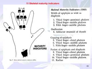
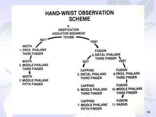
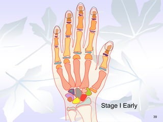
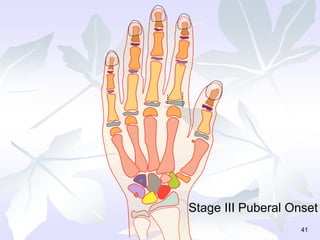
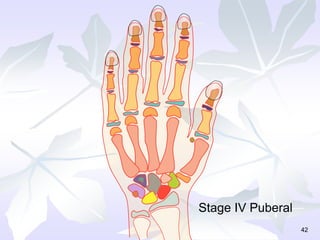
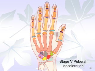
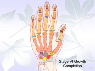
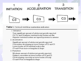
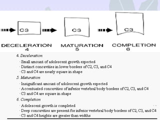
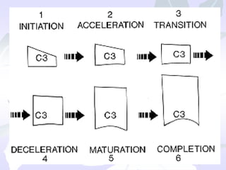
SKELETAL MATURITY
INDICATORS
Dr.SURESH KUMAR.K
Dept of Orthodontics and Dentofacial
Orthopedics
S.B. Patil intitute for Dental Sciences and
Research
Contents
• Introduction
• Skeletal age assessment methods
I) Hand wrist X-rays
• - Anatomy of the hand wrist
• - Methods of assessment
Greulich and Pyle
Grave, Brown and Bjork method
Fishman’s method
Hagg and Taranger’s method
Julian Singer’s method
II) Tooth mineralization ( canine root calcification)
III) Cervical vertebrae as maturity indicators
• Anatomy of the cervical vertebrae
• Advantages
• Disadvantages
• Methods of evaluating cervical vertebral
maturity
Introduction
• Health care professionals including
orthodontists are extremely interested in
predicting the adolescent growth spurt for
treatment planning and timing purposes.
• The issue of optimal treatment timing for
dentofacial orthopedics is linked intimately
to the identification of periods of acclerated
or intense growth (pubertal growth spurt)
that can contribute significantly to the
correction of skeletal imbalances in the
individual patient.
Introduction
• The greatest response to functional jaw
orthopedics tends to occur during the circum
pubertal growth period.
• Thus, the use of a reliable biologic indicator
aimed to detect the pubertal growth spurt in
mandibular growth represents a crucial
diagnostic tool for a rational treatment
planning especially in class II skeletal pattern
with mandibular deficiencies.
• Adolescence
It is the transitional period b/w juvenile
stage and adulthood during which the
secondary sexual characteristics appear,
the adolescent growth spurt takes place,
fertility is attained, and profound
physiologic changes occur.(PROFFIT)
- Important period
- Treatment planning & outcome of
treatment 5
Growth & Development –
Not uniform.
Periods of acceleration and deceleration.
Developmental status of the child judged
by :
i. peak height velocity
ii. menarche in case of girls
iii. voice change in boys
iv. dental development
v. skeletal ossification 6
• A spurt is defined as growth acceleration
up to a maximum where the annual
increment of growth exceeded the
previous one by at least 7mm.
-Erkstrom.
7
Growth spurts
Normal growth spurts
infantile spurt – 3years
 juvenile spurt – 7-8 years females,
8-10 years males.
Pubertal spurt – 10-13 years females,
13-15 years males.
8
9
Best indicator for the
pubertal growth spurt is
the attainment of the
peak height velocity

The clinician prefers to
start the treatment during
the acceleration phase
Earlier in girls
10
11
Assessment Of Amount Of Growth
• Various skeletal maturity indicators are
– Hand wrist radiograph.
– Cervical vertebrae.
– Canine calcification.
– Corpus index.
– Hyoid bone position.
– Third molar level.
– Frontal sinus.
– Antigonial notch.
Hand Wrist Radiographs:-
• Bone age / skeletal age presents an
indication of the physical development and
maturation of the skeleton.
• The hand, the wrist and distal epiphysis of
the Radius and Ulna present a large number
of secondary centres of ossification and
show a specific pattern of appearance,
ossification and union .Some of the centres
are present at birth.
Best indicator for the pubertal growth spurt
is the attainment of the peak height
velocity
The clinician prefers to start the treatment
during the acceleration phase
13
Hand Wrist Radiographs:-
• Assessment of bone age should be made for
boys and girls separately based on the
standards.
• Generally girls attain skeletal maturity faster.
• If the male bone age is 7 years the
equivalent female bone age is about 5-6
years.
Anatomy of the hand wrist:-
The hand wrist region is
made of 4 groups of
bones
• I) Distal ends of long
bones of the forearm
• II) Carpals
• III) Metacarpals
• IV) Phalanges
Anatomy of the hand wrist
Carpal bones:
• Were first named by Lyser in 1683. They are
short spongy bones enclosed in a thin layer
of compact bone. The 8 carpal bones are
arranged in 2 rows- proximal and distal.
• Each of the 8 carpal bones ossify from one
primary centre which appears in a
predictable pattern.
• Important bones: Hamate
Anatomy of the hand wrist
Metacarpals:
• They are 5 miniature long bones forming the
skeletal framework of the palm of the hand
(numbered from 1-5).
• Each metacarpal ossifies from one primary
centre in its shaft and a secondary centre on
the distal end (except for the first metacarpal
) where it appears at the proximal end.
Anatomy of the hand wrist
Phalanges
• They are small bones forming the fingers.
They are 3 in number in each finger except in
the thumb where only 2 phalanges are
present.
Anatomy of the hand wrist
Sesamoid bone
• Is a small nodular bone most often present
embedded in the tendons in the region of
thumb. Metacarpal I as well as V may also be
frequently associated with Sesamoids.
Methods used to assess
skeletal maturity using hand
wrist X rays:
Methods
• 1) Atlas method by Greulich and Pyle
• 2) Bjork, Grave and Brown method
• 3) Fishman’s skeletal maturity indicator
• 4) Hagg and Taranger method
• 5) Julian Singer’s method
Methods used to assess skeletal
maturity using hand wrist X rays:
• 1) Greulich and Pyle - published an atlas
containing ideal skeletal age pictures of
the hand and wrist for different
chronological ages and for each sex. The
patient’s radiograph is matched on an
overall basis with one of the photographs
in the atlas.
Greulich and Pyle method
24
2 ½ years 6 ½ years 11 years 19 years
HAND WRIST RADIOGRAPH
• All these methods divide skeletal
development into different stages. Each
of the stages represent a level of
skeletal maturity. Accordingly, the
skeletal age of the individual is
interpreted.
• The important osseous indicators are-
The important osseous indicators are-
 First signs of mineralization of the adductor
sesamoid bone – reached shortly before or at the
pubertal growth spurt.
 Capping of the middle phalanx of the third finger (MP3)-
marks the peak of the pubertal growth spurt.
 Fusion of the epiphysis & diaphysis of the distal
phalanx of the third finger (DP3)- marks the end of
pubertal growth.
 Complete union of the epiphysis & diaphysis of the
radius- skeletal growth is finished.
 First signs of
mineralization of the
adductor sesamoid
bone – reached
shortly before or at the
pubertal growth spurt.
The important osseous indicators are-
 Capping of the
middle phalanx of the
third finger (MP3)-
marks the peak of the
pubertal growth spurt
The important osseous indicators are-
 Fusion of the
epiphysis & diaphysis
of the distal phalanx of
the third finger (DP3)-
marks the end of
pubertal growth
The important osseous indicators are-
 Complete union of
the epiphysis &
diaphysis of the
radius- skeletal
growth is finished.
The important osseous indicators are-
Four stages of maturation
31
32
11 discrete adolescent
skeletal maturational
indicators (SMI’s ),
covering the entire
period of adolescent
development are found
on these six sites
33
11 Skeletal maturity indicators
34
Hagg &Taranger method
• F-onset
• FG-acceleration
• G- peak of the curve.
• H-deceleration
• I-End
35
Clinically important findings
• S- attained during acceleration period of
pubertal growth spurt
• MP3 – F – attained before onset by 40%
• MP3FG – 1 yr before or at PHV by 90%
• MP3G- at or one yr after PHV by 90%
• MP3H after PHV but before end by all
boys and 90% girls
• MP3I before or at end in all subjects
36
• DP3I – during the deceleration period of
pubertal growth spurt
• RADIUS
• I - 1 yr before or at end by about 80% of
girls and 90% of boys
• IJ & J not attained before End by any
subject
37
38
39
Stage I Early
40
Stage II Pre Puberal
41
Stage III Puberal Onset
42
Stage IV Puberal
43
Stage V Puberal
deceleration
44
Stage VI Growth
Completion
Tooth mineralization (mandibular
canine root calcification) as an
indicator of pubertal growth spurt.
• The uniformity of the rate of
development of mandibular canine and
lack of sexual differences in its rate of
calcification was the reason to choose
the mandibular canine as the indicator.
• There was also no superimposition of
the palatal shelves over the developing
root of mandibular canine unlike the
maxillary canines.
• Completion of root formation prior to
apical closure was closely related to
the appearance of other maturational
indicators.
The stages of Dermijian in relation to
ossification events in the hand wrist region
are described below
I . Stage F
• Walls of the pulp chamber
form an isosceles triangle.
• Root length equals or greater
than crown height.
• Corresponds to
1) Absence of adductor
sesamoid
2) MP3, MP5- Epiphysis equal to
in length to diaphysis
• Suggests- Initiation of growth
spurt
II) Stage G:
• Walls of the root canal
are parallel
• Apical end is partially
open
• Corresponds to
1) Presence of adductor
sesamoid
2) Capping of diaphysis
by epiphysis in Mp3,
Dp3,and Pp5
• Suggests :
• Peak height velocity
(PHV)
• (0.4years before PHV
for girls and 1.3years
before PHV for boys).
III. Stage H
• Root apex is
closed
• Corresponds to :
Fusion of
epiphysis with
their respective
diaphysis
• Suggests : End of
growth
Cervical vertebrae as skeletal
maturity indicators
Cervical vertebrae as skeletal
maturity indicators
• The cervical vertebrae are invaluable in
assessing skeletal maturity in that the
ossification events in the cervical
vertebrae begin during fetal life and
continue until adulthood. The
maturational changes can be observed
in the vertebrae during this entire
interval, which covers the period when
orthodontic/ orthopedic treatment is
typically performed in growing patients.
Anatomy of cervical vertebrae:
Cervical vertebrae
• They are the smallest of the moveable
vertebrae and can be distinguished
from the other vertebrae by the
presence of the thoracic and lumbar
transverse process. The first, second
and seventh cervical vertebrae have
certain characteristic features. The
third, fourth, fifth, and sixth vertebrae
are almost identical and can be
described as typical cervical vertebrae.
1)Lamparski
2)Hassel and Farman
3) Bacetti, Franchi and Mac Namara
Methods of evaluating
cervical vertebral
maturity
58
MATURATION
5-10%
C2
C3
C4
TRANSITION
25-65%
C2
C3
C4
Assessment Of Cervical Vertebrae
INITIATION
80-100%
C2
C3
C4
ACCELERATION
65-85%
C2
C3
C4
DECELERATION
10-25%
C2
C3
C4
COMPLETION
0%
C2
C3
C4
Advantages of using cervical
vertebrae as a maturity indicators:
• 1) There is no need for an additional
radiograph- can be visualized in the
routine lateral cephalogram.
 2) C2, C3, C4 are visible even if a
thyroid protective collar is worn.
Advantages of using cervical vertebrae as a
maturity indicators:
• 3) The ossification events begin during
fetal life and continue till adulthood
covering the period when orthopedic
treatment is instituted.
 4)Early detection of any abnormalities of
spine is possible by the Orthodontist and t
patient can be referred to the concerned
specialist.
Disadvantages of cervical vertebral
maturity indicators:
• 1) The maturity indicators of the cervical
vertebrae undergo very subtle changes
through time and are not as easily visualized
as those of the fingers seen in a hand wrist -
ray.
 2) Improper position of the neck from not
having an upright position during taking the
radiograph makes visualization very difficult.
Thank you
62

Skeletal maturity.ppt

  • 1.
    SKELETAL MATURITY INDICATORS Dr.SURESH KUMAR.K Deptof Orthodontics and Dentofacial Orthopedics S.B. Patil intitute for Dental Sciences and Research
  • 2.
    Contents • Introduction • Skeletalage assessment methods I) Hand wrist X-rays • - Anatomy of the hand wrist • - Methods of assessment Greulich and Pyle Grave, Brown and Bjork method Fishman’s method Hagg and Taranger’s method Julian Singer’s method II) Tooth mineralization ( canine root calcification) III) Cervical vertebrae as maturity indicators • Anatomy of the cervical vertebrae • Advantages • Disadvantages • Methods of evaluating cervical vertebral maturity
  • 3.
    Introduction • Health careprofessionals including orthodontists are extremely interested in predicting the adolescent growth spurt for treatment planning and timing purposes. • The issue of optimal treatment timing for dentofacial orthopedics is linked intimately to the identification of periods of acclerated or intense growth (pubertal growth spurt) that can contribute significantly to the correction of skeletal imbalances in the individual patient.
  • 4.
    Introduction • The greatestresponse to functional jaw orthopedics tends to occur during the circum pubertal growth period. • Thus, the use of a reliable biologic indicator aimed to detect the pubertal growth spurt in mandibular growth represents a crucial diagnostic tool for a rational treatment planning especially in class II skeletal pattern with mandibular deficiencies.
  • 5.
    • Adolescence It isthe transitional period b/w juvenile stage and adulthood during which the secondary sexual characteristics appear, the adolescent growth spurt takes place, fertility is attained, and profound physiologic changes occur.(PROFFIT) - Important period - Treatment planning & outcome of treatment 5
  • 6.
    Growth & Development– Not uniform. Periods of acceleration and deceleration. Developmental status of the child judged by : i. peak height velocity ii. menarche in case of girls iii. voice change in boys iv. dental development v. skeletal ossification 6
  • 7.
    • A spurtis defined as growth acceleration up to a maximum where the annual increment of growth exceeded the previous one by at least 7mm. -Erkstrom. 7 Growth spurts
  • 8.
    Normal growth spurts infantilespurt – 3years  juvenile spurt – 7-8 years females, 8-10 years males. Pubertal spurt – 10-13 years females, 13-15 years males. 8
  • 9.
    9 Best indicator forthe pubertal growth spurt is the attainment of the peak height velocity  The clinician prefers to start the treatment during the acceleration phase Earlier in girls
  • 10.
  • 11.
    11 Assessment Of AmountOf Growth • Various skeletal maturity indicators are – Hand wrist radiograph. – Cervical vertebrae. – Canine calcification. – Corpus index. – Hyoid bone position. – Third molar level. – Frontal sinus. – Antigonial notch.
  • 12.
    Hand Wrist Radiographs:- •Bone age / skeletal age presents an indication of the physical development and maturation of the skeleton. • The hand, the wrist and distal epiphysis of the Radius and Ulna present a large number of secondary centres of ossification and show a specific pattern of appearance, ossification and union .Some of the centres are present at birth.
  • 13.
    Best indicator forthe pubertal growth spurt is the attainment of the peak height velocity The clinician prefers to start the treatment during the acceleration phase 13
  • 14.
    Hand Wrist Radiographs:- •Assessment of bone age should be made for boys and girls separately based on the standards. • Generally girls attain skeletal maturity faster. • If the male bone age is 7 years the equivalent female bone age is about 5-6 years.
  • 15.
    Anatomy of thehand wrist:- The hand wrist region is made of 4 groups of bones • I) Distal ends of long bones of the forearm • II) Carpals • III) Metacarpals • IV) Phalanges
  • 16.
    Anatomy of thehand wrist Carpal bones: • Were first named by Lyser in 1683. They are short spongy bones enclosed in a thin layer of compact bone. The 8 carpal bones are arranged in 2 rows- proximal and distal. • Each of the 8 carpal bones ossify from one primary centre which appears in a predictable pattern. • Important bones: Hamate
  • 17.
    Anatomy of thehand wrist Metacarpals: • They are 5 miniature long bones forming the skeletal framework of the palm of the hand (numbered from 1-5). • Each metacarpal ossifies from one primary centre in its shaft and a secondary centre on the distal end (except for the first metacarpal ) where it appears at the proximal end.
  • 18.
    Anatomy of thehand wrist Phalanges • They are small bones forming the fingers. They are 3 in number in each finger except in the thumb where only 2 phalanges are present.
  • 19.
    Anatomy of thehand wrist Sesamoid bone • Is a small nodular bone most often present embedded in the tendons in the region of thumb. Metacarpal I as well as V may also be frequently associated with Sesamoids.
  • 20.
    Methods used toassess skeletal maturity using hand wrist X rays:
  • 21.
    Methods • 1) Atlasmethod by Greulich and Pyle • 2) Bjork, Grave and Brown method • 3) Fishman’s skeletal maturity indicator • 4) Hagg and Taranger method • 5) Julian Singer’s method
  • 22.
    Methods used toassess skeletal maturity using hand wrist X rays: • 1) Greulich and Pyle - published an atlas containing ideal skeletal age pictures of the hand and wrist for different chronological ages and for each sex. The patient’s radiograph is matched on an overall basis with one of the photographs in the atlas.
  • 23.
  • 24.
    24 2 ½ years6 ½ years 11 years 19 years HAND WRIST RADIOGRAPH
  • 25.
    • All thesemethods divide skeletal development into different stages. Each of the stages represent a level of skeletal maturity. Accordingly, the skeletal age of the individual is interpreted. • The important osseous indicators are-
  • 26.
    The important osseousindicators are-  First signs of mineralization of the adductor sesamoid bone – reached shortly before or at the pubertal growth spurt.  Capping of the middle phalanx of the third finger (MP3)- marks the peak of the pubertal growth spurt.  Fusion of the epiphysis & diaphysis of the distal phalanx of the third finger (DP3)- marks the end of pubertal growth.  Complete union of the epiphysis & diaphysis of the radius- skeletal growth is finished.
  • 27.
     First signsof mineralization of the adductor sesamoid bone – reached shortly before or at the pubertal growth spurt. The important osseous indicators are-
  • 28.
     Capping ofthe middle phalanx of the third finger (MP3)- marks the peak of the pubertal growth spurt The important osseous indicators are-
  • 29.
     Fusion ofthe epiphysis & diaphysis of the distal phalanx of the third finger (DP3)- marks the end of pubertal growth The important osseous indicators are-
  • 30.
     Complete unionof the epiphysis & diaphysis of the radius- skeletal growth is finished. The important osseous indicators are-
  • 31.
    Four stages ofmaturation 31
  • 32.
    32 11 discrete adolescent skeletalmaturational indicators (SMI’s ), covering the entire period of adolescent development are found on these six sites
  • 33.
  • 34.
  • 35.
    Hagg &Taranger method •F-onset • FG-acceleration • G- peak of the curve. • H-deceleration • I-End 35
  • 36.
    Clinically important findings •S- attained during acceleration period of pubertal growth spurt • MP3 – F – attained before onset by 40% • MP3FG – 1 yr before or at PHV by 90% • MP3G- at or one yr after PHV by 90% • MP3H after PHV but before end by all boys and 90% girls • MP3I before or at end in all subjects 36
  • 37.
    • DP3I –during the deceleration period of pubertal growth spurt • RADIUS • I - 1 yr before or at end by about 80% of girls and 90% of boys • IJ & J not attained before End by any subject 37
  • 38.
  • 39.
  • 40.
  • 41.
  • 42.
  • 43.
  • 44.
  • 45.
    Tooth mineralization (mandibular canineroot calcification) as an indicator of pubertal growth spurt.
  • 46.
    • The uniformityof the rate of development of mandibular canine and lack of sexual differences in its rate of calcification was the reason to choose the mandibular canine as the indicator. • There was also no superimposition of the palatal shelves over the developing root of mandibular canine unlike the maxillary canines. • Completion of root formation prior to apical closure was closely related to the appearance of other maturational indicators.
  • 47.
    The stages ofDermijian in relation to ossification events in the hand wrist region are described below I . Stage F • Walls of the pulp chamber form an isosceles triangle. • Root length equals or greater than crown height. • Corresponds to 1) Absence of adductor sesamoid 2) MP3, MP5- Epiphysis equal to in length to diaphysis • Suggests- Initiation of growth spurt
  • 48.
    II) Stage G: •Walls of the root canal are parallel • Apical end is partially open • Corresponds to 1) Presence of adductor sesamoid 2) Capping of diaphysis by epiphysis in Mp3, Dp3,and Pp5 • Suggests : • Peak height velocity (PHV) • (0.4years before PHV for girls and 1.3years before PHV for boys).
  • 49.
    III. Stage H •Root apex is closed • Corresponds to : Fusion of epiphysis with their respective diaphysis • Suggests : End of growth
  • 50.
    Cervical vertebrae asskeletal maturity indicators
  • 51.
    Cervical vertebrae asskeletal maturity indicators • The cervical vertebrae are invaluable in assessing skeletal maturity in that the ossification events in the cervical vertebrae begin during fetal life and continue until adulthood. The maturational changes can be observed in the vertebrae during this entire interval, which covers the period when orthodontic/ orthopedic treatment is typically performed in growing patients.
  • 52.
    Anatomy of cervicalvertebrae: Cervical vertebrae • They are the smallest of the moveable vertebrae and can be distinguished from the other vertebrae by the presence of the thoracic and lumbar transverse process. The first, second and seventh cervical vertebrae have certain characteristic features. The third, fourth, fifth, and sixth vertebrae are almost identical and can be described as typical cervical vertebrae.
  • 54.
    1)Lamparski 2)Hassel and Farman 3)Bacetti, Franchi and Mac Namara Methods of evaluating cervical vertebral maturity
  • 58.
    58 MATURATION 5-10% C2 C3 C4 TRANSITION 25-65% C2 C3 C4 Assessment Of CervicalVertebrae INITIATION 80-100% C2 C3 C4 ACCELERATION 65-85% C2 C3 C4 DECELERATION 10-25% C2 C3 C4 COMPLETION 0% C2 C3 C4
  • 59.
    Advantages of usingcervical vertebrae as a maturity indicators: • 1) There is no need for an additional radiograph- can be visualized in the routine lateral cephalogram.  2) C2, C3, C4 are visible even if a thyroid protective collar is worn.
  • 60.
    Advantages of usingcervical vertebrae as a maturity indicators: • 3) The ossification events begin during fetal life and continue till adulthood covering the period when orthopedic treatment is instituted.  4)Early detection of any abnormalities of spine is possible by the Orthodontist and t patient can be referred to the concerned specialist.
  • 61.
    Disadvantages of cervicalvertebral maturity indicators: • 1) The maturity indicators of the cervical vertebrae undergo very subtle changes through time and are not as easily visualized as those of the fingers seen in a hand wrist - ray.  2) Improper position of the neck from not having an upright position during taking the radiograph makes visualization very difficult.
  • 62.